Abstract
Systemic administration of chemotherapy for cancer often has toxic side effects, limiting the doses that can be used in its treatment. In this study, we developed methoxy poly(ethylene glycol)-poly(caprolactone) (MPEG-PCL) micelles loaded with curcumin and doxorubicin (Cur-Dox/MPEG-PCL) that were tolerated by recipient mice and had enhanced antitumor effects and fewer side effects. It was shown that these Cur-Dox/MPEG-PCL micelles could release curcumin and doxorubicin slowly in vitro. The long circulation time of MPEG-PCL micelles and the slow rate of release of curcumin and doxorubicin in vivo may help to maintain plasma concentrations of active drug. We also demonstrated that Cur-Dox/MPEG-PCL had improved antitumor effects both in vivo and in vitro. The mechanism by which Cur-Dox/MPEG-PCL micelles inhibit lung cancer might involve increased apoptosis of tumor cells and inhibition of tumor angiogenesis. We found advantages using Cur-Dox/MPEG-PCL micelles in the treatment of cancer, with Cur-Dox/MPEG-PCL achieving better inhibition of LL/2 lung cancer growth in vivo and in vitro. Our study indicates that Cur-Dox/MPEG-PCL micelles may be an effective treatment strategy for cancer in the future.
Introduction
Chemotherapy is the usual treatment strategy for cancer, and is often followed by radiation therapy or surgery to remove residual tumor tissue. Chemotherapy is administered systemically and often causes side effects, including pain, nausea and vomiting, fatigue, mouth ulcers, nerve damage, and skin reactions. These side effects can limit the doses of chemotherapy that can be given, which prevents patients from deriving optimal benefits from treatment. Therefore, reducing the side effects of chemotherapy has become a focus in the management of cancer.
Natural herbs are sometimes used in combination with conventional medication to help reduce the side effects of chemotherapy. Herbal remedies have been known to support the body holistically, strengthen the immune system, and help to maintain blood cell counts. Curcumin, a chemical constituent of turmeric, has been used in the treatment of inflammatory disorders and cancerCitation1–Citation4 for many years. There is some evidence suggesting that curcumin is an ideal chemosensitizer for chemotherapy and that it helps to protect patients from the side effects of treatment.Citation5–Citation10 Curcumin is a highly effective scavenger of reactive oxygen species and also inhibits the c-Jun NH2-terminal kinase pathway.Citation11 Both reactive oxygen species and activation of the c-Jun NH2-terminal kinase pathway are crucial elements in the success of chemotherapy.Citation7 Curcumin may inhibit tumor growth via multiple mechanisms, including antitumor angiogenesis,Citation12,Citation13 suppression of proliferation,Citation14,Citation15 induction of apoptosis,Citation8,Citation16 and prevention of metastasis.Citation17,Citation18 However, the clinical applications of curcumin remain limited because of its short biological half-life, poor solubility resulting in poor absorption, and low bioavailability via the oral route.Citation19–Citation21 Development of an intravenous preparation is a promising approach to resolving these issues in the application of curcumin.Citation22
In recent decades, many novel chemotherapeutic formulations have been developed. These formulations contain chemotherapy inside the vehicle, resulting in less toxicity and better drug penetration into tumor tissue. Biodegradable polymeric nanoparticles are often used to achieve controlled release of drugs in advanced anticancer drug delivery systems.Citation23–Citation26 Also, some biodegradable polymer-derived drug delivery systems, such as nanoparticles delivering anticancer agents, are commercially available.Citation27 Poly(caprolactone)-poly(ethylene glycol) (PCL-PEG) copolymers are biodegradable, amphiphilic, and easy to produce, and have potential application in drug delivery systems.Citation28,Citation29
Previously, many efforts have been made using nanotechnology to overcome the poor solubility of curcumin in water, including liposomal curcumin,Citation30 a PEG-curcumin conjugate,Citation31 and PCL-PEG-PCL nanofibers or micelles encapsulating curcumin.Citation32,Citation33 Recently, a novel formulation of curcumin-loaded methoxy poly(ethylene glycol) (MPEG)-PCL nanoparticles was developed in our laboratory.Citation29 We hypothesized that encapsulating curcumin in MPEG-PCL micelles might be a promising method by which to develop an improved formulation of curcumin. In this study, we loaded both curcumin and doxorubicin into MPEG-PCL (Cur-Dox/MPEG-PCL) micelles and investigated the antitumor activity of systemically administered Cur-Dox/MPEG-PCL in a mouse model of lung cancer. Our results indicate that this compound can be tolerated by recipient mice and that it has enhanced antitumor activity and fewer side effects in vivo, with potential clinical applications.
Materials and methods
Materials
Curcumin, doxorubicin, alginate, fluorescein isothiocyanate (FITC)-dextran, Dulbecco’s Modified Eagle’s Medium (DMEM) (Gibco BRL, Grand Island, NY, USA), and 3-(4,5-dimethylthiazol-2-yl)-2,5-diphenyl tetrazolium bromide (MTT) were purchased from Sigma (St Louis, MO, USA). Methanol and acetic acid (high-pressure liquid chromatography grade) were purchased from Fisher Scientific UK Ltd (Loughborough, UK). Dimethyl sulfoxide and acetone were purchased from Liaoning Kelong Fine Chemical Co., Ltd. (Liaoning, People’s Republic of China). A MPEG-PCL diblock copolymer designed to have a molecular weight of 4000 was synthesized according to our previous reports.Citation28,Citation34 The molecular number of MPEG-PCL copolymer was 4050 (determined by 1H-nuclear magnetic resonance, data not shown).
Tg(flk:EGFP) transgenic zebrafish were provided by Dr Shuo Lin (University of California, Los Angeles, CA, USA). Female BALB/c mice (aged 6–8 weeks) were purchased from the Laboratory Animal Center of Sichuan University (Chengdu, People’s Republic of China). All studies involving mice were approved by the institute’s animal care and use committee.
Preparation of Cur-Dox/MPEG-PCL micelles
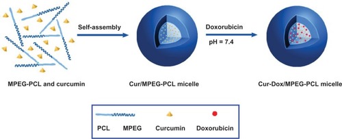
Cur-Dox/MPEG-PCL micelles were prepared in two stages. First, Cur/MPEG-PCL was prepared by a self-assembly method. Briefly, 5 mg of curcumin and 90 mg of MPEG-PCL diblock copolymer were dissolved in 2 mL of acetone solution, followed by evaporation under reduced pressure in a rotary evaporator at 55°C. Normal saline was then added to the above polymer and drug mixture, allowing self-assembly of curcumin and MPEG-PCL, creating core-shell-structured Cur/MPEG-PCL micelles with core-encapsulated curcumin. Cur-Dox/MPEG-PCL micelles were then prepared using a pH-induced self-assembly method. Briefly, 1.4 mL of Cur/MPEG-PCL micelles prepared in the previous step was placed into a tube, and 0.2 mL of phosphate-buffered solution (10×, pH 7.4) was added to this solution under stirring. Next, 0.4 mL of doxorubicin aqueous solution (5 mg/mL) was added to the above solution. Because doxorubicin has low solubility in phosphate-buffered solution at pH 7.4, it was able to self-assemble into the hydrophobic core of the MPEG-PCL micelle. Twenty minutes later, Cur-Dox/MPEG-PCL micelles were obtained. The Cur/MPEG-PCL, Dox/MPEG-PCL, and empty MPEG-PCL micelle formulations were prepared in the same way as the Cur-Dox/MPEG-PCL micelles, but without curcumin or doxorubicin in the mixtures.
Characterization of Cur-Dox/MPEG-PCL micelles
The particle size and zeta potential of the Cur-Dox/MPEG-PCL micelles was determined by dynamic light scattering (Nano-ZS 90, Malvern, Worcestershire, UK). The temperature was kept at 25°C during the measurement process. All results were the mean of three test runs. The morphology of the Cur-Dox/MPEG-PCL micelles was observed under a transmission electron microscope (H-6009IV, Hitachi, Tokyo, Japan), for which the micelles were diluted with distilled water and placed on a copper grid covered with nitrocellulose. The samples were negatively stained with phosphotungstic acid and dried at room temperature.
In vitro release study
To determine the release kinetics of curcumin from Cur/MPEG-PCL and Cur-Dox/MPEG-PCL micelles and those of doxorubicin from Dox/MPEG-PCL and Cur-Dox/MPEG-PCL micelles, 0.5 mL of Cur/MPEG-PCL, Dox/MPEG-PCL, or Cur-Dox/MPEG-PCL micelle solution was placed in a dialysis bag (molecular weight cutoff, 3.5 kDa) which was incubated in 30 mL of phosphate-buffered solution (pH 7.4) containing Tween 80 (0.5% w/w) at 37°C with gentle shaking. At predetermined time points, the spent incubation medium was replaced with fresh medium.
Cell culture
LL/2 and MS1 cell lines (American Type Culture Collection, Manassas, VA, USA) were cultured in DMEM supplemented with 10% heat-inactivated fetal bovine serum and 100 μg/mL amikacin, and maintained in a humidified chamber at 37°C in an atmosphere containing 5% CO2.
Cytotoxicity assay
To compare the effects of Cur-Dox/MPEG-PCL, Cur/MPEG-PCL, Dox/MPEG-PCL, and empty MPEG-PCL micelles on tumor cell viability, an LL/2 cell line (5 × 10³ cells/well) was plated in 96-well plates and incubated in DMEM supplemented with 10% fetal bovine serum. After 24 hours, the cells were washed with serum free medium and treated with various concentrations of the trial preparations in DMEM medium. Cell viability in response to treatment was determined using the MTT method after 24 or 48 hours of incubation in medium. Viability was measured as the percentage compared with untreated cells (showing 100% survival). The experiment was performed in triplicate, repeated at least twice, and relative cell viability was calculated by dividing the absorbance of treated cells by the absorbance of control cells. The surviving cells converted MTT to formazan that generates a blue-purple color when dissolved in dimethyl sulfoxide. The color intensity was measured at 570 nm using a plate reader (OptiMax™, Molecular Devices, San Jose, CA, USA). These experiments were repeated at least three times, and the data are expressed as the mean ± standard deviation.
Cell apoptosis assay
The extent of apoptosis in LL/2 cells was evaluated by flow cytometry (ESP Elite, Beckman-Coulter, Miami, FL, USA) using FITC-conjugated Annexin V-propidium iodide (BD PharMingen, CA, USA) staining as per the manufacturer’s instructions, as previously described.Citation35 Both early apoptotic (Annexin V-positive, propidium iodide-negative) and late apoptotic (Annexin V-positive and propidium iodide-positive) cells were included in the determinations of cell death.
Antiangiogenic activity of Cur-Dox/MPEG-PCL
A transgenic Tg(flk: EGFP) zebrafish line was raised by standard methods as previously described.Citation36 After transplantation, the embryos were put into 24-well plates (4–5 embryos per well) in 1 mL of Holtfreter’s solution (15 mM NaCl. 0.6 mM NaHCO3. 0.2 mM KCl. 0.2 mM CaCl2. 0.2 mM MgSO4⋅ 7H20), and maintained at 28°C. Forty-eight hours later, the embryos were treated with Holtfreter’s solution (control) or Cur-Dox/MPEG-PCL micelles (curcumin and doxorubicin 5 mg/mL) for 72 hours. Blood vessels from the zebrafish were then examined using a Zeiss M2Bio fluorescence microscope (Carl Zeiss Microimaging Inc, Thornwood NY, USA).
In vivo tumor model
Female C57 mice (aged 6–8 weeks) were housed in groups containing five mice each in our animal research laboratory under standard conditions in top-filtered cages. All animals were fed a regular diet and given acidified water without antibiotics. The mice were injected subcutaneously with 100 μL of LL/2 cell suspension (1 × 10⁶) into the right flank. When the mean tumor diameter reached about 6 mm, the tumor-bearing mice were randomly divided into five groups and received their assigned treatment via intravenous tail injection. The first group of mice were treated with normal saline, the second with empty MPEG-PCL nanoparticles, the third with Cur/MPEG-PCL nanoparticles (5 mg/kg), the fourth with Dox/MPEG-PCL nanoparticles (5 mg/kg), and the fifth with Cur-Dox/MPEG-PCL (curcumin to doxorubicin ratio 1:1, 5 mg/kg) every five days, for a total of three treatments. The tumor volume was measured every two days. When the control mice became weak, they were euthanized by dislocation of the cervical vertebra, and their tumors were harvested immediately and measured.
TUNEL and CD31 assays
The LL2 tumors were fixed in 4% paraformaldehyde and phosphate-buffered solution for at least 24 hours, placed in 70% ethanol overnight, and then embedded in paraffin. Sections 3–5 μm thick were cut and mounted. A commercially available TUNEL kit (Promega, Madison, WI, USA) was used to identify apoptotic cells in the tumors. This analysis was performed following the manufacturer’s protocol, and the samples were then examined using a fluorescence microscope (×400).
For blood vessel staining, the tumors were stored at −80°C to examine microvessel expression, immunostained with epithelial cell marker goat antimouse CD31 antibody (dilution 1:100; Santa Cruz Biotechnology, Santa Cruz, CA, USA) overnight at 4°C, after which rabbit-antigoat FITC (dilution 1:100; Santa Cruz Biotechnology) was added and the specimens were left in a humidified chamber protected from light at 37°C for one hour. Microvessel density was determined as the average number of small CD31-positive vessels in a high-power (×400) field.
Statistical analysis
All data were evaluated using the two-tailed unpaired Student’s t-test or compared by one-way analysis of variance, and are expressed as the mean ± standard deviation. P < 0.05 was considered to be statistically significant.
Results
Preparation and characterization of Cur-Dox/MPEG-PCL micelles
In order to improve the water solubility of curcumin, we used MPEG-PCL to codeliver curcumin and doxorubicin. Cur-Dox/MPEG-PCL micelles were prepared in two steps, as shown in . First, Cur-Dox/MPEG-PCL micelles were constructed using a self-assembly method. Briefly, curcumin and MPEG-PCL were dissolved in acetone. The organic solvent was then evaporated under reduced pressure in a rotary evaporator, forming a thin film of curcumin and MPEG-PCL mixture. Finally, distilled water was added to the mixture, allowing self-assembly of curcumin and MPEG-PCL. In the structure of MPEG-PCL, PEG is the hydrophilic segment and PCL is the hydrophobic segment, so the MPEG-PCL micelles always had a core-shell structure with a PCL core and a PEG shell. Self-assembly of curcumin and MPEG-PCL created core-shell Cur-Dox/MPEG-PCL micelles with curcumin encapsulated in the core.
Figure 1 Preparation scheme for doxorubicin-loaded MPEG-PCL nanoparticles. There were two steps involved in the preparation of Cur-Dox/MPEG-PCL. First, Cur/MPEG-PCL was prepared by a self-assembly method. Second, Cur-Dox/MPEG-PCL micelles were prepared by pH-induced self-assembly method.
Abbreviations: Cur, curcumin; Dox, doxorubicin; MPEG, methoxy poly(ethylene glycol); PCL, poly(caprolactone).
Next, the Cur-Dox/MPEG-PCL micelles were characterized in detail, and found to have a drug loading and encapsulation efficiency of 8% ± 0.1% and 98.7% ± 0.4%, respectively. shows that the freshly prepared Cur-Dox/MPEG-PCL micelles had a very narrow particle size distribution (polydispersity index 0.12) and a mean particle size of 38.4 ± 1.2 nm (determined by dynamic light scattering). shows that the Cur-Dox/MPEG-PCL micelles had the zeta potential of −0.269 mV. The morphology of these micelles was investigated by transmission electron microscopy, and the results are shown in , indicating that they were spherical with a mean diameter of approximately 27 nm. Transmission electron microscopy determines the size of dry particles, while dynamic light scattering determines the hydrodynamic diameter of particles in water. Because amphiphilic block polymeric micelles always have a loose structure in water, the particle size determined by dynamic light scattering is always slightly larger than that determined by transmission electron microscopy.
Figure 2 Characterization of Cur-Dox/MPEG-PCL micelles showing their particle size. The Cur-Dox/MPEG-PCL micelles had a very narrow particle size distribution (polydispersity index 0.12) with a mean particle size of 38.4 ± 1.2 nm, determined by dynamic light scattering (A). Cur-Dox/MPEG-PCL micelles with a zeta potential of −0.269 mV (B). Transmission electronic microscopic images of Cur-Dox/MPEG-PCL micelles showing a spherical morphology with a mean diameter of about 27 nm (C). Appearance of Dox/MPEG-PCL (D) and Cur/MPEG-PCL (E) micelles in aqueous solution. Appearance of Cur-Dox/MPEG-PCL, Dox/MPEG-PCL, and Cur/MPEG-PCL diluted in water (F). Cur-Dox/MPEG-PCL, Dox/MPEG-PCL, Cur/MPEG-PCL and water are shown from left to right. Data are shown as the mean ± standard.
Abbreviations: Cur, curcumin; Dox, doxorubicin; MPEG, methoxy poly(ethylene glycol); PCL, poly(caprolactone); NS, normal saline; M, MPEG-PCL; C, Cur/MPEG-PCL; D, Dox/MPEG-PCL; C+D: Cur-Dox/MPEG-PCL.
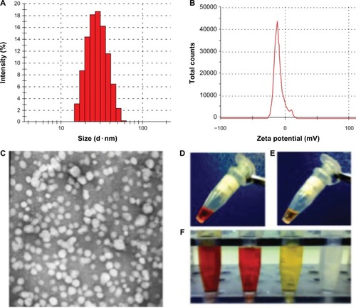
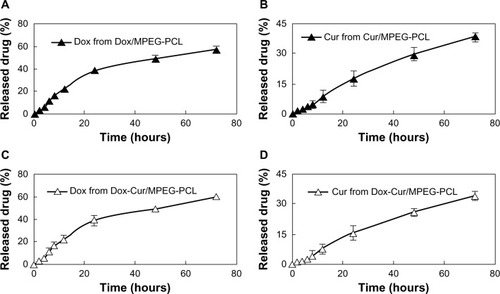
One of the main reasons for encapsulation of curcumin and doxorubicin in MPEG-PCL micelles was to render curcumin and doxorubicin completely dispersible in aqueous medium. The appearance of the Cur-Dox/MPEG-PCL micelles in aqueous solution is shown in –. Curcumin cannot be dissolved in pure water, as confirmed by the observation of a turbid yellow slurry. In contrast, the Cur-Dox/MPEG-PCL micelle solution containing equivalent amounts of curcumin and doxorubicin was transparent, indicating full dispersibility of curcumin and doxorubicin in water. The release profiles of the Cur/MPEG-PCL and Dox/MPEG-PCL micelles were studied in vitro using a dialysis method. As shown in , curcumin and doxorubicin were released from the Cur/MPEG-PCL, Dox/MPEG-PC, and Cur-Dox/MPEG-PCL micelles over an extended period.
Figure 3 Release profile of curcumin or doxorubicin micelles in vitro studied using a dialysis method. Doxorubicin was released from Dox/MPEG-PCL (A), curcumin was released from Cur/MPEG-PCL (B), doxorubicin was released from Cur-Dox/MPEG-PCL (C) and curcumin was released from Cur-Dox/MPEG-PCL (D) over an extended period. Data are shown as the mean ± standard error of the mean.
Abbreviations: Cur, curcumin; Dox, doxorubicin; MPEG, methoxy poly(ethylene glycol); PCL, poly(caprolactone).
Anticancer activity in vitro
In order to determine the synergistic antitumor effects of curcumin and doxorubicin, we used the MTT assay and flow cytometry to determine cell apoptosis. Using the MTT assay, we found that there was no difference between the curcumin alone, doxorubicin alone, and curcumin and doxorubicin in combination (curcumin to doxorubicin ratio 1:1) treatment groups at low concentrations. The combination group was more effective than the curcumin or doxorubicin alone groups at concentrations of 0.75 μg/mL, 1.5 μg/mL, and 3 μg/mL at 24 hours (), indicating that curcumin could act synergistically with doxorubicin in vitro.
Figure 4 Synergistic antitumor effect of curcumin and doxorubicin on an LL/2 tumor cell line. The MTT assay (A), showed that the combination group (ratio of curcumin to doxorubicin, 1:1) was more effective than curcumin and doxorubicin alone at concentrations of 0.75 μg/mL, 1.5 μg/mL, and 3 μg/mL at 24 hours. Flow cytometry determination of cell apoptosis (B) showed that the curcumin-induced apoptosis efficiency was 9.34%, doxorubicin-induced apoptosis was 10.55%, and apoptosis induced by combination therapy was 16.08% at a concentration of 0.75 μg/mL; the 1.5 μg/mL concentration of curcumin induced an apoptosis efficiency of 9.57%, the doxorubicin-induced apoptosis was 11.33%, and combination therapy induced apoptosis of 20.02%; at the 3 μg/mL concentration, curcumin-induced apoptosis efficiency was 11.54%, doxorubicin-induced apoptosis was 15.53%, and apoptosis induced by combination therapy was 29.13%. Data are shown as the mean ± standard error of the mean. *P < 0.05 between groups.
Abbreviations: Cur, curcumin; Dox, doxorubicin; MPEG, methoxy poly(ethylene glycol); PCL, poly(caprolactone); groups are as follow: NS, normal saline treated group; M, MPEG-PCL micelles treated group; C, Cur/MPEG-PCL micelles treated group; D, Dox/MPEG-PCL micelles treated group; C+D: Cur-Dox/MPEG-PCL micelles treated group.
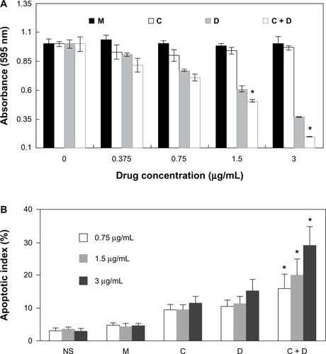
As shown in , curcumin induced 9.34% of cell apoptosis, doxorubicin induced 10.55%, and combination therapy induced 16.08% at a concentration of 0.75 μg/mL. At a curcumin concentration of 1.5 μg/mL, the efficiency of apoptosis was 9.57%, the apoptosis induced by doxorubicin was 11.33%, and that induced by combination therapy was 20.02%. At a curcumin concentration of 3 μg/mL, the efficiency of apoptosis was 11.54%, the apoptosis induced by doxorubicin was 15.53%, and that induced by combination therapy was 29.13%. These results indicate that doxorubicin and curcumin had a synergistic anticancer effect in vitro.
Inhibition of angiogenesis
Neovascularization is very important for tumor growth, making inhibition of vessel formation an excellent target for cancer therapy. We used the transgenic zebra fish model to determine the effect of Cur-Dox/MPEG-PCL on antiangiogenesis.
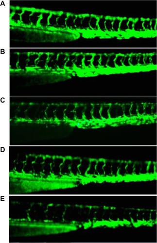
Transgenic Tg(flk: EGFP) zebra fish embryos were treated with Cur/MPEG-PCL, Dox/MPEG-PCL, Cur-Dox/MPEG-PCL micelles (5 mg/mL), or empty MPEG-PCL micelles for 72 hours. Microangiographic analysis was performed to assess the functional integrity of the vasculature of each embryo. shows that embryos treated with Cur-Dox/MPEG-PCL micelles had defective vascular formation of variable severity, with intersegmental vessels either sprouting abnormally or failing to form (arrows), which did not occur in control embryos. These findings confirm that Cur-Dox/MPEG-PCL micelles can inhibit angiogenesis in a zebra fish model.
Figure 5 Antiangiogenesis effect of Cur-Dox/MPEG-PCL. Normal embryos (A), embryos treated with empty MPEG-PCL (B), embryos treated with Cur/MPEG-PCL micelles (C), embryos treated with Dox/MPEG-PCL micelles (D), and embryos treated with Cur-Dox/MPEG-PCL micelles (E). Embryos treated with Cur-Dox/MPEG-PCL micelles showing defective vascular formation of variable severity, with intersegmental vessels either sprouting abnormally or failing to form in comparison with control embryos.
Abbreviations: Cur, curcumin; Dox, doxorubicin; MPEG, methoxy poly(ethylene glycol); PCL, poly(caprolactone).
Anticancer activity in vivo
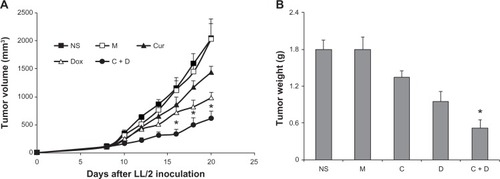
The ability of Cur/MPEG-PCL micelles to inhibit growth of LL/2 lung carcinoma in vivo was evaluated in a mouse model. Six groups of mice bearing LL/2 lung carcinoma were intravenously injected with normal saline, MPEG-PCL micelles, Cur/MPEG-PCL micelles (containing curcumin 5 mg/kg), Dox/MPEG-PCL micelles (containing doxorubicin 5 mg/kg), or Cur-Dox/MPEG-PCL micelles (curcumin and doxorubicin, 5 mg/kg) respectively.
The tumor growth curves for each group are shown in , with a representative tumor shown for each group. Tumors in the groups treated with Cur-Dox/MPEG-PCL were smaller than those receiving the other treatments. Moreover, the weight of the tumors () showed that Cur-Dox/MPEG-PCL was more effective than vehicle, curcumin, or doxorubicin at the same doses. These findings indicate that systemic application of Cur-Dox/MPEG-PCL micelles can inhibit the growth of subcutaneous LL/2 lung carcinoma in vivo and that curcumin and doxorubicin have synergistic effects in this regard.
Figure 6 Anticancer activity in vivo. A C57BL/6J mouse tumor model was established by subcutaneous injection with LL/2 cells. Mice were treated with normal saline (NS), MPEG-PCL (M) micelles, Cur/MPEG-PCL (Cur) micelles (curcumin 5 mg/kg), Dox/MPEG-PCL (Dox) micelles (doxorubicin 5 mg/kg), Cur-Dox/MPEG-PCL (C + D) micelles (curcumin and doxorubicin 5 mg/kg), respectively. A statistically significant difference was found in tumor volume (*P < 0.05) between the Cur-Dox/MPEG-PCL group and other treatments alone (A); the same result was found for tumor weights (B). Points, mean (n = 8); bars, standard deviation.
Abbreviations: Cur, curcumin; Dox, doxorubicin; MPEG, methoxy poly(ethylene glycol); PCL, poly(caprolactone).
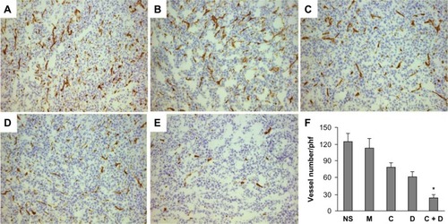
TUNEL assay and CD31 staining was used to study the mechanism associated with the anticancer activity of Cur-Dox/MPEG-PCL in vivo. As shown in , many strongly positive nuclei identified as apoptotic could be observed in tumor tissue treated with Cur-Dox/MPEG-PCL, whereas such nuclei were rare in the other groups. This implies that induction of apoptosis may be one possible mechanism by which Cur-Dox/MPEG-PCL inhibits lung cancer in vivo. As shown in , limited angiogenesis was observed in tumor tissue treated with Cur-Dox/MPEG-PCL, whereas much more angiogenesis was found in the other groups. This suggests that decreased vessel density in the tumors might also be responsible for the inhibition of lung cancer by Cur-Dox/MPEG-PCL in vivo.
Figure 7 Apoptosis of lung cancer cells was detected using TUNEL analysis. The percentage of apoptosis was determined by counting the number of apoptotic cells and dividing by the total number of cells in the field (five high power fields per slide). Normal saline group (A), MPEG-PCL group (B), Cur/MPEG-PCL group (C), Dox/MPEG-PCL group (D), Cur-Dox/MPEG-PCL group (E), and Percent apoptosis in each group (F). Treatment with Cur-Dox/MPEG-PCL resulted in significantly increased apoptosis compared with that of other groups. *P, 0.05. Bars indicate standard deviation; columns show the mean.
Abbreviations: Cur, curcumin; Dox, doxorubicin; MPEG, methoxy poly(ethylene glycol); PCL, poly(caprolactone); NS, normal saline; M, MPEG-PCL; C, Cur/MPEG-PCL; D, Dox/MPEG-PCL; C+D: Cur-Dox/MPEG-PCL.
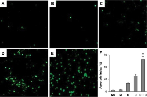
Figure 8 Normal saline group (A), MPEG-PCL group (B), Cur/MPEG-PCL group (C), Dox/MPEG-PCL group (D), Cur-Dox/MPEG-PCL group (E), and vessel density of tumor tissues in each group (F). *P<0.05. Bars indicate standard deviation; columns show the mean.
Abbreviations: Cur, curcumin; Dox, doxorubicin; MPEG, methoxy poly(ethylene glycol); PCL, poly(caprolactone); phf, per high field; NS, normal saline; M, MPEG-PCL; C, Cur/MPEG-PCL; D, Dox/MPEG-PCL; C+D: Cur-Dox/MPEG-PCL.
Discussion
Cancer is a leading cause of mortality worldwide, and chemotherapy is one of the most important strategies used in its treatment. In spite of advances in the field, chemotherapy still has serious side effects, with many patients experiencing acutely debilitating symptoms, including nausea, vomiting, diarrhea, hypersensitivity to light, and hair loss. The side effects of these cytotoxic compounds frequently limit the frequency and dose of chemotherapy that can be administered.
Lung cancer continues to be the leading cause of death in men and women. Non-small cell lung cancer accounts for approximately 85% of all lung cancers. The five-year survival rate in patients with advanced disease is less than 15%.Citation37–Citation39 More than 50% of patients with advanced non-small cell lung cancer receive chemotherapy which is often unsuccessful. Doxorubicin is a chemotherapeutic agent mainly used in the treatment of lung, breast, and liver cancer. It is a dose-intensive agent,Citation40 but side effects frequently limit the dose that can be used.
Curcumin, a biologically active component of turmeric, has been used as a herbal medicine for the treatment of inflammatory disorders, cancer, acquired immune deficiency syndrome, and other diseases for a long time.Citation41–Citation44 It has been found to have preventive and therapeutic effects in various cancers, and has been confirmed to be a potent chemosensitizer.Citation45 The problem with using curcumin is that it is poorly soluble in water and is easily degraded by the body. Thus, curcumin cannot be used via the intravenous route, where it could possibly exert a maximal pharmacological effect.
In this study, we developed Cur-Dox/MPEG-PCL micelles which could be injected intravenously, characterized the formulation in detail, and evaluated its anticancer effect both in vitro and in vivo. Two of the main reasons for encapsulation of curcumin and doxorubicin in MPEG-PCL micelles are to alleviate the side effects of curcumin and doxorubicin and to make curcumin completely dispersible in aqueous medium. Curcumin cannot be dissolved in pure water, as confirmed by the observation of a turbid yellow slurry in our study. In contrast, a Cur-Dox/MPEG-PCL micelle solution containing equivalent amounts of curcumin and doxorubicin was transparent, indicating full dispersibility of curcumin and doxorubicin in water. Doxorubicin may cause myocardial damage, but our results show that doxorubicin loaded in MPEG-PCL was less likely to cause such side effects (data not shown) because it was released from Dox/MPEG-PCL and Cur-Dox/MPEG-PCL micelles over an extended period of time.
MPEG-PCL micelles have potential application in drug delivery systems, and are already used to deliver some drugs.Citation27,Citation34 In our study, Cur-Dox/MPEG-PCL micelles were prepared using a single-step nanoprecipitation method. The MPEG-PCL copolymer self-assembled into core-shell structures encapsulating curcumin, doxorubicin, and curcumin-doxorubicin during the nanoprecipitation process. Our in vitro release experiments showed that the Cur-Dox/MPEG-PCL micelles could release curcumin and doxorubicin slowly. These micelles were spherical with a mean diameter of approximately 27 nm, which may allow Cur/MPEG-PCL micelles to circulate for a longer time in vivo after systemic injection. The longer circulation time of MPEG-PCL micelles and the slow release of curcumin and doxorubicin in vivo may help to maintain plasma drug concentrations.
In this study, we prepared injectable Cur-Dox/MPEG-PCL micelles able to be administered systemically in models of cancer. The anticancer effects of Cur-Dox/MPEG-PCL micelles were confirmed, with no side effects observed in vivo. The ability of curcumin to enhance the efficacy of existing chemotherapeutic agents is an added advantage. In summary, our findings indicate that Cur-Dox/MPEG-PCL micelles may be an ideal treatment for cancer. Systemic administration of Cur-Dox/MPEG-PCL micelles should now be investigated in clinical trials.
Conclusion
We identified advantages in the use of Cur-Dox/MPEG-PCL micelles as an anticancer therapy. They had promising effects with regard to inhibiting growth of LL/2 lung cancer both in vivo and in vitro. The mechanisms by which Cur-Dox/MPEG-PCL micelles inhibit lung cancer might include increasing apoptosis in tumor cells and decreasing the density of the tumor microvasculature. Our present research indicates that Cur-Dox/MPEG-PCL micelles may be an optimal treatment for cancer in the future.
Acknowledgments
The study was supported by a grant from the National Natural Science Foundation of China (81101603).
Disclosure
The authors declare that they have no competing interests in this work.
References
- BhartiACDonatoNSinghSAggarwalBBCurcumin (diferuloylmethane) down-regulates the constitutive activation of nuclear factor-kappa B and IkappaBalpha kinase in human multiple myeloma cells, leading to suppression of proliferation and induction of apoptosisBlood20031011053106212393461
- JobinCBradhamCARussoMPCurcumin blocks cytokine-mediated NF-kappa B activation and proinflammatory gene expression by inhibiting inhibitory factor I-kappa B kinase activityJ Immunol19991633474348310477620
- KunnumakkaraABDiagaradjanePGuhaSCurcumin sensitizes human colorectal cancer xenografts in nude mice to gamma-radiation by targeting nuclear factor-kappaB-regulated gene productsClin Cancer Res2008142128213618381954
- AnandPSundaramCJhuraniSKunnumakkaraABAggarwalBBCurcumin and cancer: an “old-age” disease with an “age-old” solutionCancer Lett200826713316418462866
- DuvoixABlasiusRDelhalleSChemopreventive and therapeutic effects of curcuminCancer Lett200522318119015896452
- MitchellTMCorrespondence re: Somasundaram et al. Dietary curcumin inhibits chemotherapy-induced apoptosis in models of human breast cancerCancer Res2002623868387512097302 Cancer Res2003635165516612941849
- SomasundaramSEdmundNAMooreDTSmallGWShiYYOrlowskiRZDietary curcumin inhibits chemotherapy-induced apoptosis in models of human breast cancerCancer Res2002623868387512097302
- van’t LandBBlijlevensNMMarteijnJRole of curcumin and the inhibition of NF-kappaB in the onset of chemotherapy-induced mucosal barrier injuryLeukemia20041827628414671640
- DuBJiangLXiaQZhongLSynergistic inhibitory effects of curcumin and 5-fluorouracil on the growth of the human colon cancer cell line HT-29Chemotherapy200652232816340194
- PatelBBSenguptaRQaziSCurcumin enhances the effects of 5-fluorouracil and oxaliplatin in mediating growth inhibition of colon cancer cells by modulating EGFR and IGF-1RInt J Cancer200812226727317918158
- ChenYRTanTHInhibition of the c-Jun N-terminal kinase (JNK) signaling pathway by curcuminOncogene1998171731789674701
- ArbiserJLKlauberNRohanRCurcumin is an in vivo inhibitor of angiogenesisMol Med1998437638310780880
- LinYGKunnumakkaraABNairACurcumin inhibits tumor growth and angiogenesis in ovarian carcinoma by targeting the nuclear factor-kappaB pathwayClin Cancer Res2007133423343017545551
- DoraiTCaoYCDoraiBButtyanRKatzAETherapeutic potential of curcumin in human prostate cancer. III. Curcumin inhibits proliferation, induces apoptosis, and inhibits angiogenesis of LNCaP prostate cancer cells in vivoProstate20014729330311398177
- KunnumakkaraABAnandPAggarwalBBCurcumin inhibits proliferation, invasion, angiogenesis and metastasis of different cancers through interaction with multiple cell signaling proteinsCancer Lett200826919922518479807
- BhaumikSAnjumRRangarajNPardhasaradhiBVKharACurcumin mediated apoptosis in AK-5 tumor cells involves the production of reactive oxygen intermediatesFEBS Lett199945631131410456330
- AggarwalSIchikawaHTakadaYSandurSKShishodiaSAggarwalBBCurcumin (diferuloylmethane) down-regulates expression of cell proliferation and antiapoptotic and metastatic gene products through suppression of IkappaBalpha kinase and Akt activationMol Pharmacol20066919520616219905
- MenonLGKuttanRKuttanGAnti-metastatic activity of curcumin and catechinCancer Lett199914115916510454257
- BishtSFeldmannGSoniSPolymeric nanoparticle-encapsulated curcumin (“nanocurcumin”): a novel strategy for human cancer therapyJ Nanobiotechnology20075317439648
- LaoCDRuffinMTIVNormolleDDose escalation of a curcuminoid formulationBMC Complement Altern Med200661016545122
- BishtKWagnerKHBulmerACCurcumin, resveratrol and flavonoids as anti-inflammatory, cyto- and DNA-protective dietary compoundsToxicology20102788810019903510
- ShahaniKSwaminathanSKFreemanDBlumAMaLPanyamJInjectable sustained release microparticles of curcumin: a new concept for cancer chemopreventionCancer Res2010704443445220460537
- ShutavaTGBalkundiSSVangalaPLayer-by-layer-coated gelatin nanoparticles as a vehicle for delivery of natural polyphenolsACS Nano200931877188519534472
- ShiehYAYangSJWeiMFShiehMJAptamer-based tumor-targeted drug delivery for photodynamic therapyACS Nano201041433144220166743
- WangTHeNPreparation, characterization and applications of low-molecular-weight alginate-oligochitosan nanocapsulesNanoscale2010223023920644799
- HeNWangTJiangLWangDHuYZhangLTherapy for cerebral ischemic injury with erythropoietin-containing nanoparticlesJ Nanosci Nanotechnol2010105320532321125890
- WangXYangLChenZGShinDMApplication of nanotechnology in cancer therapy and imagingCA Cancer J Clin2008589711018227410
- WeiXGongCGouMBiodegradable poly(epsilon-caprolactone)-poly(ethylene glycol) copolymers as drug delivery systemInt J Pharm200938111819664700
- GouMWeiXMenKPCL/PEG copolymeric nanoparticles: potential nanoplatforms for anticancer agent deliveryCurr Drug Targets2011121131115021443476
- ShiHSGaoXLiDA systemic administration of liposomal curcumin inhibits radiation pneumonitis and sensitizes lung carcinoma to radiationInt J Nanomedicine201272601261122679371
- SafavyARaischKPMantenaSDesign and development of water-soluble curcumin conjugates as potential anticancer agentsJ Med Chem2007506284628817973470
- GuoGFuSZhouLPreparation of curcumin loaded poly(ɛ-caprolactone)-poly(ethylene glycol)-poly(ɛ-caprolactone) nanofibers and their in vitro antitumor activity against glioma 9L cellsNanoscale201133825383221847493
- MenKGouMLGuoQFA novel drug and gene co-delivery system based on poly(epsilon-caprolactone)-poly(ethylene glycol)-poly(epsilon-caprolactone) grafted polyethyleneimine micelleJ Nanosci Nanotechnol2010107958796421121283
- GouMZhengXMenKSelf-assembled hydrophobic honokiol loaded MPEG-PCL diblock copolymer micellesPharm Res2009262164217319568695
- ShiHSLiDZhangJSilencing of pkm2 increases the efficacy of docetaxel in human lung cancer xenografts in miceCancer Sci20101011447145320507318
- Lopes de MenezesDEPilarskiLMAllenTMIn vitro and in vivo targeting of immunoliposomal doxorubicin to human B-cell lymphomaCancer Res199858332033309699662
- JemalASiegelRWardECancer statistics, 2008CA Cancer J Clin200858719618287387
- SherTDyGKAdjeiAASmall cell lung cancerMayo Clin Proc20088335536718316005
- DetterbeckFCBoffaDJTanoueLTThe new lung cancer staging systemChest200913626027119584208
- YanagawaHBandoHTakishitaYDose-intensive chemotherapy with cisplatin, vincristine, doxorubicin and etoposide against lung cancer: a pilot studyAnticancer Res1995156136167539241
- Di SantoRCostiRArticoMDesign, synthesis and biological evaluation of heteroaryl diketohexenoic and diketobutanoic acids as HIV-1 integrase inhibitors endowed with antiretroviral activityFarmaco20056040941715910813
- AbouzidKAKhalilNAAhmedEMZaitoneSASynthesis and biological evaluation of new heteroaryl carboxylic acid derivatives as anti-inflammatory-analgesic agentsChem Pharm Bull (Tokyo)20136122222823370197
- XuYKuBTieLCurcumin reverses the effects of chronic stress on behavior, the HPA axis, BDNF expression and phosphorylation of CREBBrain Res20061122566417022948
- ShiMCaiQYaoLMaoYMingYOuyangGAntiproliferation and apoptosis induced by curcumin in human ovarian cancer cellsCell Biol Int20063022122616376585
- SenGSMohantySHossainDMCurcumin enhances the efficacy of chemotherapy by tailoring p65NFκB-p300 cross-talk in favor of p53-p300 in breast cancerJ Biol Chem2011286422324224722013068