 ?Mathematical formulae have been encoded as MathML and are displayed in this HTML version using MathJax in order to improve their display. Uncheck the box to turn MathJax off. This feature requires Javascript. Click on a formula to zoom.
Abstract
The photothermal effect of single-walled carbon nanotubes (SWCNTs) in combination with the anticancer drug doxorubicin (DOX) for targeting and accelerated destruction of breast cancer cells is demonstrated in this paper. A targeted drug-delivery system was developed for selective killing of breast cancer cells with polyethylene glycol biofunctionalized and DOX-loaded SWCNTs conjugated with folic acid. In our work, in vitro drug-release studies showed that the drug (DOX) binds at physiological pH (pH 7.4) and is released only at a lower pH, ie, lysosomal pH (pH 4.0), which is the characteristic pH of the tumor environment. A sustained release of DOX from the SWCNTs was observed for a period of 3 days. SWCNTs have strong optical absorbance in the near-infrared (NIR) region. In this special spectral window, biological systems are highly transparent. Our study reports that under laser irradiation at 800 nm, SWCNTs exhibited strong light–heat transfer characteristics. These optical properties of SWCNTs open the way for selective photothermal ablation in cancer therapy. It was also observed that internalization and uptake of folate-conjugated NTs into cancer cells was achieved by a receptor-mediated endocytosis mechanism. Results of the in vitro experiments show that laser was effective in destroying the cancer cells, while sparing the normal cells. When the above laser effect was combined with DOX-conjugated SWCNTs, we found enhanced and accelerated killing of breast cancer cells. Thus, this nanodrug-delivery system, consisting of laser, drug, and SWCNTs, looks to be a promising selective modality with high treatment efficacy and low side effects for cancer therapy.
Introduction
Chemotherapy is used in cancer treatment to destroy cancer cells for maximum treatment efficacy, but with side effects to healthy tissues.Citation1 Although medical science and biomedical engineering have advanced to a significant extent, the therapeutic development of anticancer strategies is still limited,Citation2 due to reduced solubility, poor nonselective biodistribution, and restriction by dose-limiting toxicity. Thus, detecting cancer in its early stage in combination with controlled and targeted therapeutics may provide a more efficient and less harmful solution to the limitations of conventional techniques.Citation3,Citation4 Nanomedicine, an emerging research area that integrates nanomaterials and biomedicine, has attracted increasing interest as a novel therapeutic strategy in cancer. Nanodrug-delivery systems (NDDSs) have been developed to overcome the above limitations and to improve the pharmacological and therapeutic effects of anticancer drugs. An NDDS provides advantages like site-directed drug targetingCitation5 for improved drug efficiency, decreased side effects, early stage cancer detection,Citation6 improved drug-loading capacity, and controlled drug-release rates. A tumor targeted NDDS generally combines tumor-recognition moiety with drug-loaded nanoparticles.Citation7–Citation13 In recent years, various nanosized drug-delivery vehicles have been evaluated,Citation14–Citation16 of which carbon nanotubes (CNTs)Citation17,Citation18 have been shown to be advantageous to cancer therapy and imaging. Single-walled CNTs (SWCNTs), which are thin sheets of benzene rings rolled up into the shape of seamless cylinders with many unique physical and chemical properties, have attracted significant attention as promising drug-delivery nanovehicles for cancer diagnosis and chemotherapy, due to such advantages as remarkable cell-membrane penetrability, high drug-loading capacity, pH-dependent therapeutic unloading, and prolonged circulation half-lives.Citation19–Citation21 SWCNT based NDDSs have already been investigated as potential delivery vehicles for intracellular transport of nucleic acids,Citation22,Citation23 proteins,Citation24–Citation26 and drug molecules,Citation27–Citation30 and it has been repeatedly and independently proven by many in vitro results that multifunctional SWCNTs can greatly improve the therapeutic efficiency of drugs while reducing their toxicity.Citation30–Citation32 Thus, considering the advantages of SWCNTs, their potential as nanocarriers for effective and safe transport for drug therapy is very promising.
CNTs, especially SWCNTs consisting of quasi-one-dimensional quantum wires,Citation33 have many interesting inherent optical properties that can be useful in biomedical imaging.Citation34–Citation38 SWCNTs have strong optical absorption from ultraviolet (UV) to near-infrared (NIR) regions, which can be utilized for photothermal therapyCitation17,Citation35,Citation39,Citation40 and photoacoustic imagingCitation41,Citation42 from the heat they generate from NIR light absorption. Semiconducting SWCNTs with small band gaps of the order of 1 eV show photoluminescence in the NIR to IR-A range, which covers the tissue-transparency window, and are therefore suitable for fluorescence imaging in biological systems.Citation43,Citation44 Therefore, SWCNTs appear to be an excellent platform for biomedical molecular imaging.
Photothermal therapy for cancer has been widely investigated as an ideal, local, noninvasive treatment approach in comparison with other methods,Citation45 due to its precise energy delivery to target cells and the sensitivity of tumor cells to temperature elevation.Citation46 Laser light in the NIR region is highly beneficial for in vivo use because of the low absorbance of biological tissues in the NIR region, thus making it a more promising approach towards cancer cell destruction with negligible side effects to healthy tissues.
In bionanotechnology-based cancer therapy, nanostructures with unique photothermal properties have been considered for the destruction of cancer cells.Citation17,Citation18,Citation29,Citation47,Citation48 The intrinsic properties of SWCNTs are suitable for these techniques due to their strong optical absorbance in the NIR region, which can release significant heat and enhance the thermal destruction of cells during NIR laser irradiation.
Unmodified SWCNTs have highly hydrophobic surfaces and are not soluble in aqueous solutions. For biomedical applications, functionalization is required to solubilize SWCNTs and to achieve biocompatibility and low toxicity. Surface functionalization of SWCNTs can be made by covalent or noncovalent chemical reactions. Oxidation is one of the most common methods to functionalize SWCNTs covalently,Citation49 where the CNTs are treated with oxidizing agents like nitric acid. Noncovalent functionalization of SWCNTs can be carried out by coating the SWCNTs with amphiphilic surfactant molecules or polymers.Citation50 Since SWCNTs are insoluble in water, they aggregate in the presence of salts, and thus cannot be directly used for biological applications due to the high salt content of most of the biological solutions. Further modification can be achieved by attaching hydrophilic polymers such as polyethylene glycol (PEG) to oxidized SWCNTs, yielding SWCNT–polymer conjugates stable in biological environments.Citation32,Citation51
PEGylation is a common strategy to impart versatile functionalities, high water solubility, biocompatibility, and prolonged circulation in blood. PEG is composed of repeating ethylene glycol units –(CH2–CH2–O)n–, where the integer n is the degree of polymerization. PEG-coated SWCNTs are obtained by adsorption of amphiphilic polymer functionalized with activated PEG chains onto SWCNTs.Citation52 Polymers bind to SWCNTs through hydrophobic interactions between the lipophilic moieties and the graphitic SWCNT sidewalls, leaving the PEG chains and other hydrophilic groups projecting from the sidewall, thus imparting water solubility and biocompatibility.Citation53 PEGylated SWCNTs are highly stable in highly saline solutions and in serum. This is highly desirable for biological applications, because it reduces their nonspecific uptake by cells within the reticuloendothelial system, which diminishes their phagocytosis, thus leading to prolonged circulation time in blood.Citation54 PEGylation of SWCNTs does not disrupt the π network of SWCNTs, thus preserving their physical properties, which are promising for multiple biomedical applications, including imagining.Citation3
In our present work, harnessing the advantages of PEGylated SWCNTs, we have developed an SWCNT-based tumor-targeted NDDS that consists of PEG-modified SWCNTs functionalized with folic acid (FA) as a targeting group for the targeted delivery of the anticancer drug doxorubicin (DOX).
FA as a targeting moiety was selected because folate receptors are overexpressed on many tumors, including ovarian, breast, brain, kidney, lung, and liver.Citation55 The nanoparticle–FA conjugates have shown the ability to enter some tumor cells via the FA receptor-mediated pathway,Citation56–Citation60 and following internalization the drug is selectively released into the acidic environment of the lysosomes and endosomes.Citation3 The uptake of FA-conjugated SWCNTs into cancer cells is investigated via a confocal fluorescence-imaging route. In vitro cytotoxicity of PEGylated SWCNTs conjugated with FA as a targeting moiety and loaded with DOX was tested against MCF7 cells.
The ability to kill tumor cells by our system (DOX-FA-PEG-SWCNTs) has been further enhanced through NIR irradiation-mediated targeted cancer destruction by using the photothermal effect of the SWCNTs. This approach, which uses a combination of DOX and photothermal properties of SWCNTs, might provide a mechanism for enhanced cancer therapy and biological imaging applications.
Materials and methods
The SWCNTs (length 0.5–100 μm, diameter 1–2 nm), DSPE-PEG2000-NH2-FA (1,2-distearoyl-sn-glycero-3-phosphoethanolamine-N-[PEG2000]FA), DSPE-PEG2000-NH2 (1,2-distearoyl-sn-glycero-3-phosphoethanolamine-N-amine [PEG2000]), fluorescein-FA-PEG and fluorescein-PEG-amine were obtained from Sigma-Aldrich (St Louis, MO, USA). DOX hydrochloride was obtained from Wako Chemicals (Osaka, Japan). Concentrated acids and all other reagents were purchased from Thermo Fisher Scientific (Waltham, MA, USA). Chemicals for cell-culturing work – LysoTracker, Trypan blue, trypsin (0.25%), Dulbecco’s Modified Eagle’s Medium (DMEM), and fetal bovine serum – were purchased from Sigma-Aldrich and Life Technologies (Carlsbad, CA, USA). An Alamar blue toxicology kit was purchased from Life Technologies. All chemicals used for this work were of reagent grade.
Purification of SWCNTs
Purification of SWCNTs was carried out according to a previously reported procedure.Citation61 The SWCNTs (30 mg) were added to a solution containing 96% H2SO4 and 70% HNO3 (3:1, V/V; 120 mL) and subjected to sonication at 0°C for 24 hours. Then, the SWCNTs were thoroughly washed with deionized water and filtered through a microporous filtration membrane (0.22 μm). After filtration, they were redispersed in HNO3 (2.6 M, 200 mL) and refluxed for 24 hours, collected by filtration, and washed with ultrapure water to neutrality. The obtained product was then dried at 50°C for 24 hours.
Preparation of PEGylated SWCNTs
Purified SWCNTs (0.2 mg) were sonicated in 0.10 mL of dimethylformamide for 2 hours to give a homogeneous suspension. Oxalyl chloride (0.008 mL) was added drop-wise to the purified SWCNT suspension at 0°C under N2 atmosphere. The mixture was stirred at 0°C for 2 hours and then at room temperature for another 2 hours. Finally, the temperature was raised to 70°C and the mixture was stirred overnight on a magnetic stirrer to remove excess oxalyl chloride. FA-conjugated PEG (FA-PEG) dispersed in chloroform and methanol was used for bioconjugation. FA-PEG (0.2 mM) was added to the SWCNT suspension, and the mixture was stirred at 100°C for 5 days. After it was cooled to room temperature, the mixture was filtered through a 0.2 μm-pore membrane and washed thoroughly with ethyl alcohol and deionized water. The PEGylated SWCNTs were collected on the membrane and dried overnight under vacuum.Citation62
Drug loading onto the PEGylated SWCNTs
DOX-loaded PEGylated NTs were prepared for anticancer treatment. Drug-loading efficiency and release profile from the PEGylated NTs were studied. DOX hydrochloride (15 mg) was stirred with the PEGylated NTs (5 mg) dispersed in a phosphate-buffered saline (PBS) solution of pH 7.4 (10 mL) and stirred for 16 hours at room temperature in dark conditions to generate the targeted drug-delivery system (DOX-FA-PEG-SWCNTs). Unbound excess DOX was removed by repeated centrifugation and washing with water until the filtrate was no longer red (red color corresponds to free DOX). Then, the resulting DOX-FA-PEG-SWCNT complexes were finally centrifuged at 12,000 rpm for 10 minutes, the supernatant was decanted, and the DOX-FA-PEG-SWCNT complexes were freeze-dried.Citation63
Characterization of the modified nanotubes
Morphological features of pristine and purified SWCNTs were characterized using a field-emission transmission electron microscope (TEM; JEM 2200 FS; JEOL, Tokyo, Japan). One drop of NT suspension was placed on a carbon-coated copper grid after hydrophilizing the grid for 30 seconds in a TEM grid hydrophilizer (Datum HDT-400; JEOL) and dried thoroughly. NTs were observed using TEM at 200 kV, and the tubular nature of the SWNTs was observed and images were recorded. Surface characteristics of the NTs were analyzed using a scanning electron microscope (SEM; JSM 7400F; JEOL). NT samples were prepared on silica substrates and sputter-coated with platinum by an Auto Fine Coater (JEOL) for 50 seconds, then the silica substrates were fixed to sample stubs using double-sided carbon tape and were viewed at an accelerating voltage of 3–5 kV under SEM. For atomic force microscopy (AFM), the sample was deposited on a glass surface and vacuum-dried. The tapping mode of the cantilever was used in the AFM analysis (MFP-3D-CF AFM; Asylum Research, Goleta, CA, USA).
The presence of FA-PEG on FA-PEG-SWCNTs was confirmed by studying the characteristic absorption peaks associated with functional groups of SWCNTs, FA, and PEG using X-ray photoelectron spectroscopy (XPS) (Axis His-165 Ultra, Kratos Analytical; Shimadzu, Kyoto, Japan). Analysis was carried out under a basic pressure of 1.7 × 10⁻⁸ Torr, and the X-ray source used was anode mono-Al with pass energy of 40 (survey scan). XPS spectra for FA-PEG-SWCNTs with peaks of C, O, and N were obtained. The zeta potential (Zetasizer Nano, Malvern Instruments, Malvern, UK) of pristine SWCNTs, purified SWCNTs and PEGy-lated SWCNTs was analyzed to confirm the change in their surface potential due to proper biofunctionalization. DOX conjugation to the PEGylated SWCNTs was determined by UV-visible (UV-vis) absorption spectrophotometry (UV-3100; Shimadzu).
Drug-loading efficiency analysis
The amount of DOX loaded onto the PEGylated NTs was quantified spectrophotometrically with the help of a UV-vis absorption spectroscope at an absorbance of 490 nm based on a standard curve of DOX.Citation64 Initially, a standard absorbance curve was plotted using standard concentrations of DOX in PBS solution to determine the exact amount of the drug loaded onto the NTs (data not shown). To calculate the loading efficiency of the drug, 100 μL of the drug-loaded samples was drawn before and after the centrifugation step and analyzed. The following formula was used for calculating the drug efficiency:
where A total drug is the initial drug concentration and A free drug is the free-drug concentration in the supernatant.
In vitro drug-release studies
The in vitro drug-release profile of DOX from DOX-PEG-SWCNTs was studied at the physiological temperature of 37°C and pH of 7.4, 5.3, and 4.0 in PBS. pH values of 5.3 and 4.0 (the endosomal pH of cancer cells) and pH 7.4 (the physiological pH) were selected for in vitro drug-release studies. All experiments were performed in triplicate. Suspensions of the DOX-loaded SWCNTs (1 mg) were prepared in 5 mL of PBS solutions and maintained at 37°C under continuous shaking at 100 rpm for 3 days. At predetermined intervals, 1 mL of the sample supernatant was collected and centrifuged, and the concentration of released DOX in the supernatant was estimated by UV-vis spectrophotometry at 490 nm. At the same time, the suspension was compensated with 1 mL of fresh PBS. The concentration of drug released at a given time was calculated using a standard curve for DOX.
Synthesis of fluorescent SWCNTs
Fluorescein isothiocyanate (FITC)-FA-PEG was used to label SWCNTs. FITC-FA-PEG (1 mM) was sonicated with 0.25 mg/mL of SWCNTs in water for 1 hour, and the resulting black suspension was centrifuged at 25,000 g for 6 hours. The pellets formed at the bottom of the centrifuge tube containing aggregated CNTs and impurities were discarded. The supernatant was collected and filtered through a centrifugal filter (100 kDa molecular weight cutoff; EMD Millipore, Billerica, MA, USA). The sample was washed several times with water to remove the excess PEGylated fluorescein, resuspended in water, and stored for further studies with NIR laser.Citation65 UV-vis measurements of FITC-FA-PEG-SWCNTs, SWCNTs, and FITC-FA-PEG were carried out.
Laser measurements
For in vitro experiments, SWCNT solution was irradiated by the 800 nm [Chameleon Ultra diode Pumped Mode Locked-Sub 200 Femtosecond Laser (Coherent 80 MHz repetition rate)] at 1.726 W/cm2 for 3 minutes, and the temperature was measured with an IR thermal camera [Thermal imager test 881–2 (Testo AG, Lenzkirch Germany)]. All the experiments were conducted at room temperature.
Mammalian cell lines
Breast adenocarcinoma cells (MCF7) and mouse connective tissue (L929) fibroblast cells were procured from Riken Bioresource Center, Japan.
Cell culture
Breast cancer cell lines (MCF7) and mouse fibroblast cell lines (L929) were cultivated for in vitro experimental studies. MCF7 cells and L929 cells were cultured in T25 flasks and maintained separately in monolayers to 80% confluence using DMEM supplemented with 10% fetal bovine serum and 1% penicillin–streptomycin solution in a 5% CO2 humidified atmosphere at 37°C. For use in experiments, the respective cells were trypsinized, counted, and loaded onto their respective plates for testing. Cells were seeded into six-well plates for biocompatibility studies, in 96-well plates for cytotoxic studies, and in a 33 mm glass-base dish for confocal studies. For cytotoxicity studies, 5000 cells/well were seeded, and for confocal studies 30,000 cells/glass-base dish were plated and grown for 24 hours before treating them with the nanoparticles.
Alamar blue assay
Alamar blue assay evaluates the proliferation and metabolic activity of cells. In living cells, the mitochondrial reductase enzymes are active and reduce blue Alamar blue to a differently colored product. This reducing ability of the cells explains the active metabolism that takes place within the cells. When the samples added to the cells are toxic in nature, the reducing ability of the cells to reduce the dye decreases. The fluorescence intensity of Alamar blue assay was quantified at 590–620 nm.
Biocompatibility studies of PEGylated SWCNTs
Biocompatibility studies were carried out using phase-contrast microscopy and Alamar blue assay. Phase-contrast microscopy was studied to analyze the biocompatibility of the PEGylated NTs. MCF7 and L929 cells were plated onto six-well plates, and the plates were incubated at 37°C in CO2 incubated with 5% CO2 and allowed to grow to 70% confluence. The PEGylated NTs were added at a concentration of 0.1 mg/mL on day 2. The cells were again an incubated for 24 hours and washed before viewing under an inverted phase-contrast microscope (Eclipse TE2000-U; Nikon, Tokyo, Japan).
The biocompatibility of pristine and PEGylated NTs was also estimated by Alamar blue assay. Three different concentrations (0.1, 0.5, and 1.0 mg/mL) of pristine and PEGylated NTs were prepared with PBS and applied to MCF7 and L929 cells already grown in 96-well microplates for 24 hours, and then these plates were further incubated for 24 hours. After the addition of 10% Alamar blue dye to each well, the plates were incubated for 4 hours, and viability was assessed using a microplate reader (microplate scanner; Dainippon Sumitomo Pharma, Osaka, Japan) by measuring the absorbance and fluorescence intensity of the resultant product. Experiments were conducted in triplicate.
Selective internalization of SWCNTs into cancer cells
The internalization of the NTs with cancer MCF7 and control L929 cells was studied using confocal laser scanning microscopy. Cells were seeded in a glass-base dish with standard medium and incubated at 37°C for imaging studies. After 24 hours of growth, 0.1 mg/mL of DOX-PEG-SWCNTs and DOX-FA-PEG-SWCNTs were suspended in a medium, and from that concentration, 20 μL was taken, added to the cells, and incubated for different time intervals (1, 3, and 5 hours) at 37°C for uptake by the cells. At the end of the incubation period the media was removed and the cells were washed thrice with PBS and stained with LysoTracker as per the manufacturer’s instructions, to mark the location of lysosomes within the cells and to understand the localization of NTs within the cells. In addition, the endosome-mediated uptake of the NTs was also confirmed. All the images were taken using a 100x oil-immersion objective lens. The cells were viewed under a confocal microscope (IX81; Olympus Corp., Tokyo, Japan) with a confocal scanning unit (CSU-X1, Yokogawa Electric Corp., Tokyo, Japan) and a CCD camera (iXon DU897, Andor Technology, Belfast, UK). Emission lasers of 561/488 nm were used to observe the fluorescence emitted by the DOX-conjugated NTs and LysoTracker.
In vitro cytotoxicity studies
The in vitro cytotoxicity profile of the DOX-FA-PEG-SWCNTs in comparison with free DOX was studied using Alamar blue assay. MCF7 cells were exposed to three different concentrations (0.1, 0.5, and 1.0 mg/mL) for the above two samples for 72 hours. Experiments were conducted in triplicate.
The percentage of cell viability was calculated using the formula:
where A sample is the absorbance of the sample used, and A control is the absorbance of control sample.
Cancer destruction using the NIR effect of SWCNTs
In this study, we explored the effects of irradiation by an 800 nm laser on FITC-FA-PEG-SWCNTs. The accelerated and combined destructive effects of DOX-FA-PEG-SWCNTs on excitation with laser were analyzed. SWCNTs can efficiently convert 800 nm laser energy into heat, and selectively destroy target cells. The effect of NIR laser was studied using MCF7 cancer cell lines. Untreated cells were used as controls. Cells were seeded at a density of 1.6 × 1 0 Citation4 cells/mL in 35 mm petri dishes. After 24 hours of growth, MCF7 cells without SWCNTs, with FITC-FA-PEG-SWCNTs, and with DOX-FA-PEG-SWCNTs at a concentration of 0.1 mg/mL were added to the cells and again incubated for 3 hours, rinsed with PBS, and stained with LysoTracker as per the manufacturer’s instructions. The cells were again washed and placed in fresh medium and irradiated by 800 nm laser at 1.726 W/cm2 for 3 minutes, and the temperature changes were recorded using an IR thermal camera [Thermal imager test 881–2(Testo AG, Lenzkirch, Germany)]. All the experiments were conducted at room temperature. The cells were viewed under confocal microscope before and after the laser treatments using a 100× oil objective and 488/561 nm excitations.
In vitro cytotoxicity assays of nanotubes under laser irradiation
The in vitro cytotoxicity profile of the MCF7 cells without SWCNTs and with FITC-FA-PEG-SWCNTs and DOX-FA-PEG-SWCNTs after laser irradiation was studied using Alamar blue assay. MCF7 cells were exposed to 0.1 mg/mL of the functionalized SWCNTs for the above two samples. Untreated cells were used as controls. Experiments were conducted in triplicate. The experiments were carried out at time intervals of 6, 12, and 24 hours. The fluorescence intensity of Alamar blue assay was quantified at 590–620 nm.
Results and discussion
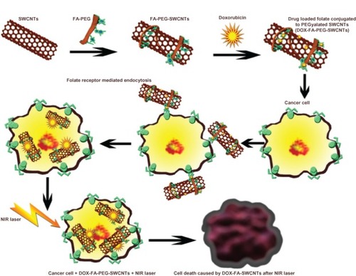
An ideal NDDS should have high drug-loading capability, strong affinity for target cells, and should release drugs triggered by a characteristic feature of the diseased cells, thus improving the efficacy of the drug and minimizing the systemic toxicity. In this study, as shown in , a targeted drug-delivery system based on SWCNTs biofunctionalized with PEG, conjugated with FA as targeting moiety, and loaded with DOX for selective killing of tumor cells was developed. Also, the photothermal effect of SWCNTs in combination with the anticancer drug DOX for targeting and selective destruction of breast cancer cells was demonstrated. Here, the SWCNTs were purified before using as delivery vehicles for chemotherapy, as the metal catalysts used for the synthesis of CNTs have been proven to be toxic. The SWCNTs can be purified or surface-modified by exposing them to oxidizing conditions (solutions containing sulfuric acid and nitric acid). This results in the formation of carboxylic groups on the surface of SWCNTs, which increases their dispersibility in aqueous solutions.
Figure 1 Schematic representation of a nanodrug-delivery system based on single-walled carbon nanotubes (SWCNTs), biofunctionalized with polyethylene glycol (PEG), conjugated with folic acid (FA), and loaded with doxorubicin (DOX), along with photothermal therapy for selective killing of tumor cells.
Abbreviation: NIR, near-infrared.
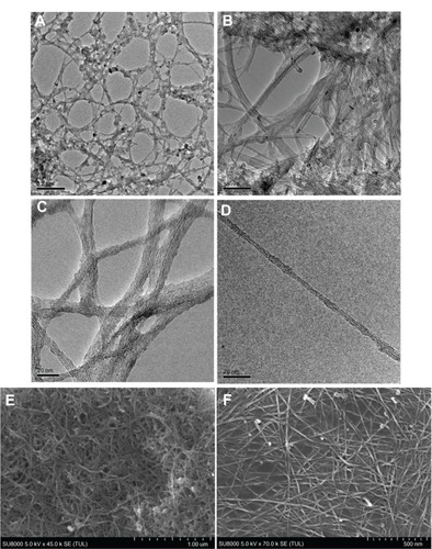
By TEM and SEM observations, we found that the purified NTs were dispersed individually or in small bundles compared with pristine SWCNTs, which were bundled, and aggregated with black metal catalyst and amorphous carbon particles. We observed a concomitant decrease in the quantity of metal particles and amorphous carbon in the purified NTs when compared to pristine SWCNTs ().
Figure 2 Transmission electron microscopy images of (A and B) pristine and (C and D) purified single-walled carbon nanotubes (SWCNTs) and scanning electron microscopy images of (E) pristine and (F) purified SWCNTs.
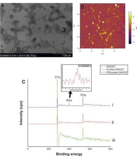
To analyze the effect of PEGylation on the morphology of SWCNTs, we carried out SEM and AFM analyses of the PEGylated SWCNTs. On SEM and AFM analyses, we observed uniformly distributed PEGylated SWCNTs (). These pictures clearly showed that PEGylated SWCNTs were well dispersed and distributed.
Figure 3 (A) Scanning electron microscopy and (B) atomic force microscopy images of polyethylene glycolated (PEGylated) single-walled carbon nanotubes (SWCNTs). (C) X-ray photoelectron spectroscopy peaks of the SWCNTs: wide-scan spectra of pristine SWCNTs (i), purified SWCNTs (ii), and PEGylated SWCNTs (iii). Inset corresponds to the N1s signal spectra of PEGylated SWCNTs.
To study the change in the surface properties of the modified SWCNTs by PEG coating, we analyzed the zeta potential of the pristine, purified, and PEGylated NTs. The zeta potential is an indicator of the stability of colloidal systems. The pristine SWCNTs had a zeta potential of −26.9 mV. The zeta potential increased to −54.2 mV for purified SWCNTs, and this may be due to the existence of many COO− groups on the sidewalls of SWCNTs.Citation63 The PEGylated SWCNTs showed a zeta potential of −34.2 mV. PEGylated SWCNTs have less negative potential than purified SWCNTs since the PEGylation converts the carboxylic acid groups into esters.Citation62 The solubility of biofunctionalized SWCNTs was increased, presumably due to the oxygen-containing glycol chain, which can form hydrogen bonds with the water molecules and capture cations present in the solution.Citation62 The shift towards more negative potential for PEGylated SWCNTs clearly proves the conjugation of PEG moieties onto the SWCNTs.
Electron spectroscopy for chemical analysis was used to confirm the presence of functional groups on the oxidized SWCNTs. The attachment of FA-PEG to oxidized SWCNTs was confirmed by the N2 peak. The wide spectrum obtained clearly shows the peaks corresponding to carbon, oxygen, and nitrogen. Nitrogen peak is absent in oxidized SWCNTs, and the presence of nitrogen peak in the PEGylated SWCNTsCitation66 confirms the PEGylation of the oxidized SWCNTs ().
DOX loading onto the PEGylated nanotubes
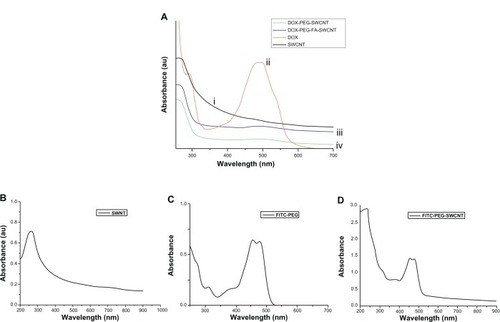
DOX loading onto the PEGylated SWCNTs was monitored by UV-vis absorption spectroscopy. shows the absorption spectra of pristine SWCNTs, plain DOX, and DOX loaded onto PEGylated SWCNTs. Plain DOX in water displays absorptions at 490 nm. The stacking of DOX onto PEGylated NTs was evident from the UV-vis spectrum, which clearly shows the characteristic absorption peaks of DOX indicative of the interaction between DOX and SWCNTs.
Figure 4 (A) Ultraviolet-visible absorbance spectra of pristine single-walled carbon nanotubes (SWCNTs) (i), plain doxorubicin (DOX) (ii), DOX-folic acid (FA)-polyethylene glycol (PEG)-SWCNTs (iii), and DOX-PEG-SWCNTs (iv). (B) Pristine SWCNTs, (C) fluorescein isothiocyanate (FITC)-PEG, (D) FITC-PEG-SWCNTs.
Drug-loading and drug-release studies
The loading of DOX onto the NTs can be determined by the analysis of the supernatant for free drug using a UV-vis spectrophotometer after ultracentrifugation of the DOX-loaded SWCNTs. We obtained a DOX loading efficiency of 58% onto the PEGylated NTs.
In vitro drug release studies
The drug-release profile of DOX from the DOX-loaded NTs was studied at 37°C in PBS at three different pH conditions – 7.4, 5.3, and 4.0 – with continuous shaking at 100 rpm for 72 hours. The temperature of 37°C was selected for drug-release response because it is close to the physiological temperature. The pH of 7.4 corresponds to physiological pH, and pH of 4.0 and 5.3 corresponds to lysosomal pH of cancer cells. The drug-release curves () indicate that the release of DOX from the PEGylated NTs is pH-triggered, and the drug-release studies were carried out till it reached the stationary phase. At pH 7.4, the drug-release curve shows that DOX loaded on SWCNTs is released at a very low and slow rate for 6 hours and attains a stationary phase in the ensuing hours, with very minimum drug release up to 24 hours. However, at pH 4.0, the DOX-release rate was significantly enhanced during the initial 6 hours. We observed an initial burst of drug release up to 4 hours, followed by a sustained-release pattern till 12 hours. This drug-release pattern was repeated with a small burst of drug after 12 hours and again followed by a sustained release till 72 hours. The drug-release profile of pH 5.3 overlapped with that of pH 4.0. These results can be ascribed to the hydrogen-bonding interaction between DOX and SWCNTs, which is stronger in neutral conditions, resulting in a controlled release. However, the drug-release pattern under acidic media indicates a higher amount of DOX release than at neutral conditions. Under acidic conditions, the amine (−NH2) groups of DOX get protonated, resulting in the partial dissociation of hydrogen-bonding interaction, hence the amount of DOX released from SWCNTs is much higher. This efficient loading and release of DOX indicates strong π–π stacking interaction between SWCNTs and DOX.Citation2,Citation29
Figure 5 In vitro drug release profile of doxorubicin from doxorubicin–folic acid–polyethylene glycol single-walled carbon nanotubes in phosphate-buffered saline at pH 7.4, 5.3, and 4.0.
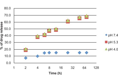
The loading and release of DOX depends upon the hydrogen-bonding interaction with SWCNTs and is a function of pH. At pH 7.4, four possibilities of hydrogen bonding were expected: (1) –COOH of SWCNTs and –OH of DOX, (2) –COOH of SWCNTs and –NH2 of DOX, (3) –OH of SWCNTs and –OH of DOX, and (4) –OH of SWCNTs and –NH2 of DOX. This overall hydrogen-bonding interaction between SWCNTs and DOX is higher at pH 7.4.Citation2,Citation58 Under acidic conditions, two kinds of hydrogen bonding can be expected: (1) –COOH of SWCNTs and –OH of DOX, and (2) between –OH of SWCNTs and –OH of DOX. Also, the –NH2 of DOX forms –NH3+ with H+, which cannot participate in hydrogen bonding. Furthermore at low pH, the H+ in solution would compete with the hydrogen bond-forming group and weaken the hydrogen-bonding interaction outlined above, which may lead to a greater release of DOX.Citation2 Around 70% of the drug was released within 72 hours in pH 4.0 buffer, whereas only 17% of the drug was released in pH 7.4 buffer, indicating a higher percentage of release of DOX under acidic conditions. In summary, the FA-PEG-SWCNTs displayed pH-sensitive release of DOX, suggesting they may be a promising delivery vehicle for the anticancer drugs and showing potential for tumor-targeting and controlled-release applications.
Characterization of the fluorescent SWCNTs
The functionalization of SWCNTs with FITC-PEG was analyzed by UV-vis absorption spectroscopy. – shows the absorption spectra of pristine SWCNTs, FITC-PEG, and FITC-PEG-SWCNTs. The absorbance peaks of FITC-PEG-SWCNTs at 250 nm and 550 nm correspond to the characteristic peaks of SWCNTs and FITC-PEG, respectively.
Temperature measurement during NIR radiation
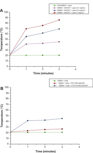
To detect the effects of 800 nm optical excitation of SWCNTs, we carried out two different sets of control experiments. The first set was carried out by irradiating DMEM without and with SWCNTs ex vitro. Three different NT concentrations (0.1, 0.5, and 1 mg/mL) were selected. We observed that irradiation of DMEM without SWCNTs caused a temperature increase from 20.1°C to 20.5°C. However, DMEM with SWCNTs at 0.1, 0.5, and 1 mg/mL concentrations irradiated by 1.726 W/cm2 800 nm laser for 3 minutes caused the temperature to elevate from 21.4°C to 45.3°C, 21.5°C to 69.2°C, and 21.1°C to 85.7°C, respectively (). In the second set of experiments, MCF7 cancer cells were seeded at a density of 1.6 × 10⁴ cells/mL in 35 mm petri dishes. After 24 hours of growth, MCF7 cells without SWCNTs and MCF7 cells with FITC-PEG-SWCNTs and FITC-FA-PEG-SWCNTs at a concentration of 0.1 mg/mL were added to the cells and again incubated for 3 hours, rinsed with PBS to remove the unbound SWCNTs, and followed by irradiation with a 800 nm laser for 3 minutes. We observed a temperature increase from 20.6°C to 20.8°C for MCF7 cells without SWCNTs, whereas temperature elevation from 21.3°C to 26°C and 21°C to 45.1°C for MCF7 cells with FITC-PEG-SWCNTs and with FITC-FA-PEG-SWCNTs, respectively, were noted (). These findings clearly demonstrated the strong light–heat transfer characteristics of the FITC-FA-PEG-SWCNTs by 800 nm light. Also, the heating efficiency of FITC-FA-PEG-SWCNTs relies strongly on time and dose, indicating that with increasing concentration and time, the temperature was significantly higher.
Figure 6 (A) Plots of temperature increase for suspensions of single-walled carbon nanotubes (SWCNTs) at various concentrations as a function of irradiation time using laser at 1.726 W/cm2 for 3 minutes. (B) In vitro temperature measurements using SWCNTs at a concentration of 0.1 mg/mL incubated with MCF7 cells during irradiation by 800 nm laser at 0.5–1 W/cm2 for 3 minutes.
Abbreviations: DMEM, Dulbecco’s Modified Eagle’s Medium; FITC, fluorescein isothiocyanate; PEG, polyethylene glycol.
Biocompatibility studies
Phase-contrast studies were carried out to analyze the biocompatibility of functionalized SWCNTs. L929 cells and MCF7 cells were plated onto six-well plates until they attained 70% confluence. Pristine SWCNTs and PEGylated SWCNTs (PEG-SWCNTs) at a concentration of 0.1 mg/mL were added to each well, and the plates were incubated for 24 hours. The biocompatibility of the functionalized SWCNTs can be seen in the phase-contrast images taken after 24 hours (). The image clearly shows the PEGylated SWCNT-treated cells growing competently at par with the control cells. However, some dead cells were observed in the images of cells treated with pristine SWCNTs. The biocompatibility of the pristine and PEGylated NTs was further studied using Alamar blue assay. These samples were incubated with L929 cells and MCF7 for 24 hours.
Figure 7 Phase-contrast microscopic images of normal (L929) and cancer (MCF7) cells treated with pristine single-walled carbon nanotubes (SWCNTs) and polyethylene glycolated (PEGylated) SWCNTs after 24 hours. (A and B) Control images of L929 and MCF7 cells; (C and D) images of cells treated with pristine SWCNTs; (E and F) images of cells treated with PEGylated SWCNTS and showing biocompatibility of cells after 24 hours of incubation.
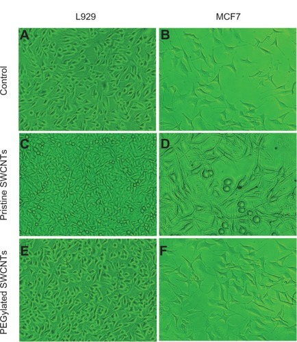
The viability of L929 and MCF7 cells when treated with the highest concentration of 1 mg/mL of pristine SWCNTs was found to be 64% and 59%, respectively. However, the viability of the cells increased to 87% and 84% in L929 and MCF7 cells, when treated with the same highest concentration, ie, 1 mg/mL of PEGylated SWCNTs, thereby indicating successful PEGylation of the SWCNTs with PEG. Thus, we can confirm that the PEGylated SWCNTs are highly biocompatible and least cytotoxic in nature.
Selective internalization of SWCNTs into cancer cells
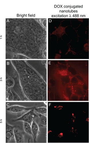
Receptor-mediated endocytosis is the most common pathway of endocytosis.Citation67 It provides a means for the selective and efficient uptake of particles that may be present in the extracellular medium. Receptors are present on the cells for the uptake of different types of ligands, such as plasma proteins, enzymes, hormones, and growth factors.Citation67 Here, we investigated the uptake of FA-conjugated NTs into MCF7 cells that overexpressed FA receptors on the surface of the cell membrane and compared the uptake in FA-negative L929 cells. The selective internalization and uptake of SWCNTs into cancer cells were recorded by confocal imaging to determine the intracellular fate of the NTs (). Time-dependent cellular uptake of the NTs was also studied at 1-, 3-, and 5-hour incubation periods. After incubating the cells with DOX-FA-PEG-SWCNTs for 1 hour, the SWCNTs were initially seen attached to the plasma membrane of the cells; also, the fluorescence intensity was very low. After 3 hours of incubation, strong fluorescence was observed in the cytoplasm, indicating the entry of SWCNTs into cells. After 5 hours, confocal images revealed decreased fluorescence inside cells, corresponding to redistribution and discharge of SWCNTs out of the cells (). No fluorescence was observed in the nucleus for any cells, indicating the lack of SWCNTs translocating into the nucleus. To further elucidate the endosome-mediated pathway of the NTs, lysosomal staining was performed with green LysoTracker. The overlapping signals of red from the DOX-FA-PEG-SWCNTs and green from the lysosomes confirmed the receptor-mediated endosomal uptake of the NTs into the cells. High targeting capability is vital for the selective destruction of cancer cells. It means that the targeting agents would bind to cancer cells at a much higher rate than to normal cells. To demonstrate the targeted delivery of DOX by SWCNTs, we conjugated FA as the targeting moiety that targets FA receptors in cancer cells. Increased DOX fluorescence was observed in MCF7 cells with DOX-FA-PEG-SWCNTs compared to L929 cells, which showed minimum internalization of the NTs. The selective uptake of DOX-PEG-FA-SWCNTs inside cancer cells clearly indicates that FA receptor-mediated endocytosis is more selective and efficient than nonspecific endocytosis.
Figure 8 (A–I) Confocal laser scanning microscopy images showing the selective internalization of single-walled carbon nanotubes (SWCNTs) by L929 and MCF7 cells. (A, D and G) Bright-field images of the cells treated with SWCNTs; (B, E and H) green fluorescence of the lysosomal staining of the cells with LysoTracker dye; (C, F and I) red fluorescence images of cells internalized with doxorubicin (DOX)-conjugated SWCNTs.
Abbreviations: FA, folic acid; PEG, polyethylene glycol.
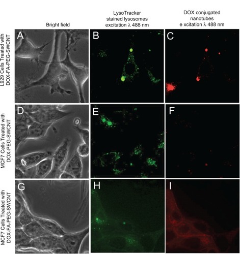
Figure 9 (A–F) Confocal images of MCF7 cells treated with doxorubicin (DOX)-folic acid (FA)-polyethylene glycol (PEG) single-walled carbon nanotubes (SWCNTs) at different incubation time intervals. (A–C) Bright-field images of MCF7 cells; (D–F) fluorescence images of MCF7 cells treated with DOX-FA-PEG-SWCNTs for 1, 3, and 5 hours.
In vitro cytotoxicity studies
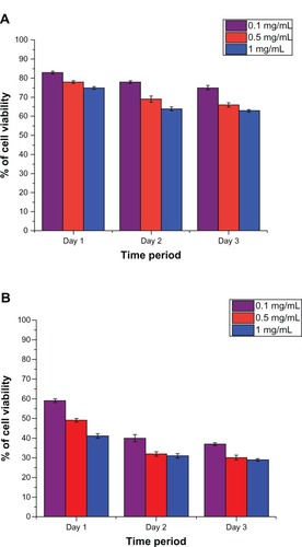
The in vitro cytotoxicity profile of the DOX-FA-PEG-SWCNTs in comparison with free DOX was studied using Alamar blue assay. MCF7 cells were used for the cytotoxicity analyses. Three different concentrations each of the DOX-FA-PEG-SWCNTs and DOX as test sample were used. The assays were carried out for 72 hours, and the fluorescence and absorbance readings were taken for analyses. shows the percentage of cell viability measured for free DOX and DOX-FA-PEG-SWCNTs, respectively, using Alamar blue assay. In the case of DOX-FA-PEG-SWCNTs, we observed that cell viability decreased with increasing concentration for the initial 24 hours. After 24 hours, there was sustained release of drug, resulting in a slower mortality rate. Viability was abruptly reduced to 59% even at the lowest concentration (0.1 mg/mL) in 24 hours. The viability of the cancer cells was further decreased to 29% in 72 hours at the same minimum concentration. DOX-FA-PEG-SWCNTs induced serious cytotoxicity, even at a dose much lower than free DOX. In the following study, when 1 mg/mL of free DOX was used, cell viability was 75% after 24 hours, and 63% of cells were viable after 72 hours. This is because the permeability of cells for free DOX is very poor, and so it cannot effectively kill the cancer cells. We also found that with the increase in concentration, the viability of DOX-FA-PEG-SWCNT-treated cells apparently decreased, indicating that the therapeutic efficiency of DOX-FA-PEG-SWCNTs was dosage-dependent. The high efficiency of DOX-FA-PEG-SWCNTs may be due to the following reasons: (1) the FA-attached SWCNTs could target the DOX-FA-PEG-SWCNT conjugates to the targeted sites, while free DOX is nonspecific and damages normal tissues, leading to serious side effects,Citation2 (2) DOX could easily enter cancer cells after conjugation onto SWCNTs because of their high cell-membrane permeability,Citation68,Citation69 (3) the release of DOX from the SWCNTs is pH-triggered, which explains the abrupt decrease in cancer cell viability at 24 hours and which then follows a slow drug-release pattern, slow drug release pattern and therefore, slower killing rates up to 72 h. From the results obtained, it can be concluded that DOX-FA-PEG-SWCNTs have proved to be the most cytotoxic and without any damage to normal tissues when compared to free DOX.
Figure 10 (A and B) Results of cytotoxicity assay. (A) Three days’ study of plain doxorubicin on MCF7 cells; (B) 3 days’ study of doxorubicin–folic acid–polyethylene glycol single-walled carbon nanotubes on MCF7 cells.
Cancer destruction using the NIR effect of SWCNTs
We investigated the effects of SWCNTs on cancer cells during NIR laser treatment. After achieving confluence, MCF7 cells were incubated with FITC-PEG-SWCNTs and FITC-FA-PEG-SWCNTs for 3 hours, followed by irradiation with an 800 nm laser for 3 minutes. (1A–1C) shows the confocal images of MCF7 cells treated with FITC-PEG-SWCNTs before and after laser irradiation. The images show that the cells survived even after 3 minutes’ laser exposure; these results can be attributed to the low uptake of FITC-PEG-SWCNTs into the MCF7 cells. From the confocal images of cancer cells with FITC-FA-PEG-SWCNT uptake before and after laser treatment, as shown in (2A–2C), we could easily observe the breaking of cancer cells due to the hyperthermic effects in FITC-FA-PEG-SWCNT-treated cells under laser excitation. Before laser treatment, the MCF7 cells had a clear dividing line between the nucleus and the cytoplasm, and the cells remained essentially intact. The SWCNTs mainly localized in the cytoplasm, as evidenced by the presence of green fluorescence in the cytoplasm. After the laser treatment, it was difficult to distinguish between the cytoplasm and nucleus, since all cancer cells showed the distorted morphology of cells undergoing apoptosis. Also, green fluorescence in the cells undergoing apoptosis could be seen inside whole cells, thus indicating the damage of the nuclear envelope caused by the hyperthermic effect by SWCNTs under laser irradiation. These results clearly state the high selectivity of FITC-FA-PEG-SWCNTs on the NIR destruction of cancer cells. The selective destruction of cancer cells was further analyzed by Alamar blue assay. The studies were carried out in three sets: (1) cancer cells + laser, (2) cancer cells + FITC-PEG-SWCNTs + laser, and (3) cancer cells + FITC-FA-PEG-SWCNTs + laser. Untreated cells were used as controls. All cells were irradiated with an 800 nm laser for 3 minutes. The experiments were carried out at time intervals of 6, 12, and 24 hours. We observed that the cell viability of FITC-FA-PEG-SWCNTs with laser treatment was 54%, 27%, and 5% at 6, 12, and 24 hours, respectively, at a concentration of 0.1 mg/mL (). The rate of viability of cells with only laser treatment remained high, showing no obvious difference from the control group, indicating the NIR property of the laser where biological tissues are highly transparent. In the case of cells treated with FITC-PEG-SWCNTs, a high cell-viability rate was observed. However, cell viability was drastically decreased for FITC-FA-PEG-SWCNTs.
Table 1 Results of cytotoxicity assay of cancer cells treated with various modified single-walled carbon nanotubes at a concentration of 0.1 mg/mL after laser irradiation by 800 nm laser at 1.726 W/cm2 for 3 minutes and studied up to 24 hours
Figure 11 Targeted destruction of cancer cells by single-walled carbon nanotubes (SWCNTs) photothermal effect. Confocal images of cancer cells (MCF7) treated with fluorescein isothiocyanate (FITC)-polyethylene glycol (PEG)-SWCNTs (1), FITC-folic acid (FA)-PEG-SWCNTs (2), and DOX-FA-PEG-SWCNTs (3). Before laser treatment (A), confocal image after 3 minutes’ laser treatment and viewed after 12 hours (B), confocal image after 3 minutes’ laser treatment and viewed after 24 hours (C).
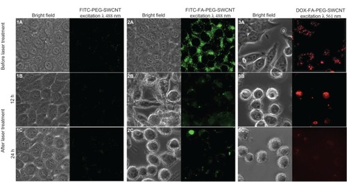
We also studied the combined cytotoxic effect of laser and DOX-loaded SWCNTs. When the MCF7 cells were treated with DOX-FA-PEG-SWCNTs in the presence of laser irradiation for 3 minutes, cell viability was reduced significantly. The confocal images clearly show apoptosis in the cancer cells treated with DOX-FA-PEG-SWCNTs after 3 minutes’ laser exposure ( [3A–C]). The reason for this may be that cell tolerance drops dramatically at a certain temperature during heat treatment.Citation70 Also, laser-treatment application might have triggered the release of drug from the DOX-PEG-FA-SWCNTs, resulting in increased cell death. The cytotoxic effect of DOX-PEG-FA-SWCNTs in combination with laser on MCF7 cells was further analyzed by Alamar blue assay. From the results shown in , significant reduction in cell viability was observed, and cell viability was 37%, 11% and 2% for 6, 12, and 24 hours, respectively. The inhibition rate of the cells under this mode was greater when compared to that of the cells treated with DOX-free SWCNTs under laser. These results show that SWCNTs have a significant photothermal effect, and when combined with chemotherapy they are ideal for cancer treatment, without causing toxicity to normal cells.
Conclusion
An ideal NDDS against cancer is expected to enter and destroy cancer cells while minimizing the side effects to normal tissues. We successfully synthesized a highly effective targeted NDDS based on PEG-coated SWCNTs functionalized with a targeting moiety – FA receptor – for recognizing and targeting tumor cells conjugated with an anticancer drug (DOX). Our results show that FA-functionalized SWCNTs could be selectively internalized into cancer cells via an FA–FA receptor-mediated pathway, without internalization into normal cells. The obtained system (DOX-FA-PEG-SWCNTs) displays excellent stability under physiological conditions. It was found that it could also effectively release DOX at reduced pH typical of the tumor environment of intracellular lysosomes and endosomes. We further demonstrated a photothermal technique for targeted cancer destruction by using the photothermal effect of SWCNTs. SWCNTs have a high optical absorbance in the NIR region, where biological tissues are highly transparent. From the observation of our data, it is clear that SWCNTs act efficiently to convert laser energy into heat after exposure to 800 nm laser irradiation in vitro. This advantage was used in selective photothermal therapy, for killing only cancer cells while sparing normal cells. Our results also showed that both concentration of SWCNTs and duration of laser are controlling factors for thermally induced cytotoxicity. Also, the combined effect of targeted drug-loaded DOX-FA-PEG-SWCNTs with photothermal therapy was studied, and it was observed that the combined effect synergistically killed almost 95% of cancer cells at an accelerated rate. We conclude that this NDDS is more selective and effective than the free drug, and results in enhanced therapeutic effects, when combined with photothermal therapy and reduced general toxicity. In light of these promising in vitro drug-delivery results, the application of DOX-FA-PEG-SWCNTs combined with NIR laser could be extended to enhance the efficiency of cancer therapy in the near future.
Acknowledgments
Prashanti Jeyamohan thanks the Otsuka Toshimi Scholarship Foundation for financial support given as a scholarship. Part of this study was supported by a grant for the program of the Strategic Research Foundation at Private Universities (S1101017), organized by the Ministry of Education, Culture, Sports, Science and Technology (MEXT), Japan, since April 2012. We thank Athulya Aravind, Ankur Baliyan, Srivani Veeranarayanan, Aby Cheruvathoor Poulose, and Saino Hanna Varghese for their support and help in this work.
Disclosure
The authors report no conflicts of interest in this work.
References
- WongHLBendayanRRauthAMLiYWuXYChemotherapy with anticancer drugs encapsulated in solid lipid nanoparticlesAdv Drug Delivery Rev200759491504
- DepanDShahJMisraRDKShahJControlled release of drug from folate-decorated and graphene mediated drug delivery system: Synthesis, loading efficiency, and drug release responseMater Sci Eng C Mater Biol Appl20113113051312
- JiZLinGLuQTargeted therapy of SMMC-7721 liver cancer in vitro and in vivo with carbon nanotubes based drug delivery systemJ Colloid Interface Sci201236514314921974923
- AlexisFRheeJWRichieJPRadovic-MorenoAFLangerRFarokhzadOCNew frontiers in nanotechnology for cancer treatmentUrol Oncol Semin O I2008267485
- AndoEKuromatsuRTanakaMSurveillance program for early detection of hepatocellular carcinoma in japan results of specialized department of liver diseaseJ Clin Gastroentrol200640942948
- JaraczSChenJKuznetsovaLVOjimaIRecent Advances in Tumor-targeting Anticancer Drug ConjugatesBioorg Med Chem2005135043505415955702
- MamotCDrummondDCNobleCOEpidermal growth factor receptor–targeted immunoliposomes significantly enhance the efficacy of multiple anticancer drugs in vivoCancer Res200565116311163816357174
- ChenXWangXWangYImproved tumor-targeting drug delivery and therapeutic efficacy by cationic liposome modified with truncated bFGF peptideJ Control Release2010145172520307599
- WangXYangLChenZShinDMApplication of nanotechnology in cancer therapy and imagingCA Cancer J Clin2008589711018227410
- WeitmanSDLarkRHConeyLRDistribution of the folate receptor GP38 in normal and malignant cell lines and tissuesCancer Res199252339634011596899
- AntonyACFolate receptorsAnn Rev Nutr1996165015218839936
- LuYLowPSFolate-mediated delivery of macromolecular anticancer therapeutic agentsAdv Drug Deliv Rev20025467569312204598
- MaziarzKMMonacoHLShenFRatnamMComplete mapping of divergent amino acids responsible for differential ligand binding of folate receptors α and βJ Biol Chem1999274110861109110196192
- PeerDKarpJMHongSFarokhzadOCMargaliteRLangerRNanocarriers as an emerging platform for cancer therapyNat Nanotechnol2007275176018654426
- MoghimiSMHunterACMurrayJCNanomedicine; current status and future prospectsThe FASEB Journal200519311330
- YounsMHoheiselJDEfferthTTherapeutic and diagnostic applications of nanoparticlesCurrent Drug Targets20111235736520955146
- KamNWSO’ConnellMWisdomJADaiHJCarbon nanotubes as multifunctional biological transporters and near-infrared agents for selective cancer cell destructionProc Natl Acad Sci USA20051021160016087878
- ShaoNLuSWickstromEPanchapakesanBIntegrated molecular targeting of IGF1R and Her2 surface receptors and destruction of breast cancer cells using carbon nanotubesNanotechnology200718
- Kostarelos1KBiancoAPratoMPromises, facts and challenges for carbon nanotubes in imaging and therapeuticsNature Nanotechnology20094627633
- TranPAZhangLWebsterTJCarbon nanofibres and carbon nanotubes in regenerative medicineAdv Drug Deliv Rev2009611097111419647768
- YangWThordarsonPGoodingJRingerSPBraetFCarbon nano-tubes for biological and biomedical applicationsNanotechnology200718412001112
- LacerdaLBiancoAPratoMKostarelosKCarbon nanotube cell translocation and delivery of nucleic acids in vitro and in vivoJ Mater Chem2008181722
- LiuZWintersMHolodniyMDaiHsiRNA delivery into human T cells and primary cells with carbon-nanotube transportersAngew Chem Int Ed Engl2007462023202717290476
- KamNWSJessopTCWenderPADaiHNanotube Molecular Transporters: Internalization of Carbon Nanotube-Protein Conjugates into Mammalian CellsJ Am Chem Soc20041266850685115174838
- ChinSFBaughmanRHDaltonABAmphiphilic helical peptide enhances the uptake of single-walled carbon nanotubes by living cellsExp Biol Med (Maywood)20072321236124417895532
- KamNWDaiHCarbon nanotubes as intracellular protein transporters: generality and biological functionalityJ Am Chem Soc20051276021602615839702
- LiuZTabakmanSWelsherKDaiHCarbon nanotubes in biology and medicine: In vitro and in vivo detection, imaging and drug deliveryNano Research200928512020174481
- SinghRLillardJWNanoparticle-based targeted drug deliveryExp Mol Pathol20098621522319186176
- LiuZSunXNakayama-RatchfordNDaiHSupramolecular chemistry on water-soluble carbon nanotubes for drug loading and deliveryACS Nano20071505619203129
- ChenJChenSZhaoXKuznetsovaLVWongSSOjimaIFunctionalized single-walled carbon nanotubes as rationally designed vehicles for tumor-targeted drug deliveryJ Am Chem Soc2008130167781678519554734
- SchipperMLNakayama-RatchfordNDavisCRA pilot toxicology study of single-walled carbon nanotubes in a small sample of miceNature Nanotechnology20083216221
- PastorinGWuWWieckowskiSDouble functionalization of carbon nanotubes for multimodal drug deliveryChem Commun20061111821184
- TansSJDevoretMHDaiHJIndividual single-wall carbon nanotubes as quantum wiresNature1997386474477
- LiuZDavisCCaiWHeLChenXDaiHCirculation and long-term fate of functionalized, biocompatible single-walled carbon nanotubes in mice probed by raman spectroscopyProc Natl Acad Sci USA20081051410141518230737
- MoonHKLeeSHChoiHCIn vivo near-infrared mediated tumor destruction by photothermal effect of carbon nanotubesACS Nano200933707371319877694
- WelsherKLiuZSherlockSPA route to brightly fluorescent carbon nanotubes for near-infrared imaging in miceNat Nanotechnol2009477378019893526
- LiuZTabakmanSSherlockSMultiplexed five-color molecular imaging of cancer cells and tumor tissues with carbon nanotube raman tags in the near-infraredNano Res2010322222321442006
- LiuZPengRInorganic nanomaterials for tumor angiogenesis imagingEur J Nucl Med Mol Imaging201037147163
- GhoshSDuttaSGomesEIncreased heating efficiency and selective thermal ablation of malignant tissue with dna-encased multiwalled carbon nanotubesACS Nano200932667267319655728
- KangBYuDDaiYChangSChenDDingYCancer-cell targeting and photoacoustic therapy using carbon nanotubes as bomb agentsSmall2009111292130119274646
- de La ZerdaAZavaletaCKerenSCarbon nanotubes as photoacoustic molecular imaging agents in living miceNat Nanotechnol2008355756218772918
- XiangLZYuanYXingDOuZMYangSHZhouFFPhotoacoustic molecular imaging with antibody-functionalized single-walled carbon nanotubes for early diagnosis of tumorJ Biomed Opt20091402100819405721
- WelsherKLiuZDaranciangDDaiHSelective probing and imaging of cells with single walled carbon nanotubes as near-infrared fluorescent moleculesNano Lett2008858659018197719
- O’ConnellMJBachiloSMHuffmanCBBand gap fluorescence from individual single-walled carbon nanotubesScience200229759359612142535
- AminZDonaldJJMastersAHepatic metastases: interstitial laser photocoagulation with real-time US monitoring and dynamic CT evaluation of treatmentRadiology, Easton, Pa1993187339347
- AnghileriLJRobertJHyperthermia in cancer treatmentCRC PressBoca Raton, FL1986
- HuangXHEL-SayedIHQianWEL-SayedMACancer cells assemble and align gold nanorods conjugated to antibodies to produce highly enhanced, sharp, and polarized surface raman spectra: a potential cancer diagnostic markerNano Lett200771591159717474783
- KamNWSLiuZADaiHJCarbon nanotubes as intracellular transporters for proteins and dna: an investigation of the uptake mechanism and pathwayAngew Chem Int Ed200645577581
- RoscaIDWatariFUoMAkasakaTOxidation of multiwalled carbon nanotubes by nitric acidCarbon20054331243131
- MooreVCStranoMSHarozEHHaugeRHSmalleyREIndividually suspended single-walled carbon nanotubes in various surfactantsNano Lett2003313791382
- BottiniMRosatoNBottiniNPEG-Modified Carbon Nanotubes in Biomedicine: Current Status and Challenges AheadBiomacromolecules2011123381339321916410
- MamonMAItkisMENiyogiSEffect of rehybridization on the electronic structure of single-walled carbon nanotubesJ Am Chem Soc2001123112921129311697973
- SudimackJJLeeRJTargeted drug delivery via the folate receptorAdv Drug Deliv Rev20004114716210699311
- ElnakatHRatnamMRole of folate receptor genes in reproduction and related cancersFrontiers in Bioscience20061150651916146749
- BasalEEghbali-FatourechiGZKalliKRFunctional Folate Receptor Alpha is Elevated in the Blood of Ovarian Cancer PatientsPLoS ONE20094e629219617914
- MathiasCJWangSLeeRJWatersDJLowPSGreenMATumor-selective radiopharmaceutical targeting via receptor-mediated endocytosis of gallium-67-deferoxamine-folateJ Nucl Med199637100310088683292
- YooHSParkTGFolate-receptor-targeted delivery of doxorubicin nano-aggregates stabilized by doxorubicin–PEG–folate conjugateJournal of Controlled Release200410024725615544872
- HuangHYuanQShahJSMisraRDKA new family of folate-decorated and carbon nanotube-mediated drug delivery system: Synthesis and drug delivery responseAdvanced Drug Delivery Reviews2011631332133921514336
- VeeranarayananSPouloseACMohamedMSSynergistic targeting of cancer and associated angiogenesis using triple-targeted dual-drug silica nanoformulations for theragnosticsSmall2012834763489
- SivakumarBAswathyRGNagaokaYMultifunctional carboxymethyl cellulose-based magnetic nanovector as a theragnostic system for folate receptor targeted chemotherapy, imaging, and hyperthermia against cancerLangmuir2012293453346623409925
- LiuJRinzlerAGDaiHFullerene pipesScience1998280125312569596576
- ZhaoBHuHYuAPereaDHaddonRCSynthesis and characterization of water soluble single-walled carbon nanotube graft copolymersJ Am Chem Soc20051278197820315926849
- ZhangXKMengLJLuQGFeiZFDysonPJTargeted delivery and controlled release of doxorubicin to cancer cells using modified single wall carbon nanotubesBiomaterials2009306041604719643474
- GuYJChengJJinJChengSHWongWTDevelopment and evaluation of pH-responsive single-walled carbon nanotube-doxorubicin complexes in cancer cellsInt J Nanomedicine201162889289822131835
- Nakayama-RatchfordNBangsaruntipSSunXWelsherKDaiHFunctionalization of carbon nanotubes by fluorescein-polyethylene glycol: supramolecular conjugates with pH-dependent absorbance and fluorescenceJ Am Chem Soc20071292448244917284037
- ChenSJiangYWangZZhangXDaiLSmetMLight-controlled single-walled carbon nanotube dispersions in aqueous solutionLangmuir2008249233923618672920
- SinghRDPuriVValiyaveettilJTMarksDLBittmanRPaganoRESelective caveolin-1-dependent endocytosis of glycosphingolipidsMol Biol Cell2003143254326512925761
- KostarelosKLacerdaLPastorinGCellular uptake of functionalized carbon nanotubes is independent of functional group and cell typeNat Nanotechnol2007210811318654229
- ChengJPFernandoKASVecaLMReversible accumulation of pegylated single-walled carbon nanotubes in the mammalian nucleusACS Nano220082085209419206455
- WangLZhangMZhangNSynergistic enhancement of cancer therapy using a combination of docetaxel and photothermal ablation induced by single-walled carbon nanotubesInt J Nanomedicine201162641265222114495