Abstract
Titanium dioxide (TiO2) nanoparticles are among the top five nanoparticles used in consumer products, paints, and pharmaceutical preparations. Given that exposure to such nanoparticles is mainly via the skin and inhalation, the present study was conducted in male Wistar albino rats (Rattus norvegicus). Our aim was to investigate the effect of TiO2 nanoparticles on hepatic tissue in an attempt to understand their toxicity and the potential effect of their therapeutic and diagnostic use. To investigate the effects of TiO2 nanoparticles on liver tissue, 30 healthy male Wistar albino rats were exposed to TiO2 nanoparticles at doses of 63 mg, 126 mg, and 252 mg per animal for 24 and 48 hours. Serum glutamate oxaloacetate transaminase and alkaline phosphatase activity was altered. Changes in hepatocytes can be summarized as hydropic degeneration, cloudy swelling, fatty degeneration, portal and lobular infiltration by chronic inflammatory cells, and congested dilated central veins. The histologic alterations observed might be an indication of hepatocyte injury due to the toxicity of TiO2 nanoparticles, resulting in an inability to deal with accumulated residues from the metabolic and structural disturbances caused by these nanoparticles. The appearance of cytoplasmic degeneration and destruction of nuclei in hepatocytes suggests that TiO2 nanoparticles interact with proteins and enzymes in hepatic tissue, interfering with antioxidant defense mechanisms and leading to generation of reactive oxygen species which, in turn, may induce stress in hepatocytes, promoting atrophy, apoptosis, and necrosis. More immunohistochemical and ultrastructural investigations are needed in relation to TiO2 nanoparticles and their potential effects when used as therapeutic and diagnostic tools.
Introduction
Nanotechnology is a promising new field with potential applications in the domestic, industrial, and biomedical fields.Citation1 Due to the growing number of applications, there is an increasing risk to animals from environmental exposure to nanomaterials. Their potential toxicologic impact is still under investigation and our actual knowledge about the effects of nanosized contaminants on biological systems remains incomplete.Citation2,Citation3 These effects need to be assessed in order to provide a scientific basis for safe development of nanotechnologies. Use of nanotechnology has seen exponential growth in the areas of health care, consumer products, clothes, electronics, and sporting goods.Citation2 This is due to the unique chemical, mechanical, optical, magnetic, and biological properties of nanomaterials that make them desirable for commercial and medical applications.Citation4 According to a recent survey, the number of nanotechnology-based consumer products available on the world market now exceeds 1000.Citation5 TiO2 nanoparticles have several industrial applications, and as such, come in different sizes, shapes, chemical compositions, and crystalline structures.Citation6 TiO2 occurs in four crystalline polymorphic forms, of which rutile and anatase are the most common.Citation7 Rutile is considered to be a more inert form whereas anatase is an active form of TiO2. Some studies indicate that anatase TiO2 nanoparticles are more cytotoxic than rutile TiO2 nanoparticles.Citation8 Yeo et alCitation9 have reported that different TiO2 nanoparticles induce different toxicities during embryogenesis of the zebra fish due to their different sizes and crystalline phases. The increased use of nanoparticles is a matter of great concern to health professionals and environmental scientists because of the potential risks to humans and to the environment.Citation10 Of the possible exposure routes, inhalation and skin contact are considered to be the most important for nanoparticles. The toxic effects of nanoparticles can be attributed to their small size and hence large surface area, which increases their chemical reactivity and penetration into living cells.Citation11 TiO2 nanoparticles also induce reactive oxygen species, leading to toxicity.Citation12
The dimensions of TiO2 nanoparticles are critical from the toxicity point of view, given that TiO2 nanoparticles have more pronounced toxicity than conventional TiO2 particles.Citation13 TiO2 nanoparticles have been shown to impair the function of macrophages, to cause persistent inflammatory reactions, and to increase pulmonary retention compared with fine TiO2 particles.Citation14 TiO2 nanoparticles can be absorbed into the body by inhalation, ingestion, and dermal penetration, and are distributed to important organ systems, including lymph, brain, lung, liver, and kidney.Citation15–Citation17 Of note, it has been observed in vivo that anatase TiO2 nanoparticles increased inflammatory indicators, cell proliferation, and histopathology in bronchoalveolar lavage fluid.Citation18
Characteristics of TiO2 nanoparticles can be modified by several methods to improve their performance. Due to their small size, nanoparticles may cross biological barriers to reach a number of organs, and according to their size and surface properties, accumulation of metal nanoparticles has been observed previously in all organs in vivo.Citation19 Generation of free oxygen radicals and oxidative stress triggers a host of cellular events, including DNA damage and apoptosis.Citation20 Therefore, in the present study, an attempt was made to assess the toxicity and apoptotic potential of TiO2 nanoparticles in the liver tissue of male rats.
Materials and methods
Chemicals and animals
Titanium (IV) oxide (TiO2) nanopowder (99.7% anatase, CAS 1317-70-0) was obtained from Sigma-Aldrich (St Louis, MO, USA). A terminal transferase-mediated biotinylated 16-deoxy-uridine-triphosphate (dUTP) nick-end labeling (TUNEL) apoptosis detection kit (catalog number L00300) with anti-fluorescein antibody conjugated peroxidase (FITC-labeled POD) for paraffin-embedded tissue sections was purchased from GenScript Biology CRO (Piscataway NJ, USA). All other chemicals were obtained locally in Saudi Arabia and were of analytical reagent grade. Forty healthy male Wistar albino rats (Rattus norvegicus) aged eight weeks with a mean weight of 126 g were obtained from the Animal Care Center, College of Pharmacy, King Saud University.
Preparation and characterization of TiO2 nanoparticles
TiO2 NPs were suspended in Milli-Q water (Millipore Corporation, Billerica, MA, USA) at a concentration of 1 mg/mL. A stock suspension was probe-sonicated at 40 W for 15 minutes. Samples for analysis by transmission electron microscopy (TEM) were prepared by drop coating a TiO2 nanoparticle solution on carbon-coated copper TEM grids. The films formed on the TEM grids were allowed to dry prior to measurement. TEM measurements were performed using a JEOL instrument (model 1101F, JEOL Ltd Tokyo, Japan) operated at an accelerating voltage at 200 kV.
Experimental design
The rats were housed in groups under standard lighting conditions with free access to water and food. Humidity and temperature (22°C ± 1°C) were controlled in ventilated cages on a 12-hour day/night cycle. Five animals from each group were anesthetized and euthanized by cervical dislocation after 24 and 48 hours of exposure to TiO2 nanoparticles. All experiments were conducted in accordance with the guidelines approved by the local animal care and use committee at King Saud University.
TiO2 nanoparticle doses were calculated based on average body weight.Citation21,Citation22 The study was conducted to compare the toxicity of the nanoparticles at three different doses. The animals were divided into four groups of ten rats each, which were injected intraperitoneally as follows for two days:
group 1, normal animals, injected with Milli-Q water
group 2, injected with 63 mg of TiO2 nanoparticles per animal
group 3, injected with 126 mg of TiO2 nanoparticles per animal
group 4, injected with 252 mg of TiO2 nanoparticles per animal.
Histopathology
The animals were exposed to different doses of TiO2 nanoparticles intraperitoneally for 24 and 48 hours. After euthanizing the animals, fresh portions of the lateral lobes of the liver from each rat were cut rapidly, fixed in neutral buffered formalin (10%), then dehydrated using grades of ethanol (70%, 80%, 90%, 95%, and 100%). Dehydration was followed by clearing the samples in two changes of xylene. The samples were then impregnated with two changes of molten paraffin wax, embedded, and blocked out. The tissue sections (4–5 μm) were stained according to the method described by Bancroft and StevensCitation23 using conventional histologic stains. Stained sections from the control and treated rats were observed and photographs were taken using an optical microscope (Olympus, Tokyo, Japan) for alterations in architecture, hepatocytes, and sinusoids, and for the presence of degeneration, necrosis, fatty changes, and portal fibrosis.
Estimation of GOT and ALP
After exposure for 48 hours, blood samples were taken from five rats per group, and the serum was separated out for estimation of alkaline phosphatase (ALP) and glutamate oxaloacetate transaminase (GOT) activity using already reported methods.Citation24,Citation25
TUNEL assay
Formalin-fixed, paraffin-embedded tissue sections were dewaxed in xylene, rehydrated through graded ethanol, and pretreated with proteinase-K 20 μg/mL in phosphate-buffered saline for 15 minutes at 37°C in a humidified chamber. After washing twice in distilled water and rinsing in Tris-buffered saline (pH 7.6), the sections were incubated for 60 minutes at 37°C with a reaction mixture containing 0.3 U/μL terminal deoxynucleotidyl transferase (GenScript), terminal transferase buffer (GenScript) 100 mM cacodylate buffer (pH 6.8), 1 mM cobalt chloride, and 0.5 mM DL-dithiothreitol with biotinylated 16-dUTP added (GenScript). The reaction was terminated by rinsing twice in Tris-buffered saline. Next, the sections were covered with 2% bovine serum albumin in Tris-buffered saline for 15 minutes and then incubated with avidin-biotin-conjugated ALP (GenScript) at 1:100 for 30 minutes. Staining was done using 5-bromo-chloro-indoxyl phosphate/nitro blue tetrazolium (GenScript) with 1 mM levamisole added to inhibit endogenous ALP activity. The result imparted an orange/brown color to the nuclei of apoptotic cells.
Positive control slides was created by staining sections of rat liver tissue with 50 μL of DNase I solution (GenScript). For the negative control, terminal deoxynucleotidyl transferase was omitted from the reaction buffer. The slides for the control and treated rat hepatocytes were observed and the images captured using an optical Olympus microscope.
Statistical analysis
One independent experiment was carried out for evaluation. Data were expressed as the mean ± standard error and tested by one-way analysis of variance. A P-value less than 0.01 was considered to be statistically significant.
Results and discussion
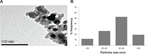
The TiO2 nanoparticles were characterized first for size and found to be in the nanoscale range, but formed small agglomerates in aqueous solution. TEM imaging revealed the morphology of the nanoparticles (). The average size measured by TEM was 50.40 ± 5.60 nm (). The typical TEM image shown in suggests that most of the TiO2 nanoparticles had a polyhedral morphology. Like other materials, when normal-scale TiO2 is converted into nanoscale TiO2, the physicochemical properties change. The special physicochemical properties of nanoparticles come from their high surface-to-volume ratio. They also have a considerable higher percentage of atoms on their surface compared with bulk particles, which makes them more reactive.
Figure 1 Characterization of titanium oxide nanoparticles. (A) Transmission electron microscopic image and (B) size distribution histogram generated using transmission electron micrography.
We measured blood chemistry parameters, including enzymes, to evaluate organ function in our experimental animals. There was a significant (P < 0.05) increase in GOT and ALP levels (). GOT and ALP levels are indicative of the functional efficiency of the liver, and are very sensitive to any disease process of the liver.Citation26 The histologic changes observed in the liver and the accompanying increase in ALP and GOT levels indicate compromised liver function. In comparison with the control group, histologic changes were detected in the liver tissue of rats treated with TiO2 nanoparticles (). Apoptosis was also seen in the hepatocytes of rats treated with these nanoparticles. The appearance of inflammatory cells in hepatic tissue suggests that the TiO2 nanoparticles can interact with proteins and enzymes in the interstitial tissue of the liver, interfering with the antioxidant defense mechanism and leading to generation of reactive oxygen species, which in turn may imitate an inflammatory response.Citation27 Distortion and swelling of hepatocytes together with dilatation of the central vein and blood sinusoids indicate that these nanoparticles may affect permeability of the cell membrane in hepatocytes and the endothelial lining of blood vessels. Swelling of hepatocytes on exposure to nanoparticles as seen in the present study might lead to adaptation of cell transporters.Citation28 Binucleation is a consequence of cell injury and a type of chromosomal hyperplasia usually seen in regenerating cells.Citation29 Cloudy swelling might be seen as a result of disturbed membrane function, leading to a massive influx of water and sodium due to the effects of nanoparticles. Cell swelling might be accompanied by leakage of lysosomal hydrolytic enzymes, leading to degeneration of the cytoplasm and macromolecular crowding.Citation30 Hydropic degeneration is a result of ion and fluid homeostasis, and leads to an increase in intracellular water.Citation31 The vacuolated swelling seen in the cytoplasm of hepatocytes from rats exposed to TiO2 nanoparticles indicates acute liver injury.
Figure 2 Levels of (A) alkaline phosphatase and (B) glutamate oxaloacetate transaminase after exposure of titanium oxide nanoparticles in the different experimental groups at 48 hours. Each value represents the mean ± standard error of three experiments. *P < 0.05 versus control.
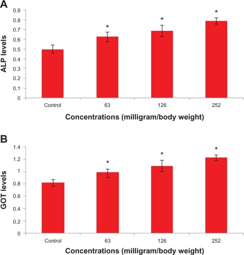
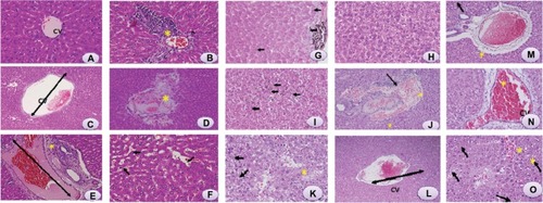
Figure 3 Light microphotographs of liver tissue.
Notes: (A) Male rats injected with Milli-Q water for 48 hours demonstrating normal histologic architecture. Hematoxylin and eosin, 400×. (B) Male rats 24 hours postexposure to titanium oxide nanoparticles (252 mg per animal) by intraperitoneal administration demonstrating lymphocytic infiltration (*) in the hepatic portal space. Hematoxylin and eosin, 400×. (C) Male rats 24 hours post-exposure to titanium oxide nanoparticles (252 mg per animal) by a single oral administration demonstrating marked dilatation of central vein. Hematoxylin and eosin, 400×. (D) Male rats 24 hours post-exposure to titanium oxide nanoparticles (252 mg per animal) by intraperitoneal administration demonstrating marked necrosis (*) and scattered hemorrhages. Hematoxylin and eosin, 400×. (E) Male rats 24 hours post-exposure to titanium oxide nanoparticles (252 mg per animal) by intraperitoneal administration demonstrating dilatation of congested portal vein with edema (*) around the blood vessel in the portal triad. Hematoxylin and eosin, 400×. (F) Male rats 24 hours post-exposure to titanium oxide nanoparticles (252 mg per animal) by intraperitoneal administration demonstrating dilatation and congestion of blood sinusoids (arrows) and binucleation of hepatocytes (circles). Hematoxylin and eosin, 400×. (G) Male rats 48 hours post-exposure to titanium oxide nanoparticles (252 mg per animal) by intraperitoneal administration demonstrating swelling of hepatocytes (arrows) and presence of nanoparticle beneath the capsule (*). Hematoxylin and eosin, 400×. (H) Male rats 48 hours post-exposure to titanium oxide nanoparticles (252 mg per animal) by intraperitoneal administration demonstrating vacuolization of hepatocytes. Hematoxylin and eosin, 400×. (I) Male rats 48 hours post-exposure to titanium oxide nanoparticles (252 mg per animal) by intraperitoneal administration, hydropic degeneration (ballooning) of hepatocytes and presence of nanoparticle in blood sinusoids (arrows). Hematoxylin and eosin, 400×. (J) Male rats 24 hours post-exposure to titanium oxide nanoparticles (126 mg per animal) by intraperitoneal administration demonstrating dilatation of congested portal vein with hemorrhage and edema (*) around the blood vessel and lymphocytic infiltration (arrow) in the portal triad. Hematoxylin and eosin, 400×. (K) Male rats 24 hours post-exposure to titanium oxide nanoparticles (126 mg per animal) by intraperitoneal administration demonstrating focal necrosis (*) and hydropic degeneration of hepatocytes (arrows). Hematoxylin and eosin, 400×. (L) Male rats 48 hours post-exposure to titanium oxide nanoparticles (126 mg per animal) by intraperitoneal administration demonstrating marked dilatation of congested central vein. Hematoxylin and eosin, 400×. (M) Male rats 48 hours post-exposure to titanium oxide nanoparticles (126 mg per animal) by intraperitoneal administration demonstrating dilatation of congested portal vein with edema (*) around the blood vessel in the portal triad. Hematoxylin and eosin, 400×. (N) Male rats 24 hours post-exposure to titanium oxide nanoparticles (63 mg per animal) by intraperitoneal administration demonstrating marked dilatation of congested central vein. Hematoxylin and eosin, 400×. (O) Male rats 48 hours post-exposure to titanium oxide nanoparticles (63 mg per animal) by intraperitoneal administration demonstrating focal necrosis (*) and hydropic degeneration of hepatocytes (arrows). Hematoxylin and eosin, 400×.
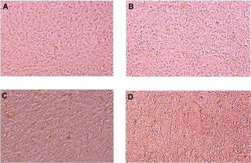
We also observed that apoptosis in hepatocytes exposed to TiO2 nanoparticles increased in a dose-dependent and time-dependent manner (). Sporadic, spotty, and well defined necrosis was also noted in some hepatocytes from rats exposed to TiO2 nanoparticles, which might have been because of oxidative stress triggered by depletion of glutathione in these cells. Park et alCitation32 reported that TiO2 nanoparticles induced oxidative stress and apoptosis in cultured BEAS-2B cells. In the present study, after intraperitoneal administration of high-dose TiO2 nanoparticles, the difficulty encountered in clearance of these nanoparticles in vivo may have resulted in deposition of particles in the liver and a hepatic lesion.Citation33 The International Programme on Chemical SafetyCitation34 shows that most ingested titanium is excreted via the urine and is not absorbed by the organism. The liver, being the main detoxification organ in the body, is activated to eliminate the side effects induced by the ingested mass of TiO2 nanoparticles, and a proportion of these nanoparticles should be excreted by the kidneys. Because of their small size and difficult clearance, TiO2 nanoparticles were retained in vivo, and liver damage occurred after intraperitoneal exposure to a high dose.
Figure 4 Photomicrograph of apoptosis in liver tissue after exposure of titanium oxide nanoparticles. (A) Control, (B) 63 mg per animal, (C) 126 mg per animal, and (D) 252 mg per animal.
Conclusion
In conclusion, our results indicate that TiO2 nanoparticles induce histologic changes in hepatocytes, which may be mediated by generation of reactive oxygen species and stress to induce atrophy and apoptosis. Long-term biological safety is another issue that will need clarification in future investigations.
Acknowledgments
The authors extend their appreciation to the Deanship of Scientific Research at King Saud University for funding this work through research group project RGP-VPP-180.
Disclosure
The authors report no conflicts of interests in this work.
References
- Peralta-VideaJRZhaoLLopez-MorenoMLde la RosaGHongJGardea-TorresdeyJLNanomaterials and the environment: a review for the biennium 2008–2010J Hazard Mater201118611521134718
- SinghNManshianBJenkinsGJSNanoGenotoxicology: the DNA damaging potential of engineered nanomaterialsBiomaterials2009303891391419427031
- SkocajMFilipicMPetkovicJNovakSTitanium dioxide in our everyday life: is it safe?Radiol Oncol20114522724722933961
- NelAXiaTMadlerLLiNToxic potential of materials at the nanolevelScience200631162262716456071
- Project of the Emerging Nanotechnologies 2009 Available from: http://www.nanotechproject.orgAccessed June 14, 2013
- LiJJMuralikrishnanSNgCTYungLYBayBHNanoparticle-induced pulmonary toxicityExp Biol Med (Maywood)20122351025103320719818
- MadlAKPinkertonKEHealth effects of inhaled engineered and incidental nanoparticlesCrit Rev Toxicol20093962965819743943
- HirakawaKMoriMYoshidaMOikawaSKawanishiSPhoto-irradiated titanium dioxide catalyzes site specific DNA damage via generation of hydrogen peroxideFree Radic Res20043843944715293551
- YeoMKKangMThe biological toxicities of two crystalline phases and differential sizes of TiO2 nanoparticles during zebra fish embryogenesis developmentMol Cell Toxicol20128317326
- OberdorsterGOberdorsterEOberdorsterJNanotoxicology: an emerging discipline evolving from studies of ultrafine particlesEnviron Health Perspect200511382383916002369
- PanZLeeWSlutskyLClarkRAPernodetNRafailovichMHAdverse effects of titanium dioxide nanoparticles on human dermal fibroblasts and how to protect cellsSmall2009551152019197964
- BarnardASOne-to-one comparison of sun screen efficacy, aesthetics and potential nanotoxicityNat Nanotechnol2012527127420208548
- DriscollKEMaurerJKCytokine and growth factor release by alveolar macrophages: potential biomarkers of pulmonary toxicityToxicol Pathol1991193984051667554
- BaggsRBFernJOberdorsterGRegression of pulmonary lesions produced by inhaled titanium dioxide in ratsVet Pathol1997345925979396140
- BermudezEMangumJBWongBAPulmonary responses of mice, rats, and hamsters to subchronic inhalation of ultrafine titanium dioxide particlesToxicol Sci20047734735714600271
- ThomasTThomasKSadriehNSavageNAdairPBronaughRResearch strategies for safety evaluation of nanomaterials, part VII: evaluating consumer exposure to nanoscale materialsToxicol Sci200691141916476686
- WangJZhouGChenCAcute toxicity and bio distribution of different sized titanium dioxide particles in mice after oral administrationToxicol Lett200716817618517197136
- WarheitDBWebbTRReedKLFrerichsSSayesCMPulmonary toxicity study in rats with three forms of ultrafine TiO2 particles: differential responses related to surface propertiesToxicology20072309010417196727
- LiYFChenCFate and toxicity of metallic and metal-containing nanoparticles for biomedical applicationsSmall201172965298021932238
- OstrovskySKazimirskyGGedankenABrodieCSelective cytotoxic effect of ZnO nanoparticles on glioma cellsNano Res20092882890
- ParkEJYoonJChoiKYiJParkKInduction of chronic inflammation in mice treated with titanium dioxide nanoparticles by intratracheal instillationToxicology20092601–3374619464567
- ZhangXDWuHYWuDToxicologic effects of gold nanoparticles in vivo by different administration routesInt J Nanomedicine2010577178121042423
- BancroftJDStevensATheory and Practice of Histological Techniques4th edLondon, UKChurchill-Livingstone1999
- KingEJArmstrongARIn vitro determination of alkaline phosphataseCan Med Assoc J19343137637920319659
- GirayBGurbayAHinealFCypermethrin induced oxidative stress in rat brain and liver is prevented by vit-E or allopurinolToxicol Lett201111813914611137320
- TietzNWFundamentals of Clinical Chemistry4th edPhiladelphia, PAWB Saunders Company1996
- JoharDRothJCBayGHWalkerJNKroczakTJLosMInflammatory response, reactive oxygen species, programmed (necrotic-like and apoptotic) cell death and cancerRocz Akad Med Bialymst200449313915631311
- JohnsonCEEffects of fluid imbalancesConnPMNeurosciences in MedicineNew York, NYJB Lippincott Company1995
- GerlyngPÅbyholmAGrotmolTBinucleation and polyploidization patterns in developmental and regenerative rat liver growthCell Prolif2008265575659116122
- Del MonteUSwelling of hepatocytes injured by oxidative stress suggests pathological changes related to macromolecular crowdingMed Hypotheses20056481882515694703
- SchrandAMRahmanMFHussainSMMetal-based nanoparticles and their toxicity assessmentNanomed Nanobiotechnol20102544568
- ParkEJYiJChungKHRyuDYChoiJParkKOxidative stress and apoptosis induced by titanium dioxide nanoparticles in cultured BEAS-2B cellsToxicol Lett200818022222918662754
- JohnstonHJHutchisonGRChristensenFMPetersSHankinSStoneVIdentification of the mechanisms that drive the toxicity of TiO2 particulates: the contribution of physicochemical characteristicsPart Fibre Toxicol200963320017923
- World Health OrganizationInternational Programme on Chemical Safety. Environmental Health Criteria 24-TitaniumGeneva, SwitzerlandWorld Health Organization1982 Available from: http://www.inchem.org/documents/ehc/ehc/ehc24.htmAccessed June 13, 2013