 ?Mathematical formulae have been encoded as MathML and are displayed in this HTML version using MathJax in order to improve their display. Uncheck the box to turn MathJax off. This feature requires Javascript. Click on a formula to zoom.
Abstract
In this paper, we report a simple, rapid, and robust method to synthesize surface-enhanced Raman-scattered gold nanoparticles (GNPs) based on green chemistry. Vitis vinifera L. extract was used to synthesize noncytotoxic Raman-active GNPs. These GNPs were characterized by ultraviolet-visible spectroscopy, dynamic light-scattering, Fourier-transform infrared (FTIR), transmission electron microscopy (TEM), X-ray diffraction (XRD), and Raman spectroscopy. The characteristic surface plasmon-resonance band at ~ 528 nm is indicative of spherical particles, and this was confirmed by TEM. The N–H and C–O stretches in FTIR spectroscopy indicated the presence of protein molecules. The predominant XRD plane at (111) and (200) indicated the crystalline nature and purity of GNPs. GNPs were stable in the buffers used for biological studies, and exhibited no cytotoxicity in noncancerous MIO-M1 (Müller glial) and MDA-MB-453 (breast cancer) cell lines. The GNPs exhibited Raman spectral peaks at 570, 788, and 1,102 cm−1. These new GNPs have potential applications in cancer diagnosis, therapy, and ultrasensitive biomarker detection.
Video abstract
Point your SmartPhone at the code above. If you have a QR code reader the video abstract will appear. Or use:
Keywords:
Introduction
Gold nanoparticles (GNPs) have been used for more than 2–3 decades for imaging and targeted therapy. GNPs are of interest due to their unique intrinsic size-dependent properties, such as surface energy and light absorption or scattering, which are attributed to surface plasmon resonance (SPR) and surface-enhanced Raman scattering (SERS). The synthesis of GNPs with a SERS signature involves multiple steps. Plasmonic nanoparticles coated with reporter molecules and covered with protective layer of molecules reduce the toxicity of plasmonic nanoparticles in living cells.Citation1 The application of nanomaterials in therapy and diagnosis of cancerous tissue is of scientific interest due to their unique size-dependent properties. Among all nanomaterials, metals are advantageous due to their SPR and SERS properties.Citation2,Citation3 In addition to the plasmonic property of metal nanoparticles, GNPs have added advantages in cancer therapy and diagnosis due to their biocompatibility and easily controllable shape and size.Citation4,Citation5 Two intravenous preparations – Aurimune™ and AuroLase® – have already been used for clinical applications.Citation6,Citation7 GNPs have been used for more than 50 years for the treatment of rheumatoid arthritis, and are also approved by the FDA for biomedical applications.Citation8 Uni-Gold Recombigen® has been approved for the detection of HIV-1 in plasma, serum, and whole blood.Citation9 Recently, insulin-coated ultrathin GNPs were approved for clinical trials in humans.Citation10
The synthesis of GNPs using green nanotechnology has an advantage over chemical methods in terms of reducing the toxicity of chemicals used in the synthesis process.Citation11,Citation12 Therefore, natural plant products like polysaccharides, phenolics and phytochemicals are commonly used for the synthesis of nanoparticles.Citation13 GNPs have been synthesized using phytochemicals from Terminalia chebula, Breynia rhamnoides, Memecylon edule, Cinnamomum verum, Macrotyloma uniflorum, Rosa hybrid, ginseng soybean and Aloe vera.Citation14–Citation18 Furthermore, Vitis vinifera L. was used for the synthesis of silver nanoparticles.Citation19 These authors showed that phytochemicals in the plant show unique kinetic property to reduce gold salts to form GNPs. Polyphenols such as flavonoids and catechins from tea have been used as reducing agents to synthesize GNPs.Citation20 The subcellular organelle chloroplast reduced the Au salts to produce the GNPs.Citation21
GNPs with SERS signatures are used extensively in biomedical applications due to their inert biocompatible properties and high sensitivity in imaging application.Citation22 Localized SPR and SERS are due to the resonance of free electrons present in the GNPs.Citation23 The introduction of noble metal substrates led to Raman intensities enhanced by as much as 1014–1015 times compared to the weak Raman signal exhibited inherently by the molecules.Citation24 Since its discovery, SERS-based technologies have been used for various biomedical applications, such as live-cell probing and diagnosis of diseased tissue.Citation25,Citation26 The broad-range application of SERS includes spectral specificity, long-term stability compared to fluorescence, and tagging of multiple molecular markers for multiplexing capabilities.Citation27,Citation28 The promising properties of GNPs and an emerging nanotechnology enabled the target-specific and efficient use of this nanocarrier in biomedical applications.Citation29 The general strategy for designing SERS-encoded nanoparticles or SERS nanotags involves the attachment of one or multiple organic dyes as signature reporters onto a metal enhancer in the form of a gold or silver nanoparticle, which is further encapsulated by a polymer biomolecule or a glass shell for protection against aggregation and biocompatibility.Citation30,Citation31 Since the fabrication process reported earlier involves multiple complicated steps, there is a need for rapid synthesis of GNPs with SERS signature.Citation30 The present rapid and simple method uses phytochemicals from fruits of V. vinifera L. to reduce sodium tetrachloroaurate and provide Raman reporter molecules instead of using toxic chemicals for the synthesis and stabilization of metallic nano-particles. The facile synthesis route further demonstrates the great potential of GNPs as a new class of SERS nanoparticles in biomedical applications.
Materials and methods
Chemicals
Sodium tetrachloroaurate (III) dehydrate (99%), L-histidine (>99% purity), L-cysteine (97% purity), cell-culture media Roswell Park Memorial Institute (RPMI) 1640 medium and Dulbecco’s Modified Eagle’s Medium (DMEM), fetal bovine serum (FBS) and poly-L-lysine were procured from Sigma-Aldrich (St Louis, MO, USA). Trypsin–ethylenediaminetet-raacetic acid (EDTA) and antibiotic antimycotic solution were procured from Hi-Media (Mumbai, India). MTT (3[4,5-dimethylthiazol-2-yl] − 2,5-diphenyl tetrazolium bromide) reagent, dimethyl sulfoxide (DMSO), and V. vinifera L. (black grapes) were obtained from (Chennai, India). Gum arabic was purchased from MP Biomedicals (Santa Ana, CA, USA).
Instrumentation
An ultraviolet (UV)-visible spectrophotometer (DU-800; Beckman Coulter, Brea, CA, USA), dynamic light scattering (DLS) analyzer (Zetasizer Nano ZS equipped with 4.0 mW, 633 nm laser, model ZEN3600; Malvern Instruments, Malvern, UK), SpectraMax M4 multimode microplate reader (Molecular Devices, Sunnyvale, CA, USA) at Sankara Nethralaya (Chennai, India), high-resolution transmission electron microscope (HRTEM; Tecnai G14, 140 kV; FEI, Hillsboro, OR, USA), Fourier-transform infrared (FTIR) spectrophotometer (580B; PerkinElmer, Waltham, MA, USA), Raman spectrophotometer (Alpha 300 confocal Raman system equipped with a 532 nm Nd:YAG laser; WITec, Ulm, Germany) at the Indian Institute of Technology (Madras, India), and an X-ray powder diffractometer (PW 1830; Philips, Amsterdam, the Netherlands) at the Indian Institute of Technology (Kanpur, India) were used in the studies.
Cell lines
The breast cancer MDA-MB-453 cell line was procured from the American Type Culture Collection (ATCC), Manassas, VA, USA, and the Müller glial MIO-M1 cell line was a kind gift from Professor Astrid Limb, London, UK.
Preparation of Vitis vinifera L
The V. vinifera L. fruit extract was washed with Milli-Q (MQ; EMD Millipore, Billerica, MA, USA) water and ground in a pestle and mortar to obtain the extract. The extract was centrifuged at 1,000 rpm for 5 minutes. The supernatant containing the extract was filtered through a 0.2 μm syringe filter (EMD Millipore) and used for the synthesis of the GNPs.
Synthesis of GNPs
GNPs were synthesized by the reduction of sodium tetrachlo-roaurate by V. vinifera L. fruit extracts. MQ (36 mL of 0.2 μm-filtered) water was heated to 80°C in a boiling flask, followed by the addition of 72 mg of gum arabic and 600 μL of V. vinifera L. fruit extract with constant stirring for 5 minutes. Then, 600 μL (0.1 M NaAuCl4) was added dropwise to the mixture. The color of the reaction mixture changed to red, indicating the synthesis of GNPs. The reaction was stopped by immediate cooling on ice. GNPs with different sizes were obtained by changing the reaction parameters.
Characterization of GNPs
The prepared GNPs were characterized by the UV-visible spectrophotometer to observe the plasmon absorption, with water as blank. The spectra, taken in the scanning range of 400–800 nm, were used to characterize the SPR of the GNPs, using water as a reference. The size distribution and zeta potential of the synthesized GNPs in suspension were measured using DLS. The parameters used in the measurements included viscosity of 0.34 cP, reflective index of 1.054, and temperature of 25°C by the Zetasizer Nano ZS.
TEM analysis
GNPs shape and size range was confirmed by TEM. The samples were prepared by drop-coating on copper grids, followed by drying overnight. The TEM analysis was carried out at an accelerating voltage of 140 kV using HRTEM (Tecnai G14).
XRD measurements
X-ray diffraction (XRD) analysis was performed to identify different phases present in the GNPs with the Philips PW 1830. The instrument was operated at 40 KV voltage and 40 MV current using Cu-Kα radiation (λ = 1.5405 Å, scan rate 1°/minute).
FTIR measurements
FTIR spectroscopy was performed in the range of 500–4,000 cm−1 to study the characteristic functional groups present in as-prepared GNPs and V vinifera L. extract. The FTIR analysis was carried out in the transmittance mode using KBr crystal. Protein estimation was done using the standard Bradford method to confirm the presence of proteins on the surface of GNPs.
Stability of GNPs in various buffers
The stability of the GNPs in different buffers was tested using UV-visible spectrometry. GNPs (1 mL) were added to 0.5 mL of RPMI + FBS RPMI, 0.25% histidine, 0.5% cysteine, borate buffer, and 1% NaCl, followed by incubation for 30 minutes at room temperature. Stability and identity were confirmed by UV-visible spectrophotometry. After 15 days of synthesis, GNPs were again tested for monodispersity as well as long-term stability. The plasmon-resonance band was measured in vitro to confirm the stability of the GNPs in all the solutions.
Cell culture
Human breast cancer (MDA-MB-453) and Müller glial (MIO-M1) cell lines were used to assess the cytotoxicity of GNPs. The cryovials received from the ATCC were rapidly thawed and subsequently cultured in DMEM, supplemented with 10% FBS and 1% antibiotic cocktail (10,000 IU penicillin, 10 mg streptomycin, and 25 μg amphotericin/mL) at 37°C in a 5% CO2 humidified atmosphere. The exponentially growing monolayer cells, upon attaining 90% confluence, were trypsinized using 0.25% trypsin–EDTA solution and used for further experiments.
Cell-toxicity determination using MTT assay
Mitochondrial activity was assessed by MTT assay. The assay was performed to determine the cytotoxicity of the GNPs. Cell viability was measured by trypan blue exclusion in a hemocytometer. Briefly, 5 × 10³ cells were seeded in a poly-L-lysine-coated 96-well polystyrene plate. The cells were cultured in a CO2 incubator overnight. The growth medium was removed, and a series of varying concentrations of GNPs − 10, 25, 50, and 100 μM – were added to the culture plate. After incubation for 6, 12, and 24 hours, 10 μL MTT (5 mg/mL) was added to each well. The culture plate was incubated until formazan crystals were formed. The MTT solution was removed followed by the addition of 140 μL DMSO to each well to dissolve the crystals. The optical density was then read by enzyme-linked immunosorbent assay at 570 nm. Percentage cell viability was calculated with respect to control.
Raman spectroscopy for SERS analysis
Raman spectra were recorded using an argon-ion laser as excitation source and scanned from the wave-number region of 2,000–500 cm−1 at a laser power of 8 mW. A 532 nm nm-excitation wavelength was used for recording Raman spectra. The GNPs of various sizes were characterized by Raman spectroscopy. The GNPs were drop-coated on the glass slide, and the Raman spectra were recorded. The spectrum from the V. vinifera L. was used as control. The SERS effect of the dye-doped GNPs was measured according to a previously reported protocol.Citation21 Briefly, 1 g of GNPs was dissolved in 500 μL of rhodamine 6G (R6G; 20 μm) solution and incubated overnight. The doped GNPs were centrifuged and washed twice with water. The dye-doped GNPs were resuspended in 50 μL MQ water. Aliquot (10 μL) was used for the SERS measurement. The R6G solution was used as a control. The Raman enhancement (RE) was calculated for R6G-doped GNPs with respect to R6G dye respectively according to the equation:Citation32
where IR6G GNP and NR6G GNP are Raman intensities of R6G dye-doped GNPs at Raman peak 1,360 cm−1 and number of R6G molecules on GNPs, respectively. IR6GN/R6G is the Raman intensity of R6G and number of dye molecules. The number of molecules was calculated using Avogadro’s number for dye-doped GNPs and R6G with known concentration of R6G. The RE for the GNPs without dye doping was calculated by using Raman spectral intensities from the prominent peak (1,102 cm−1) of the Raman spectrum normalized to the intensities from the V. vinifera L. extract alone. RE (1,102 cm−1) = peak height of enhanced 1,102 cm−1/peak height of enhanced R6G dye molecule × RE of R6G × dilution factor.
Statistical analysis for in vitro cytotoxicity assay
The in vitro assay was run in triplicate with three independent measurements, and statistical analysis was performed by SPSS version 17 (IBM Corporation, Armonk, NY, USA) software. Significant differences between samples were analyzed by one way analysis of variance, and Dunnett’s test performed for post hoc analysis. Significance was set at P < 0.05.
Results and discussion
Synthesis of GNPs
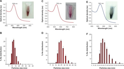
A schematic process for GNPs synthesis is presented in . In brief, the synthesis involved the addition of gum arabic,V. vinifera L. extract and NaAuCl4 into preheated water at 45°C and 80°C to NaAuCl4, followed by rapid cooling on ice. The V. vinifera L. extract alone could reduce NaAuCl4 salt to produce GNPs, but these GNPs were not stable and formed aggregates on long-term storage. Therefore, gum arabic was used as a stabilizer to avoid aggregation of GNPs. However, gum arabic alone could not reduce NaAuCl4 to produce the GNPs. Gum arabic is a highly branched polysaccharide structure containing arabinogalactan and glycoproteins. The glycoprotein coating is known to reduce the toxicity of metal nanoparticles.Citation33 The potential adverse effects of nanomaterials on human health are expected to reduce to a greater extent with the use of GNPs synthesized from natural plant compounds, such as polyphenols and phytochemicals, that are known to have abundant antioxidant and anticancer properties.Citation34,Citation35 In the current method, the organic acids, phenolic compounds, and polysaccharides might have reduced the NaAuCl4 into the GNPs.Citation33,Citation34 The GNPs of various sizes were synthesized, as different sizes could have significant effect on cellular signaling processes.Citation36 GNPs of various sizes were synthesized by changing the temperature of the reaction, the concentration of NaAuCl4, and the reducing agent (). GNPs with a diameter of 14 ± 1 nm () were synthesized at 80°C. Increasing the time of reduction resulted in the synthesis of the 28 ± 2 nm GNPs (). The 61 ± 2 nm GNPs () were synthesized at 45°C using higher Au salt concentration. The sizes of the GNPs are dependent on two independent processes, ie, growth and nucleation of the Au atoms during particle synthesis.Citation37,Citation38 The longer the duration of growth and nucleation processes, the bigger the particles synthesized. At higher temperatures with less Au salt, the atoms vibrate strongly at their lattice positions and exchange energy with neighboring atoms.Citation39 The lower temperatures resulted in the reduction in the diffusion and the higher strain energy that led to the formation of bigger particles. The effect of annealing temperature was studied previously by Goh et al with ZnO sacrificial template and polymethylsilsesquioxanes.Citation40 The size of GNPs is inversely proportional to the annealing temperature on ZnO sacrificial template. The size of GNPs decreases with increasing annealing temperature. The zeta potential of the 14 ± 1 nm particles was −32 mV. The bigger GNPs had reduced zeta potential of −22 mV, indicating lower stability of the bigger particles compared to the 14 ± 2 nm GNPs (). However, the negative zeta potential for all the GNPs indicates the relative stability of these particles in the dispersion medium.
Table 1 Important parameters that effect particle size and zeta potential
Figure 1 Schematic illustrating the GNPs synthesis procedure and coating of protein and Raman-active molecules on prepared GNPs.
Abbreviations: GNPs, gold nanoparticles; SERS, surface-enhanced Raman scattering; Au, gold; S, sulphur containing proteins.
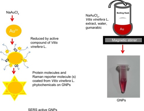
Figure 2 UV-vis spectra of the different sizes of GNPs: (A) 14 nm; (C) 28 nm; (E) 61 nm. Dynamic light-scattering histograms showing particle distribution of GNPs: (B) 14 ± 1 nm; (D) 28 ± 2 nm; (F) 61 ± 2 nm.
Abbreviation: GNPs, gold nanoparticles; UV-vis, ultraviolet-visible; au, arbitrary unit.
Characterization of GNPs
The maximum absorption at 528 nm for the GNPs with a diameter 14nm indicates that particles are homogeneous isotropic spheres in nature.Citation21,Citation41 Furthermore, the λ maximum of the SPR band can also be used to predict the size of the GNPs.Citation42 The absorption maxima of the SPR band of GNPs depends on the size of the nanoparticles, whereas the absorption intensity depends on the concentration of GNPs of same size. The wavelength maximum at 520–540 nm can be used to indicate the spherical size of the GNPs. The SPR band at 528 nm () is indicative of spherical particles less than 20 nm in diameter. Consistent with the prediction of particle size from SPR analysis, the average size measured by the DLS number distribution was 14 ± 1 nm (mean ± standard deviation) (). Single clusters of GNPs showed monodispersity of particles. The λ maximum (SPR band) of 28 ± 2 nm and 61 ± 2 nm showed a red shift at 530 nm and 541 nm, respectively () with respect to 14 ± 1 nm (). A similar red shift of the bigger nanoparticles was reported earlier.Citation43 According to Mie theory, the change in the SPR band is due to the adsorption of any material on the surface of GNPs and a change in the dielectric properties of the medium.
Previous studies found GNPs of less than 20 nm are more suitable for biological application compared to particles of bigger sizes, as their cellular uptake is higher and they can cross the blood–brain barrier in animal models.Citation44,Citation45 Therefore, for the present study, we selected GNPs sized 14 ± 1 nm for characterizations and in vitro study. In accordance with the predicted shape of the particles from absorption measurements, TEM () analysis showed spherical GNPs <20 nm. The GNPs size calculated from the TEM analysis was 16 ± 2 nm (). This value is closer to the size measured by DLS (14 ± 1 nm). The GNPs were characterized by XRD to understand the crystalline nature of the particles. The XRD spectrum exhibited peaks at 38° and 44°, which is indicative of (111) and (200) planes of face-centered cubic gold. The presences of (111) confirm the crystalline nature of the GNPs (). The indexing of the peaks using International Centre for Diffraction Data files confirmed that the characteristic X-ray peaks could be attributed to face-centered cubic Au structure. Earlier, reports of GNPs synthesis exhibited additional diffraction peaks at 64.8° and 78.8°, and these were ascribed to (214) and (311) planes, respectively.Citation21 The diffraction intensity of the (111) plane was higher compared to that of the (200) plane, indicating that (111) planes were predominant in GNPs synthesis. An overwhelmingly strong diffraction peak at the (111) plane compared to other facets was observed in an earlier report on GNPs synthesis.Citation21 The predominance of (111) could be due to lower surface energy compared to other planes, which results in weak bonding ability and chemical reactivity compared to other planes.Citation46,Citation47 The biomolecules from V. vinifera L. fruit extract preferentially adsorbed lower surface energy of the (111) plane and enabled the growth of the Au crystal facets in this plane. The adsorption of V. vinifera L. influenced the surface property and biocompatibility of GNPs. The fibronectin protein nanofibrillar Ag particles synthesized earlier had a predominant (111) plane in XRD indicates the high SERS ability.Citation47
Figure 3 (A–E) Characterization of GNPs (16 ± 2 nm). (A) TEM showing GNPs shape and size. The analysis was carried out at an accelerating voltage of 200 kV. The particles were measured on two different scales: 20 nm and 100 nm. (B) Histogram showing average size of GNPs measured by TEM. (C) X-ray diffraction spectra from the particles. The particles were recorded at 40 kV voltage and 40 MV current using Cu-Kα radiation (λ = 1.5405 Å, scan rate 1°/minute). (D and E) Fourier-transform infrared spectra of Vitis vinifera L. and GNPs. The spectrum was recorded from 500 to 4,000 cm−1 from the GNPs in the transmittance mode using KBr crystal.
Abbreviations: GNPs, gold nanoparticles; TEM transmission electron microscopy; SD, standard deviation; KBr, potassium bromide; au, arbitrary unit.
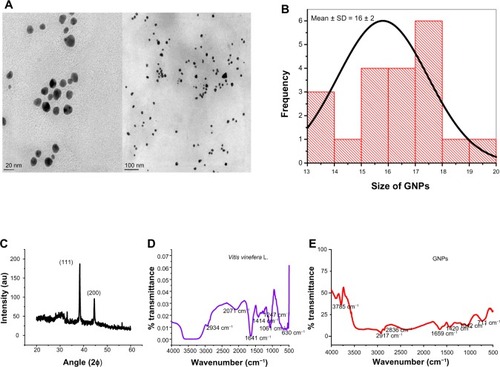
V. vinifera L. contains mainly sugar, glucose and fructose, organic acid, tartaric, malic, and citric acid, phenolic compound anthocyanins, tannins, and nitrogenous compound amino acid, peptides, and proteins.Citation48 FTIR analysis was performed to detect the vibration mode of chemical compounds and infer biochemical composition of V. vinifera L. and compound present on GNPs (). The sharp peak of GNPs at 3,785 cm−1 is probably an O−H stretch, indicating the presence of phenols from the V. vinifera L. fruit extract. The fruit extract consists of a variety of polyphenols, aldehydes, peptides, and proteins.Citation49,Citation50 The medium peaks of 2,934 cm−1 and 2,071 cm−1 indicate the C−H and C≡H of alkanes and alkynes, respectively (). The GNPs peaks at 2,917 cm−1 and 2,836 cm−1 are indicative of C−H and H−C=O bonds of aldehydes from V. vinifera L. coated on the GNPs. The peaks at 1,641 cm−1 () and 1,659 cm−1 () are indicative of N−H bond vibrations from amide groups of the proteins present in V. vinifera L. fruit extract as well as in the GNPs.Citation51,Citation52 The protein content was estimated by the Bradford test found to be 0.056 μg/2 × 107 particles for 61 ± 2 nm- and 0.045 μg/7 × 108 particles for 14 ± 1 nm-sized GNPs. The peak at 1,420 cm−1 is indicative of a C−C stretch from the polyphenols of the V. vinifera L. present on the surface of GNPs. On the other hand, peaks at 1,247 cm−1, 1,061 cm−1, and 1,042 cm−1 might be aliphatic amines with C−N stretches. The peaks at 630 cm−1 and 711 cm−1 could be from alkanes () present in the V. vinifera L. fruit extract. Furthermore, the 711 cm−1 peak could be from the rocking bond of the alkanes present on the GNPs (). Earlier reports have attributed this peak to the presence of alkanes.Citation53,Citation54 The overlapping peak of the functional group of V. vinifera L. present on the GNPs also corroborates our results in that chemical compounds from the V. vinifera L. fruit extract had been coated on the surface of GNPs and formed chemical bond with GNPs.
Stability and cytotoxicity of GNPs
The stability of nanoparticles is an indispensable parameter for biomedical applications. The in vitro stability of the GNPs () was evaluated by monitoring the plasmon wavelength (λ maximum) in borate buffer, RPMI, NaCl, RPMI + FBS, cysteine and histidine, and NaCl at 0 days, and 15 days after the synthesis of GNPs. The plasmon wavelength and bandwidth in the above buffers did not show a shift in SPR band at 0 days, whereas after 15 days the SPR band exhibited a small shift of less than 5 nm. The constant SPR band indicated that GNPs were stable in all buffers and cell-culture media. The data indicated that there was no difference in the SPR band in any of the buffers with respect to GNPs alone (as a control). Our results are consistent with other studies on the stability of GNPs synthesized from plant compounds.Citation14,Citation15
Figure 4 Stability and cytotoxicity of GNPs (top panel). UV-vis spectra of the GNPs in various buffers at various time periods: (A) 1 day; (B) 15 days. Cytotoxicity measurements of GNPs in different cell lines at various concentrations (bottom panel): (C) MIO-M1 (Müller glial, noncancerous); (D) MDA-MB 453 (breast cancer). The cell viability (% of treated cells with respect to untreated cells) of different cell lines treated with different concentrations: 10, 25, 50, and 100 μM of GNPs at various time periods.
Notes: *Significant difference with respect to control at P< 0.05; error bars represent the standard error of mean.
Abbreviations: GNPs, gold nanoparticles; RPMI+FBS, Roswell Park Memorial Institute medium plus fetal bovine serum; RPMI, Roswell Park Memorial Institute medium; Nacl, sodium chloride; UV-vis, ultraviolet-visible; au, arbitrary unit.
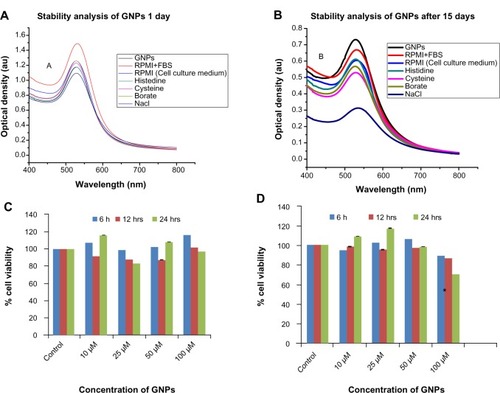
A key parameter in evaluating biocompatibility is centered on the evaluation of potential cytotoxicity of the materials. The in vitro cytotoxicity effect of nanomaterials is most often assessed from mitochondrial enzyme activity using tetrazo-lium salt (MTT assay), with respect to the untreated cell as a control.Citation55,Citation56 Cytotoxicity of the GNPs tested using MTT assay in noncancerous Müller glial (MIO-M1) and breast cancer (MDA-MB-453) cells (). The GNPs were nontoxic to the cells up to 100 μM concentration. Cell viability did not decrease after 6 and 12 hours of treatment with increasing concentrations of GNPs from 10 to 100 μM (P < 0.05) in either normal or breast cancer cell lines when compared to the untreated control cells. The cytotoxicity of the GNPs was previously reported to be dependent on GNP concentration, time of treatment, and cell type.Citation16–Citation18 GNPs synthesized using synthetic and toxic chemicals such as LiBH4 penetrate the membranes at high concentrations and decrease cell proliferation via generating reactive oxygen species, in turn decreasing the mitochondrial enzyme activity adenosine triphosphate content of the cell, causing cell damage.Citation57 In contrast, GNPs synthesized using green nanotechnology were nontoxic even at higher concentrations.Citation15 The percentage cell viability was more than 85% until 12 hours of treatment in the Müller glial (MIO-M1) and the breast cancer (MDA-MB-453) cells. However, 100 μM of GNP treatment at 24 hours showed 70.28% cell viability in MDA-MB-453 cells (). These cells showed a significant difference (P < 0.05) when compared to the control at 24 hours treatment with GNPs. On the other hand, Müller glial (MIO-M1) cells showed no significant decrease (P = 0.2) in cell viability after 24 hours of GNP treatment (). According to the ISO-10993-5 guideline,Citation58 a material can be used for biomedical application if the material shows more than 70% cell viability. The GNPs from 10 to 100 μM concentration are safe to use for in vitro studies.
Moreover the toxicity of the metal nanoparticles is reduced by surface coating.Citation59 The GNPs were coated with gum arabic in the present study. This coating might have been responsible for the observed low toxicity in the normal and breast cancer cell lines, as it prevents the direct contact of the metal nanoparticles with the cells and makes these particles more biocompatible than uncoated particles. Gum arabic-coated GNPs have been previously tested in animal models and found to be nontoxic.Citation60
SERS signature on GNPs
SERS is a unique property of metal nanoparticles that can be used for imaging applications. Therefore, in the current work, GNPs of various sizes (14 ± 1 nm and 61 ± 2 nnm) were tested for the SERS signature without addition of known synthetic Raman-reporter molecules. Synthetic chemicals as Raman reporters have been used before for SERS.Citation29 In our study, Raman intensity was measured from 500 to 2,000 cm−1 (). Raman spectral peak intensities were observed at 570, 788, and 1,102 cm−1. The peak intensity of the different sizes revealed that the intensity of the peak increased with increasing GNPs size. In agreement with the present work, earlier reports indicated that SERS intensity increases with increased particle size.Citation24,Citation61 Raman spectral peak intensities from the V. vinifera L. fruit extract were also observed at 570, 788, and 1,102 cm−1, signifying that the Raman-reporter molecules from the extract were retained on the GNPs during the synthesis process. Interestingly, Raman-signal intensity was higher in GNPs compared to V. vinifera L. fruit extract alone, indicating a SERS effect (). The GNPs were doped with the well-known Raman reporter R6G to study the RE. This enhancement is comparable to the RE attained with Raman reporters from the V. vinifera L. extract. The R6G-doped GNPs showed Raman peak signature for the R6G dye (). A similar signature was reported in an earlier study.Citation21 The RE was calculated as described by Chang et al using the formula given in the Materials and methods section.Citation32
Figure 5 (A–C) Synthesized GNPs function as SERS nanotag. (A) Raman spectra from GNPs of different sizes: black, grape extract; blue, spectrum from 14 ± 1 nm nanoparticles; pink, spectrum from 61 ± 2 nm particles. (B) Raman spectrum of R6G-doped GNPs: red, R6G-doped particles; black, R6G dye alone. (C) Stability of GNPs after long-term storage: black, 0 days; red, 30 days.
Abbreviations: GNPs, gold nanoparticles; SERS, surface-enhanced Raman scattering; R6G, rhodamine 6G; au, arbitrary unit.
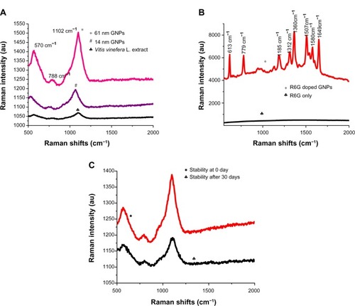
The number of R6G dye molecules in the GNPs used to obtain Raman spectra () was 120.4 × 10¹⁴. The number of dye molecules used as control was 6.23 × 10²⁰. The RE was calculated using the peak heights from the base of the 1,360 cm−1 peak for R6G. The RE for R6G dye-doped GNPs was R6G 3.4 × 10⁷. The RE of 108 has been previously reported for R6G-doped GNPs.Citation21 The peak height from the base of the most prominent peak – 1,102 cm−1 – was used for calculations of RE of GNPs without dye doping. The enhanced Raman intensity for the Raman reporters in GNPs compared to the V. vinifera L. fruit extract was used for calculations (). The RE of R6G-doped GNPs was extrapolated for the reporter molecules from V. vinifera L. fruit extract using Raman peak intensities. The RE for the 61 nm-sized GNPs was 4 × 10³. The R6G molecule exhibited higher SERS enhancement compared to the Raman-reporter molecules from V. vinifera L. (). SERS activity is known to be dependent on two key factors: (1) resonant surface-plasmon excitation of a metal substrate, and (2) close proximity of analytic molecules to the metal substrate surface.
The surface chemistry of metal substrate was considered as a critical factor for SERS activity because the analytes must be located within 0–4 nm of the substrate surface or the electromagnetic field. Therefore, we proposed that the GNPs had Raman reporter molecule(s) in the close proximity of the Au metal surfaces, resulting in SERS enhancements.Citation62 We hypothesized that the aromatic compounds from the V. vinifera L. extract act as SERS nanotags. The higher SERS activity of R6G may be due to the closer proximity of R6G molecules compared to the Raman reporters from V. vinifera L. fruit extract. The Raman peaks at 570 cm−1 could be due to in-plane bending from carbon and oxygen from the phenolic compounds or aromatic acids, such as p-coumaric acid.Citation63,Citation64 This phenol is present in V. vinifera L. fruit extract and resins. The vibrational peak at 1,102 cm−1 could be from the sugar molecules present in V. vinifera L. extract.Citation65 De Gelder et al reported that the 1,102 cm−1 band is indicative of a trehalose sugar.Citation63 The vibrational peak at 788 cm−1 could be from the histidine amino acid.Citation60 The SERS activity of such biological molecules as histidine, cysteine, and lysine using gold substrate has been reported previously.Citation21,Citation60,Citation63
The long-term stability of the GNPs was assessed for their potential application after storage of the particles. The lack of shift in Raman peaks after 1 month compared to the freshly synthesized particles indicated the stability of the Raman-reporter molecules on GNPs (). On the other hand, stored V. vinifera L. extract was incapable of reducing Au salt to form GNPs with SERS signature. The newly synthesized and stored GNPs can be used for biomedical applications. Earlier studies used SERS active gold nanoparticles for in vivo tumor-cell targeting and sensitive cancer detection.Citation27,Citation29 However, these studies employed GNPs synthesis using NaBH4 as reducing agent and synthetic dyes as SERS-reporter molecules. Our method of synthesis of SERS nanotags is faster and safer than recent syntheses using NaBH4 and ascorbic acid.Citation31
Conclusion
In summary, a single reaction-based, rapid, economical, and environment-friendly method of biosynthesizing SERS nano-particles has been successfully established using V. vinifera L. extract. The synthesized GNPs were spherical in shape and coated with aromatic compounds (proteins/peptides) from V. vinifera L. The phytochemicals from the V. vinifera L. extracts acted both as reducing agents for the synthesis of GNPs and as reporter molecules for SERS. One major advantage of GNPs is SERS ability without the addition of synthetic chemicals or the need for complicated dye-doping procedures, previously reported for the synthesis of SERS nanotags. The identity of the Raman-reporter molecule(s) is presently unknown, and work is being carried out to study the polyphenols, aldehydes, peptides, and protein compounds present in V. vinifera L. extract. As GNPs are nontoxic to cells, the authors believe that the SERS ability of GNPs has great potential in many areas, including cancer diagnosis, therapy, and ultrasensitive biomarker detection.
Acknowledgments
The authors thank the Department of Science and Technology – Nano Mission India (grant no. SR/NM/NS-83/2010) for financial support and Deakin University, Australia for scholarship (Sushma Kalmodia, ID 211823217). The authors also thank Professor Ashutosh Sharma, Indian Institute of Technology, for providing the facility of the DST Centre for Nanotechnology. They also thank Ms Shyama for critical review of manuscript for language proofreading.
Disclosure
The authors report no conflicts of interest in this work.
References
- MericanZSchillerTLHawkerCJFredericksPMBlakeyISelf-assembly and encoding of polymer-stabilized gold nanoparticles with surface-enhanced Raman reporter moleculesLangmuir200723105391054517824719
- WilletsKAVan DuyneRPLocalized surface plasmon resonance spectroscopy and sensingAnn Rev Phys Chem20075826729717067281
- LuLKobayashiATawaKOzakiYSilver nanoplates with special shapes: controlled synthesis and their surface plasmon resonance and surface-enhanced Raman scattering propertiesChem Mater20061848944901
- SteinmetzNReview of “Cancer Nanotechnology: methods and Protocols (Methods in Molecular Biology)” by Stephen R. Grobmyer (editor), Brij M. Moudgil (editor)Biomed Eng Online2010955
- ZhangXXingJZChenJEnhanced radiation sensitivity in prostate cancer by gold-nanoparticlesClin Invest Med200831E160E16718544279
- DobrovolskaiaMAMcNeilSEImmunological properties of engineered nanomaterialsNat Nanotechnol2007246947818654343
- ChenPCMwakwariSCOyelereAKGold nanoparticles: from nanomedicine to nanosensingNanotechnol Sci Appl20081456624198460
- ThakorASJokerstJZavaletaCMassoudTFGambhirSSGold nanoparticles: a revival in precious metal administration to patientsNano Lett2011114029403621846107
- Centers for Disease Control and PreventionUni-Gold Recombigen™ HIV: FDA news release2003 Available from: http://www.cdc.gov/hiv/topics/testing/resources/press_releases/FDArelease.htmAccessed August 2, 2013
- KotokATrial of insulin-coated gold nanoparticles approved2011 Available from: http://sciencebusiness.technewslit.com/?p=6957Accessed August 2, 2013
- PacardoDBSethiMJonesSENaikRRKnechtMRBiomimetic synthesis of Pd nanocatalysts for the Stille coupling reactionACS Nano200931288129619422199
- NelAXiaTMädlerLLiNToxic potential of materials at the nanolevelScience200631162262716456071
- LeeKWBodeAMDongZMolecular targets of phytochemicals for cancer preventionNat Rev Cancer20111121121821326325
- ElavazhaganTArunachalamKDMemecylon edule leaf extract mediated green synthesis of silver and gold nanoparticlesInt J Nanomedicine201161265127821753878
- AromalSAVidhuVKPhilipDGreen synthesis of well-dispersed gold nanoparticles using Macrotyloma uniflorumSpectrochim Acta A Mol Biomol Spectrosc2012859910422018585
- KattiKChandaNShuklaRGreen nanotechnology from cumin phytochemicals: generation of biocompatible gold nanoparticlesInt J Green Nanotechnol Biomed20091B39B5219890490
- ShuklaRNuneSKChandaNSoybeans as a phytochemical reservoir for the production and stabilization of biocompatible gold nanoparticlesSmall200841425143618642250
- ChandranSPChaudharyMPasrichaRAhmadASastryMSynthesis of gold nanotriangles and silver nanoparticles using Aloe vera plant extractBiotechnol Prog20062257758316599579
- ChaudharySPaulSSagarSBiosynthesis of silver nanoparticles using Vitis vinifera extract and evaluation of their antimicrobial activityInt J Biotechnol Res20122112
- NuneSKChandaNShuklaRGreen nanotechnology from tea: phytochemicals in tea as building blocks for production of biocompatible gold nanoparticlesJ Mater Chem2009192912292020161162
- ZhangYXZhengJGaoGBiosynthesis of gold nanoparticles using chloroplastsInt J Nanomedicine201162899290622162651
- MaitiKKSamantaAVendrellMSohKSOlivoMChangYTMultiplex cancer cell detection by SERS nanotags with cyanine and triphenylmethine Raman reportersChem Commun (Camb)2011283514351621308123
- LeeKSEl-SayedMAGold and silver nanoparticles in sensing and imaging: sensitivity of plasmon response to size, shape, and metal compositionJ Phys Chem B2006110192201922517004772
- NieSEmorySRProbing single molecules and single nanoparticles by surface-enhanced raman scatteringScience1997275110211069027306
- KneippJKneippHMcLaughlinMBrownDKneippKIn vivo molecular probing of cellular compartments with gold nanoparticles and nanoaggregatesNano Lett200662225223117034088
- KneippKWangYKneippHSingle molecule detection using surface-enhanced Raman scattering (SERS)Phys Rev Lett19977816671670
- LeeSChonHYoonSYFabrication of SERS-fluorescence dual modal nanoprobes and application to multiplex cancer cell imagingNanoscale2012412412922080302
- KhoKWFuCYDinishUSOlivoMClinical SERS: are we there yet?J Biophotonics2011466768421922673
- JokerstJVColeAJVan de SompelDGambhirSSGold nanorods for ovarian cancer detection with photoacoustic imaging and resection guidance via Raman imaging in living miceACS Nano20126103661037723101432
- LiJFHuangYFDingYShell-isolated nanoparticle-enhanced Raman spectroscopyNat Nanotechnol2010464392395
- BocaSRuginaDPinteaALeopoldNAstileanSDesigning gold nanoparticle-ensembles as surface enhanced Raman scattering tags inside human retinal cellsJ Nanotechnol20122012110
- ChangCWLiaoJDChangHCLinLKLinYYWengCCFabrication of nano-indented cavities on Au for the detection of chemically-adsorbed DTNB molecular probes through SERS effectJ Colloid Interface Sci201135838439121463869
- GarcíaIMarradiMPenadésSGlyconanoparticles: multifunctional nanomaterials for biomedical applicationsNanomedicine (Lond)2010577779220662648
- VisioliFDe La LastraCAAndres-LacuevaCPolyphenols and human health: a prospectusCrit Rev Food Sci Nutr20115152454621929330
- RussoMSpagnuoloCTedescoIRussoGLPhytochemicals in cancer prevention and therapy: truth or dare?Toxins (Basel)2010251755122069598
- JiangWKimBYRutkaJTChanWCNanoparticle-mediated cellular response is size-dependentNat Nanotechnol2008314515018654486
- TurkevichJStevensonPCHillierJA study of the nucleation and growth processes in the synthesis of colloidal goldDiscuss Faraday Soc1951115575
- KimlingJMaierMOkenveBKotaidisVBallotHPlechATurkevich method for gold nanoparticle synthesis revisitedJ Phys Chem B2006110157001570716898714
- MittemeijerEJFundamentals of Materials Science: The Microstructure–Property Relationship Using Metals as Model SystemsHeidelbergSpringer2011
- GohLPRazakKARidhuanNSCheongKYOoiPCAwKCDirect formation of gold nanoparticles on substrates using a novel ZnO sacrificial templated-growth hydrothermal approach and their properties in organic memory deviceNanoscale Res Lett2012756323046949
- MieGBeitrage zer Optik trüber Meiden speziell kolloidaler MetallösungenAnn Phys190825377445
- Le RuEEtchegoinPPrinciples of Surface-Enhanced Raman Spectroscopy: And Related Plasmonic EffectsAmsterdamElsevier2009
- HeSTYaoJNJiangPFormation of silver nanoparticles and self-assembled two-dimensional ordered superlatticeLangmuir20011715711575
- Semmler-BehnkeMKreylingWGLipkaJBiodistribution of 1.4- and 18-nm gold particles in ratsSmall200842108211119031432
- JinHHellerDASharmaRStranoMSSize-dependent cellular uptake and expulsion of single-walled carbon nanotubes: single particle tracking and a generic uptake model for nanoparticlesACS Nano2009314915819206261
- SunYGMayersBXiaYNTransformation of silver nanospheres into nanobelts and triangular nanoplates through a thermal processNano Lett20033675679
- WangLSunYJCuiYCWangJKLiZSynthesis of silver nanoplates with fibronectin nanofibril template and their SERS applicationsBull Korean Chem Soc201334443446
- XiaEQDengGFGuoYJLiHBBiological activities of polyphenols from grapesInt J Mol Sci20101162264620386657
- López-VázquezCOrriolsIPerellóMCde RevelGDetermination of aldehydes as pentafluorobenzyl derivatives in grape pomace distillates by HS-SPME-GC/MSFood Chem201213011271133
- AliKMalteseFChoiYHVerpoorteRMetabolic constituents of grapevine and grape-derived productsPhytochem Rev2010935737820835385
- RajasekharreddyPRaniPUSreedharBQualitative assessment of silver and gold nanoparticle synthesis in various plants: a photobiological approachJ Nanopart Res20101217111721
- BeattieIRHaverkampRGSilver and gold nanoparticles in plants: sites for the reduction to metalMetallomics2011362863221611658
- VasitaRKattiDSStructural and functional characterization of proteins adsorbed on hydrophilized polylactide-co-glycolide microfibersInt J Nanomedicine20127617122275823
- DharmadhikariMComposition of grapesNewsletter: Vineyard and Vintage View1994938
- KhlebtsovNDykmanLBiodistribution and toxicity of engineered gold nanoparticles: a review of in vitro and in vivo studiesChem Soc Rev2011401647167121082078
- MosmannTRapid colorimetric assay for cellular growth and survival: application to proliferation and cytotoxicity assaysJ Immunol Methods19831655636606682
- AshaRaniPVLow Kah MunGHandeMPValiyaveettilSCytotoxicity and genotoxicity of silver nanoparticles in human cellsACS Nano2009327929019236062
- Association for the Advancement of Medical Instrumentation, International Organization for StandardizationBiological Evaluation of Medical Devices Part 5: Tests for in Vitro CytotoxicityArlington (VA)AAMI2009
- ArnidaMaluginAGhandehariHCellular uptake and toxicity of gold nanoparticles in prostate cancer cells: a comparative study of rods and spheresJ Appl Toxicol20103021221719902477
- FentGMCasteelSWKimDYBiodistribution of maltose and gum arabic hybrid gold nanoparticles after intravenous injection in juvenile swineNanomedicine2009512813519480048
- SchwartzbergAMGrantCDWolcottAUnique gold nanoparticle aggregates as a highly active SERS substrateJ Phys Chem B20041081919119197
- ClaridgeSALiangHWBasuSRFréchetJMAlivisatosAPIsolation of discrete nanoparticle-DNA conjugates for plasmonic applicationsNano Lett200881202120618331002
- De GelderJDe GussemKVandenabeelePMoensLReference database of Raman spectra of biological moleculesJ Raman Spectrosc20073811331147
- ZukMDymińskaLKulmaAIR and Raman studies of oil and seedcake extracts from natural and genetically modified flax seedsSpectrochim Acta A Mol Biomol Spectrosc2011781080108921237701
- KolbeATiessenASchluepmannHPaulMUlrichSGeigenbergerPTrehalose 6-phosphate regulates starch synthesis via posttranslational redox activation of ADP-glucose pyrophosphorylaseProc Natl Acad Sci U S A2005102111181112316046541