
 ?Mathematical formulae have been encoded as MathML and are displayed in this HTML version using MathJax in order to improve their display. Uncheck the box to turn MathJax off. This feature requires Javascript. Click on a formula to zoom.
Abstract
Nanoparticle sulphated zirconia with Brønsted acidic sites were prepared here by an impregnation reaction followed by calcination at 600°C for 3 hours. The characterization was completed using X-ray diffraction, thermal gravimetric analysis, Fourier transform infrared spectroscopy, Brunner-Emmett-Teller surface area measurements, scanning electron microscopy with energy dispersive X-ray spectroscopy, and transmission electron microscopy. Moreover, the anticancer and antimicrobial effects were investigated for the first time. This study showed for the first time that the exposure of cancer cells to sulphated zirconia nanoparticles (3.9–1,000 μg/mL for 24 hours) resulted in a dose-dependent inhibition of cell growth, as determined by (4,5-dimethylthiazol-2-yl)-2,5-diphenyltetrazolium bromide assays. Similar promising results were observed for reducing bacteria functions. In this manner, this study demonstrated that sulphated zirconia nanoparticles with Brønsted acidic sites should be further studied for a wide range of anticancer and antibacterial applications.
Introduction
Previously, a number of organic synthesis methods have been carried out with heterogeneous solid acids, leading to better regio- and stereoselectivity.Citation1–Citation3 It has been found that it is not just the strength of the acid but also the type of acidity (Brønsted or Lewis) that matters for improved activity and selectivity. The inclusion of superacidity in solids has attracted great attention.Citation4 Among superacid nanoparticles, sulphated zirconia has found several applications and holds large promise in a number of reactions of importance, which would only increase if synthesized into nanoparticles.Citation5 The activity of nanoparticle sulphated zirconia depends on the successful inclusion of sulphoxy moieties into the zirconium oxide skeleton.Citation6 Such materials show excellent performance toward α-pinene isomerization in a moderate setting.Citation7 Production of the drug itself in nanoparticulate form has also been introduced as an approach for the increased release of hydrophobic drugs because of their distinctive advantages over colloidal drug carriers.Citation8
In general, metal oxides have been used widely either as such or as supports in conjunction with other active components for many oxidation, reduction, and acid-base-catalyzed reactions.Citation9–Citation12 For instance, nearly all developed catalysts use an oxide as the energetic phase, which reveals both proton and electron transfer capacities and could be used as a catalyst in neutralization as well as in reduction-oxidation reactions. These metal oxide redox properties are employed in catalytic decontamination schemes for the total oxidation of contaminated constituents.Citation13–Citation20 The metal oxide scheme with intrinsic redox features has also been used for discriminatory oxidation of organic materials and to produce vital fine chemicals, such as acids, nitriles, and aldehydes.Citation21–Citation23
The surface nature of the metal oxides has been considered advantageous in discerning organic conversions. Because the rate-determining step on the surface of the catalyst can be determined by the density of active centers and surface area, improved enhancers should have high surface area and smaller sizes of particles. Therefore, in the developing field of nanotechnology, the production of metal oxide nanoparticles has received considerable attention because of the unique physicochemical characteristics obtained for nano-sized compared with micron (or conventional)-sized particles.Citation24,Citation25
The surface of zirconium oxide is known to possess all of these catalytic activities. Zirconium oxide, or zirconia, when modified with anions such as sulphate ions, gives a highly acidic or superacidic catalyst that has superior activity to catalyze many reactions.Citation26–Citation28 Zirconia has been widely used to catalyze many engineering reactions.Citation29,Citation30 Thus, sulphated zirconia and modified sulphated zirconia form an important class of catalysts, as is evident from the large amount of research that has become visible during the past decade.Citation31 Furthermore, these catalysts show a promising future, in that they can offer environmentally clean routes for the chemical and pharmaceutical industries into the next millennium.
Despite their potential biomedical applications, very few studies have reported on the role of zirconia nanoparticles as anticancer materials. As far as the authors know, this is the first report of the in vitro anticancer effect of sulphated zirconia nanoparticles against three cancer cell lines. Specifically, the toxicity of sulphated zirconia nanoparticles against human colon cancer HT29, human breast cancer MCF-7 and human liver cancer HepG2 cell lines was assessed, showing promising results.
Moreover, it is reported here that these novel nanoparticles hold promise not just for anticancer applications but also for anti-infection applications. The steady increase in the antimicrobial resistance of microorganisms represents a great public health concern. This requires the search for new unconventional antimicrobial agents. Nanotechnology provides promising materials to fight infectious diseases such as nanoparticles with antimicrobial activities. In the present study, the antimicrobial properties of the as-synthesized sulphated zirconia nanoparticles were determined using the agar diffusion method against different Gram-positive and Gram-negative bacteria of clinical significance.
A survey of the literature has also demonstrated that no previous work has been done so far on the investigation of anticancer and antimicrobial effects of sulphated zirconia nanoparticles. The main purpose of this study was to prepare sulphated zirconia superacidic nanoparticles for use as a chemotherapeutic agent against human colon cancer HT29, human breast cancer MCF-7, and human liver cancer HepG2 cell lines. Moreover, the antimicrobial activity of sulphated zirconia nanoparticles against Candida albicans was also examined. The sulphated zirconia nanoparticles were prepared by an impregnation method and characterized using X-ray diffraction (XRD), thermal gravimetric analysis (TGA), Fourier transform infrared spectroscopy (FT-IR), Brunner-Emmett-Teller (BET), scanning electron microscopy with energy dispersive X-ray spectroscopy (SEM-EDS), and transmission electron microscopy (TEM).
Experimental
Materials
Ammonium sulphate, zirconium oxy nitrate, and ammonium hydroxide (28%–30%) were obtained from Sigma-Aldrich (St Louis, MO, USA). A trypsin/ethylenediamine tetraacetic acid solution was purchased from Invitrogen (Carlsbad, CA, USA). Dimethylsulfoxide, phosphate-buffered saline, 3-(4,5-dimethylthiazol-2-yl)-2,5-diphenyltetrazolium bromide (MTT), and Dulbecco’s Modified Eagle’s Medium were purchased from Sigma-Aldrich.
Preparation of the sulphated zirconia nanoparticles
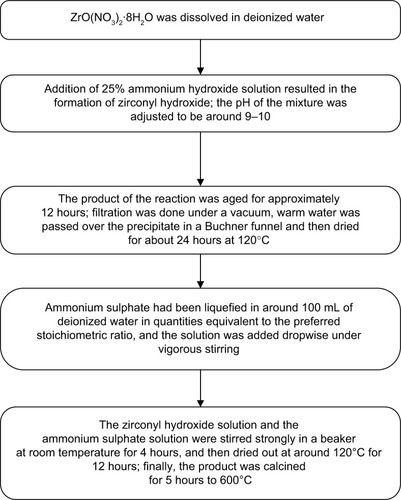
A schematic representation of the preparation of sulphated zirconia nanoparticles is shown in .
Figure 1 Flow chart for preparation of sulphated zirconia nanoparticles via impregnation method.
Physicochemical characterization of sulphated zirconia
Powder XRD analysis was carried out using a Shimadzu diffractometer model XRD 6000. The diffractometer employing Cu-Kα radiation was used to generate diffraction patterns from powder crystalline samples at ambient temperature. The Cu-Kα radiation was generated by a Philips glass diffraction X-ray tube broad focus at 2.7 kW. The crystallite size D of the samples was calculated using the Debye-Scherrer’s relationship:Citation32–Citation34
The TGA analysis was carried out on a Mettler Toledo Thermo-Gravimetric-Simultaneous Differential Thermal Analysis, Platinum and Rhodium apparatus (Pt crucibles, Pt/Pt–Rh thermocouple) with the purge gas (nitrogen) flow rate of 30 mL minute−1 and a heating rate of 10°C minute−1 from room temperature to 1,000°C.
Fourier transform infrared analysis was carried out with a PerkinElmer spectrometer model 100 series (sample preparation Universal Attenuated Total Reflectance).
SEM-EDS was used to obtain information about the morphology and chemical composition of the samples. The morphology study of the nanoparticles was carried out using a JEOL scanning electron microscope model JSM-6400, whereas the quantitative chemical composition of the prepared ferric-manganese promoted sulphated zirconia acid catalyst was characterized using EDS for elemental chemical analysis.
TEM (Hitachi H-7100, Japan) was also used to examine the crystal shape. For TEM analysis, the powder was dispersed in deionized water, dropped onto carbon-cover copper grids placed on filter paper, and dried at room temperature.
The zeta potential of the sulphated zirconia nanoparticle dispersions (1 μg of sulphated zirconia nanoparticles dispersed in 1 mL ultradeionized water) were characterized using a ZetaSizer Nano ZS (Malvern Instruments Ltd, Malvern, UK) with dynamic light scattering.
The total surface area of the nanoparticles was obtained using a BET method with nitrogen adsorption at −196°C. Analysis was conducted using a Thermo Fisher Scientific S.P.A. nitrogen adsorption-desorption analyzer (SURFER ANALYZER).
Cell culture
The following human cell lines were obtained from the American Type Culture Collection (Manassas, VA, USA) and were used in the study: human breast cancer (MCF-7), human colon cancer (HT29), human liver cancer (HepG2), and normal human breast (MCF-10a) cells, which were all characterized as virus-negative. They grow as an adherent monolayer of tightly knit epithelial cells. These cells were grown in Dulbecco’s Modified Eagle’s Medium, which was supplemented with 10% fetal bovine serum. The media contained penicillin (100 U/mL) and streptomycin (100 g/mL). The cells were grown at 37°C in a humidified 5% CO2 incubator.
Cytotoxicity MTT assay
HT29, MCF-7, MCF-10a, and HepG2 cells lines were plated at 1×10³ cells/well by adding 200 μL of a 5×10³ cells/mL suspension to each well of a 96-well tissue culture plate. The plates were incubated for sufficient time to ensure attachment at 30%–40% confluence. The media was aspirated off and replaced with fresh media (200 μL) containing sulphated zirconia nanoparticles at different concentrations (3.9–250 μg/mL) and chemotherapeutic agents at 0.156–10.0 μg/mL (oxaliplatin for HT29 cells, doxorubicin for both MCF-7 and MCF-10a cells, and tamoxifen for HepG2 cells). The last row was left as an untreated control. The plates were incubated at 37°C, 5% CO2, for 24 hours. After incubation with the compounds, the media was aspirated off and the cells were washed by phosphate-buffered saline buffer three times to ensure all drugs were removed, and then the buffer was replaced with fresh media. The MTT solution (20 μL) at a total volume of 200 μL was added to every well and mixed gently with the media, which was later incubated for 4–6 hours at 37°C with 5% CO2. The MTT-containing medium was then removed carefully and replaced with dimethylsulfoxide (200 μL per well) to dissolve the formazan crystals. The plates were read in a microtiter plate reader at 570 nm. The concentration of drug needed to inhibit cell growth by 50% (IC50) was generated from the dose-response curves for each compound and each cell line.
In vitro antimicrobial susceptibility test
Four bacterial strains were used to determine the antibacterial activity for the synthesized sulphated zirconia nanoparticles: two Gram-positive bacteria (methicillin-resistant Staphylococcus aureus and Bacillus subtilis) and two Gram-negative bacteria (Salmonella choleraesuis and Pseudomonas aeruginosa). In addition, the yeast Candida albicans was used to screen the antifungal activity of the synthesized nanoparticles. The microorganisms were obtained from the microbial culture collection unit, Institute of Bioscience, University Putra Malaysia. The bacterial cultures were maintained on Mueller Hinton agar slants (Sigma-Adrich). The bacterial cultures were incubated overnight in 5 mL Mueller Hinton broth (Sigma-Aldrich) in a Certomat BS-T incubation shaker (Sartorius Stedim Biotech, Aubagne, France) at 37°C and 150 rpm until the culture reached an optical density of 600 of 1.0 (Spekol UV VIS 3.02, Analytic Jena, Jena, Germany), which corresponds to 108 colony-forming units per ml.
The antimicrobial activities of the synthesized sulphated zirconia nanoparticles were evaluated against the abovementioned microorganisms, using the agar diffusion (cup diffusion) method according to the guidelines of the Clinical and Laboratory Standards Institute. In brief, 20 mL liquid-autoclaved Mueller Hinton agar (pH 7.3±0.2 at 25°C) was poured onto the disposable sterilized Petri dishes and solidified. The solidified agar plate surfaces were dried in an incubator before streaking of the microorganisms onto the surface of the agar plate. Next, 100 μL of the microbial suspensions in the Mueller Hinton broth was streaked over the dried surface of the agar plates and spread uniformly, using a sterilized glass rod. The plates were then dried. Wells were then created in the agar plates, using a sterilized cork borer; the wells were filled with the nanoparticle suspensions in distilled water; and the control wells were filled with ampicillin (for Gram-negative bacteria), streptomycin (for Gram-positive bacteria), and nystatin (for Candida albicans) solutions as controls. The experiment was performed in triplicate, and the diameters of the zone of inhibition were measured to the nearest millimeter, using a caliper after a 24-hour incubation at 37°C. Differences between means were determined using standard analysis of variance, followed by Student t-tests.
Results and discussion
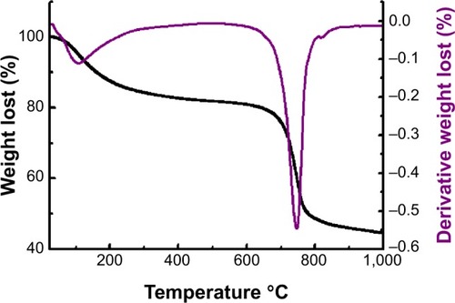
shows the thermogravimetric curve of sulphated zirconia, and the mass losses found from the TGA measurements agreed fairly well with those expected for the decomposition of hydrated sulphated zirconia.Citation35 Therefore, the heating events for this sample below 620°C are attributed to the removal of adsorbed water and the dehydroxylation process of ZrO(OH)2, whereas the second major weight loss at a higher temperature began at about 673°C and ended at 926°C and is referred to as the decomposition of sulphate groups.Citation36
Figure 2 Thermogravimetric curve of sulphated zirconia calcined at 600°C for 3 hours.
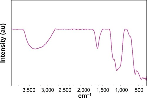
depicts the Fourier transform infrared spectroscopy spectra of sulphated zirconia from these spectra, and the presence of sulphate groups was confirmed by the band at the range of 1,225–1,070 cm−1, a result of the asymmetric and symmetric stretching frequency of the O=S=O and O–S–O groups.Citation37,Citation38 The absence of peaks around 1,450 cm−1 confirmed the presence of polynuclear sulphate in the sample irrespective of the high sulphate loading. The band around 1,625 crn−1 and 3,325 crn−1 corresponds to the bending and stretching modes of the –OH groups of water molecules present in the sample. The presence of these bands even after high temperature calcination points to the existence of Brønsted acidity in the samples.
Figure 3 Fourier transform infrared spectroscopy spectra of calcined sulphated zirconia.
The specific surface area for the sulphated zirconia and zirconia obtained using the BET method was 38.0 and 34.6 m2/g, respectively. It was also observed that the incorporation of the sulphate dopant in the structure of zirconia caused an increase in specific surface area and pore size of sulphated zirconia. This allowed reactants to be in contact with more acid sites, and the nanoparticles would have better activity.
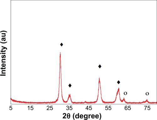
The powder XRD patterns of sulphated zirconia calcined at 600°C for 3 hours is illustrated in . The XRD patterns of sulphated zirconia show peaks assigned to both the tetragonal phase at 2θ (30.2°, 35.4°, 50.2°, 60.3°) (JSPDS file 00-014-0534: zirconium oxide) and diffraction lines around 2θ (43.8°, 62.9°, 75.1°) (JSPDS file 01-086-1449ZrO2) of the monoclinic phase of zirconia.Citation1,Citation39 By using the Scherrer’s equation, the average size of sulphated zirconia was estimated to be 40 nm.
Figure 4 X-ray diffraction patterns of sulphated zirconia calcined at 600°C for 3 hours.
Notes: ○, monoclinic phase; ♦, tetragonal phase of zirconia.
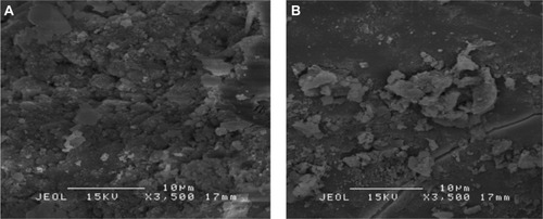
The morphology of the sulphated zirconia was studied by SEM. SEM images of zirconia and sulphated zirconia are presented in . In contrast with pure zirconia, SEM images of the sulphated zirconia showed a lower agglomeration. This supports the retarding effect of sulphate doping on the prevention of agglomeration via sulphation. EDS was used to analyze the percentage of elements present in the sample.Citation40,Citation41 The quantitative percentage of sulphated zirconia elements were found to be sulphur, at 1.66%; zirconium, at 51.95%; and oxygen, at 46.39%.
Figure 5 Scanning electron microscopy images of zirconia (A) and sulphated zirconia (B) calcined at 600°C for 3 hours.
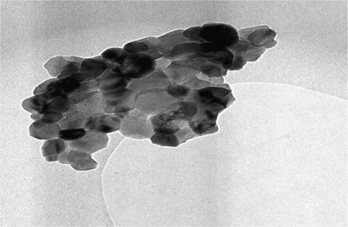
The morphology and the extent of dispersion of our nanoparticles were determined with TEM. The TEM images of pure sulphated zirconia nanoparticles are shown in . It is apparent that the nanoparticles are approximately tetragonal in shape, with diameters ranging from 37 to 54 nm, with an average size at 43 nm. In addition, most of the nanoparticles agglomerated, and a few detached.Citation42,Citation43
Figure 6 The transmission electron microscopy images of pure sulphated zirconia nanoparticles.
The zeta potential for the sulphated zirconia nanoparticle suspensions was −8.41 mV, as shown in . This finding may suggest that sulphated zirconia nanoparticles tend to aggregate in deionized and double-distilled water; their low zeta potential of −8.41 mV indicates these nanoparticles have poor electrostatic repulsion characteristics and are very unstable.
Figure 7 Surface charge characterization of sulphated zirconia nanoparticles.

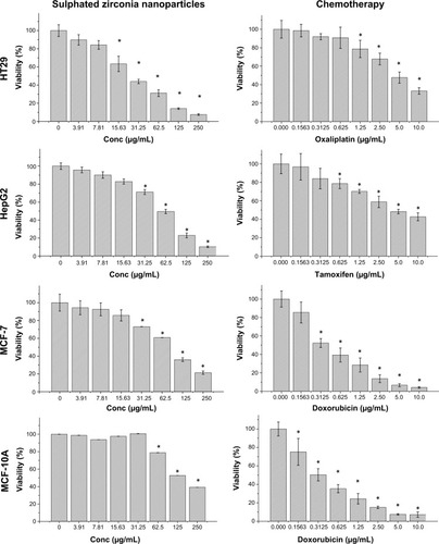
Most important, the cytotoxicity of sulphated zirconia nanoparticles on MCF-7, HT29, HepG2, and MCF-10a cells was determined by MTT assays.Citation44,Citation45 shows that sulphated zirconia nanoparticles have significant cytotoxic effects against colon cancer HT29 cells at most concentrations tested (P<0.05). For the case of both MCF-7 and HepG2 cells, 15.6 μg/mL had no significant effect on cell growth, whereas the nanoparticles inhibited the growth of all cell lines tested in a dose-dependent manner. The IC50 values of sulphated zirconia nanoparticles calculated from the dose-response curves are shown in . In contrast, the normal breast MCF-10a cells were the least sensitive to sulphated zirconia nanoparticles, with an IC50 value of approximately 151.6 μg/mL, whereas the breast cancer MCF-7 cells were found to be more sensitive, with an IC50 value of 89.9 μg/mL. The IC50 value of sulphated zirconia nanoparticles in normal breast MCF-10a cells was almost 1.7 times higher than that in breast cancer MCF-7 cell lines (). Furthermore, compared with sulphated zirconia nanoparticles, doxorubicin showed higher cytotoxicity in the normal breast MCF-10a cell line. The results obtained from the MTT assay showed significant changes in the viability of MCF-10a cells treated with nanoparticles at concentrations of 62.5 and 125 μg/mL for 24 hours when compared with untreated cells.
Table 1 Concentration of drug needed to inhibit cell growth by 50% of sulphated zirconia nanoparticles, oxaliplatin, doxorubicin, and tamoxifen on HT29, MCF-7, MCF-10a, and HepG2
Figure 8 Sulphated zirconia nanoparticles and chemotherapeutic effects on the viability of treated cells, which were evaluated through mitochondrial activity using an (4,5-dimethylthiazol-2-yl)-2,5-diphenyltetrazolium bromide assay.
Notes: Mean ± standard deviation (n=3 wells/treatment). *P<0.05 compared with the untreated cells.
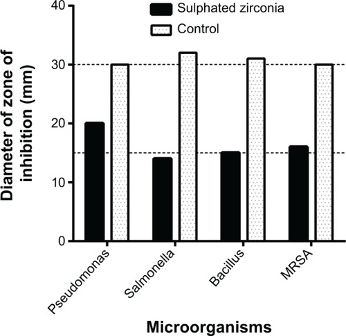
As shown in and , the sulphated zirconia nanoparticles showed high antimicrobial activity against both Gram-positive and Gram-negative bacteria. It was found that the nanoparticles showed the highest activity against Pseudomonas aeruginosa and methicillin-resistant S. aureus, followed by Bacillus subtilis and Salmonella choleraesuis. In contrast, the sulphated zirconia nanoparticles did not show any activity against Candida albicans, suggesting a lack of antifungal activity. Other fungal species may be used to detect any possible antifungal effect of the sulphated zirconia nanoparticles.
Figure 9 Antimicrobial activity of sulphated zirconia nanoparticles (SZ), against bacteria and yeast, using disk agar diffusion method.
Notes: Photographs of SZ nanoparticles: (A) Pseudomonas aeruginosa, (B) Salmonella choleraesuis, (C) Bacillus subtilis, (D) methicillin-resistant Staphylococcus aureus, and (E) Candida albicans. Control antimicrobial agents (C) (ampicillin for Gram-negative, streptomycin for Gram-positive, and nystatin for Candida albicans).
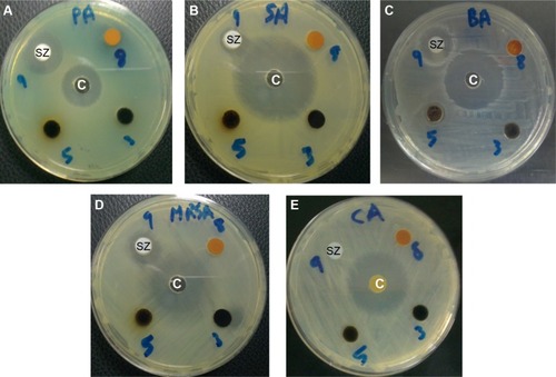
Figure 10 Diameter of zones of inhibition of sulphated zirconia nanoparticles against bacteria and yeast, along with the control antimicrobial agents (ampicillin for Gram-negative, streptomycin for Gram-positive).
Note: For Candida albicans, zone was 0 mm, and no activity was reported.
All of these microorganisms are responsible for a range of serious infections in human and animal populations. Therefore, the sulphated zirconia nanoparticles could find various biomedical applications of therapeutic importance to counteract such highly resistant microorganisms. Moreover, these sulphated zirconia nanoparticles should be further studied because of their noted anticancer properties.
Conclusion
The present study showed that sulphated zirconia nanoparticles have significant cytotoxic effects against colon cancer HT29 cells at most concentrations tested and inhibited the growth of all cancer cell lines studied here. Normal human breast MCF-10a cells exhibited the highest resistance to sulphated zirconia nanoparticles. Furthermore, the sulphated zirconia nanoparticles also showed high antimicrobial activity against both Gram-positive bacteria and Gram-negative bacteria. In conclusion, the sulphated zirconia nanoparticles could find numerous biomedical applications of therapeutic importance to counteract the colon cancer, as well as highly resistant microorganisms.
Disclosure
The authors report no conflicts of interest in this work.
References
- JentoftFCHahnAKröhnertJIncorporation of manganese and iron into the zirconia lattice in promoted sulfated zirconia catalystsJ Catal20042241124137
- ReddyBMPatilMKOrganic syntheses and transformations catalyzed by sulfated zirconiaChem Rev200910962185220819408954
- YadavGDNairJJSulfated zirconia and its modified versions as promising catalysts for industrial processesMicroporous Mesoporous Mater1999331148
- BrownACHargreavesJJSulfated metal oxide catalystsGreen Chem1999111720
- ClarkJHSolid acids for green chemistryAcc Chem Res200235979179712234209
- DelahayGEnsuqueECoqBFiguérasFSelective Catalytic Reduction of Nitric Oxide byn-Decane on Cu/Sulfated-Zirconia Catalysts in Oxygen Rich Atmosphere: Effect of Sulfur and Copper ContentsJ Catal19981751715
- EcormierMAWilsonKLeeAFStructure–reactivity correlations in sulphated-zirconia catalysts for the isomerisation of α-pineneJ Catal200321515765
- DateAAPatravaleVCurrent strategies for engineering drug nanoparticlesCurr Opin Colloid Interface Sci200493–4222235
- PissuwanDValenzuelaSMCortieMBTherapeutic possibilities of plasmonically heated gold nanoparticlesTrends Biotechnol2006242626716380179
- HafnerABraeseSEfficient Trifluoromethylation of Activated and Non Activated Alkenyl Halides by Using (Trifluoromethyl) trimethylsilaneAdv Synth Catal20113531630443048
- Thibault-StarzykFStanIAbellóSQuantification of enhanced acid site accessibility in hierarchical zeolites–The accessibility indexJ Catal200926411114
- BondGCMetal-catalysed reactions of hydrocarbonsNew YorkSpringer2006
- KašparJFornasieroPNanostructured materials for advanced automotive de-pollution catalystsJ Solid State Chem200317111929
- TrovarelliAZamarFLlorcaJLeitenburgCdDolcettiGKissJTNanophase fluorite-structured CeO2–ZrO2 catalysts prepared by high-energy mechanical millingJ Catal19971692490502
- ZhouLXuJLiXWangFMetal oxide nanoparticles from inorganic sources via a simple and general methodMater Chem Phys2006971137142
- DongSHanCZhengBZhangMHuangFPreparation of two new [2] rotaxanes based on the pillar [5] arene/alkane recognition motifTetrahedron Lett2012532836683671
- ChizalletCRaybaudPPseudo-bridging silanols as versatile Brønsted acid sites of amorphous aluminosilicate surfacesAngew Chem Int Ed Engl200948162891289319153974
- YanJYuDLiHSunPHuangHNaY zeolites modified by La3+ and Ba2+: the effect of synthesis details on surface structure and catalytic performance for lactic acid to acrylic acidJ Rare Earths2010285803806
- CarnitiPGervasiniAMarzoMAbsence of expected side-reactions in the dehydration reaction of fructose to HMF in water over niobic acid catalystCatal Commun2011121211221126
- KuhnJNHuangWTsungC-KZhangYSomorjaiGAStructure sensitivity of carbon-nitrogen ring opening: impact of platinum particle size from below 1 to 5 nm upon pyrrole hydrogenation product selectivity over monodisperse platinum nanoparticles loaded onto mesoporous silicaJ Am Chem Soc200813043140261402718834126
- LiuCWangJLiYOne-pot synthesis of 3,4-dihydropyrimidin-2 (1H)-(thio) ones using strontium (II) nitrate as a catalystJ Mol Catal Chem20062581–2367370
- AghapoorKEbadi-NiaLMohsenzadehFMohebi MoradMBalavarYDarabiHRSilica-supported bismuth (III) chloride as a new recyclable heterogeneous catalyst for the Paal–Knorr pyrrole synthesisJ Organomet Chem2012708–7092530
- ZouX-XLiG-DWangY-NDirect conversion of urea into graphitic carbon nitride over mesoporous TiO2 spheres under mild conditionChem Commun (Camb)20114731066106821079825
- YanWWangRXuZA novel, practical and green synthesis of Ag nanoparticles catalyst and its application in three-component coupling of aldehyde, alkyne, and amineJ Mol Catal Chem20062551–28185
- KumarDReddyVBMishraBGRanaRNadagoudaMNVarmaRSNanosized magnesium oxide as catalyst for the rapid and green synthesis of substituted 2-amino-2-chromenesTetrahedron2007631530933097
- ZhangXDuAJLeePSunDDLeckieJOGrafted multifunctional titanium dioxide nanotube membrane: separation and photodegradation of aquatic pollutantAppl Catal B2008841–2262267
- NaikMAMishraBGDubeyACombustion synthesized WO3–ZrO2 nanocomposites as catalyst for the solvent-free synthesis of coumarinsColloids Surf A Physicochem Eng Asp20083171–3234238
- SamantaraySHotaGMishraBPhysicochemical characterization and catalytic applications of MoO3–ZrO2 composite oxides towards one pot synthesis of amidoalkyl naphtholsCatal Commun2011121312551259
- SongXSayariASulfated zirconia-based strong solid-acid catalysts: recent progressCatal Rev1996383329412
- KatadaNEndoJ-iNotsuK-iYasunobuNNaitoNNiwaMSuperacidity and catalytic activity of sulfated zirconiaJ Phys Chem B2000104441032110328
- MeghshyamKSynthesis of Novel Zirconia Based Solid Superacids Their Characterization and Application in Organic Synthesis and Transformation Reactions [thesis]Hyderabad, IndiaOsmania University2009
- AlhassanFHRashidUAL-QubaisiMSRasedeeATaufiq-YapYHThe effect of sulphate contents on the surface properties of iron-manganese doped sulphated zirconia catalystsPowder Technol2014253809813
- Al-QubaisiMSRasedeeAFlaifelMHCytotoxicity of nickel zinc ferrite nanoparticles on cancer cells of epithelial originInt J Nanomedicine201382497250823885175
- Al-QubaisiMSRasedeeAFlaifelMHInduction of apoptosis in cancer cells by NiZn ferrite nanoparticles through mitochondrial cytochrome C releaseInt J Nanomedicine201384115412924204141
- BiMLiHPanW-PLloydWGDavisBHThermal studies of metal promoted sulfated zirconiaAm Chem Soc Fuel Div Preprints1996417781
- KhalafHATextural properties of sulfated iron hydroxide promoted with aluminumMonatsh Chem20091406669674
- ClearfieldASerretteGKhazi-SyedANature of hydrous zirconia and sulfated hydrous zirconiaCatal Today1994202295312
- MekhemerGAHKhalafHAMansourSAANohmanAKHSulfated alumina catalysts: consequences of sulfate content and sourceMonatsh Chem20051361220072016
- YamamotoTTanakaTTakenakaSStructural analysis of iron and manganese species in iron-and manganese-promoted sulfated zirconiaJ Phys Chem B19991031323852393
- ZhangLYangJZhuJProperties and liquefaction activities of ferrous sulfate based catalyst impregnated on two Chinese bituminous coalsFuel2002817951958
- YangLZhangAZhengXShrimp shell catalyst for biodiesel productionEnergy Fuels200923838593865
- ButtH-JCappellaBKapplMForce measurements with the atomic force microscope: Technique, interpretation and applicationsSurf Sci Rep2005591–21152
- LiYSomorjaiGANanoscale advances in catalysis and energy applicationsNano Lett20101072289229520524636
- Al-QubaisiMRosliRSubramaniTGoniothalamin selectively induces apoptosis on human hepatoblastoma cells through caspase-3 activationNat Prod Res201327232216221823767409
- Al-QubaisiMRozitaRYeapS-KOmarA-RAliA-MAlitheenNBSelective cytotoxicity of goniothalamin against hepatoblastoma HepG2 cellsMolecules20111642944295921471934