
 ?Mathematical formulae have been encoded as MathML and are displayed in this HTML version using MathJax in order to improve their display. Uncheck the box to turn MathJax off. This feature requires Javascript. Click on a formula to zoom.
Abstract
Magnetic resonance imaging (MRI) has become one of the most widely used and powerful tools for noninvasive clinical diagnosis owing to its high degree of soft tissue contrast, spatial resolution, and depth of penetration. MRI signal intensity is related to the relaxation times (T1, spin–lattice relaxation and T2, spin–spin relaxation) of in vivo water protons. To increase contrast, various inorganic nanoparticles and complexes (the so-called contrast agents) are administered prior to the scanning. Shortening T1 and T2 increases the corresponding relaxation rates, 1/T1 and 1/T2, producing hyperintense and hypointense signals respectively in shorter times. Moreover, the signal-to-noise ratio can be improved with the acquisition of a large number of measurements. The contrast agents used are generally based on either iron oxide nanoparticles or ferrites, providing negative contrast in T2-weighted images; or complexes of lanthanide metals (mostly containing gadolinium ions), providing positive contrast in T1-weighted images. Recently, lanthanide complexes have been immobilized in nanostructured materials in order to develop a new class of contrast agents with functions including blood-pool and organ (or tumor) targeting. Meanwhile, to overcome the limitations of individual imaging modalities, multimodal imaging techniques have been developed. An important challenge is to design all-in-one contrast agents that can be detected by multimodal techniques. Magnetoliposomes are efficient multimodal contrast agents. They can simultaneously bear both kinds of contrast and can, furthermore, incorporate targeting ligands and chains of polyethylene glycol to enhance the accumulation of nanoparticles at the site of interest and the bioavailability, respectively. Here, we review the most important characteristics of the nanoparticles or complexes used as MRI contrast agents.
Introduction
Imaging is widely used in scientific and technological applications because of the interface it provides between vision and intuition. In particular, biological imaging is a rapidly growing field, not only in fundamental biology but also in medical science.Citation1 Recently, biomedical imaging has received enormous attention in view of its capacity to aid analysis and diagnosis through images at the molecular and cellular levels.Citation2 As a result, a new discipline, known as “molecular imaging” (MI) has emerged, which combines molecular biology and in vivo imaging.Citation3 The aim of MI is to monitor and measure biological processes in living subjects via spectral data. The measurement and monitoring of biological processes provides information similar to that from a biopsy, but it is noninvasive and performed in real time, thereby offering the possibility of sequential and longitudinal monitoring. The use of MI techniques permits molecular changes associated with the onset and development of pathologic states to be quantified, and the approach can provide early diagnosis and prognosis of diseases such as cancer. Other applications include the evaluation of the response to therapy, and the study of biological processes in living subjects.Citation4–Citation6 Traditional MI modalities include X-ray computed tomography (CT), optical imaging (OI), positron emission tomography (PET), single-photon emission CT (SPECT), ultrasound, and magnetic resonance imaging (MRI).Citation7,Citation8 Several promising new imaging modalities, such as fluorescence-mediated tomography and photoacoustic tomography, are currently under development.Citation9 Each of these diagnostic modalities has its advantages and disadvantages. For example, MRI and CT have high spatial resolution and are able to provide detailed anatomical information, but they lack sensitivity. In contrast, PET and SPECT are highly sensitive, but have limited resolution and cannot provide anatomical information.
Through the development of highly specialized and efficient contrast agents, MRI has evolved into a versatile technique with multiple functions and has become one of the most powerful noninvasive imaging tools in the biomedical toolbox. High resolution and excellent soft tissue contrast are its main advantages over other in vivo imaging techniques. MRI relies on large magnetic fields and radio frequencies (RFs), and makes use of the relaxation times of protons in mobile molecules such as water, lipids, and proteins that are present in organs at different concentrations, to produce high-resolution soft tissue anatomical images with good endogenous contrast.Citation10 In the following sections, we review many of the most innovative approaches that have been adopted in the recent history of MRI contrast agents based on nanoparticles; mainly on superparamagnetic iron oxide nanoparticles. In this mini-review, we also include polynuclear or particulate contrast agents that are the result of progression from previous ionic agents. This is the case of the chelates of gadolinium and manganese oxide that were developed from experience with previous Gd3+- and Mn2+-based agents respectively. In contrast, we have excluded from this review diamagnetic chemical exchange saturation transfer (CEST) and paramagnetic CEST (PARACEST) agents, although they can be included in nanoparticulate systems such as liposomes and polymers.Citation11–Citation13
The use of nanoparticles as imaging probes has several advantages over conventional imaging agents. Loadability is one of the advantages where the concentration of the imaging agent can be controlled within each nanoparticle during the synthesis process. Another advantage is the tunability of the surface of the nanoparticles that can potentially extend the circulation time of the agent in the blood or target a specific location within the body. Finally, nanoparticles can act as multifunctional MI agents, since they have two or more properties that can be used simultaneously in multiple imaging techniques, and especially in MRI.Citation14
Magnetic resonance imaging: origin of the contrast
When a strong magnetic field is applied to a sample (in clinical diagnosis, magnetic fields of 1.5 or 3 T are usually used), the magnetic field aligns the magnetic moments of protons in the sample, producing an equilibrium magnetization along the longitudinal axis. A RF pulse, at a resonant frequency (5–100 MHz) capable of transferring energy to protons, can then rotate their magnetic moments away from the longitudinal axis, in phase, to an angle called the flip angle. Upon removal of the radiation, the magnetic moments of the protons relax to equilibrium.Citation15 In MRI, this process is repeated in a quick succession of RF pulses. The time taken by the magnetic moments to return to their original alignment with the magnetic field is called the relaxation time, and it is tissue dependent. This relaxation can be divided into two different, independent processes: 1) longitudinal relaxation (characterized by the parameter T1) and 2) transverse relaxation (characterized by the parameter T2).
T1, called the spin–lattice relaxation time, relates to how fast the magnetization parallel to the static magnetic field recovers after a perturbation is applied to the system. Protons that relax rapidly (short T1) recover full magnetization along the longitudinal axis quickly and produce high signal intensities. For protons that relax more slowly (long T1), full magnetization along the longitudinal axis is not recovered before subsequent RF pulses, and so they inherently produce a lower intensity signal.Citation15
T2 relates to how rapidly the magnetization in the plane perpendicular to the static magnetic field loses coherence. During an RF pulse, proton nuclei spin in phase with each other, whereas after the pulse, the magnetic fields of all the nuclei interact with each other, and energy is exchanged between them. As a consequence, the nuclei lose their phase coherence and tend to spin in a random fashion.Citation16 Because T2 decay is the result of the exchange of energy between spinning protons, it is referred to as spin–spin relaxation.
Longitudinal and transverse relaxation processes are executed independently and simultaneously, although T2 is usually much shorter than T1, and this difference allows tissues to be differentiated.Citation17 In most cases, the combination of the intrinsic molecular interactions of neighboring molecules and extrinsic magnetic field inhomogeneities means that the observed transverse relaxation time (T2*) is even shorter than the natural T2 that would be caused by pure spin–spin interactions. To eliminate external magnetic field effects and generate the real T2-weighted images based purely on molecular interactions, a spin–echo sequence is used. This uses 90° pulses to excite the magnetization and one or more 180° pulses to refocus the spins and generate signal echoes named spin echoes.Citation15
The endogenous MRI contrast in soft tissue comes from local differences in the proton density (water concentration) resulting in different values of T1 and T2. On this basis, endogenous contrast depends on the chemical and physical nature of the tissues.Citation18 Despite the relatively high quality of such images of soft tissues, in some cases there is not enough image contrast to diagnose the pathology of interest. In these circumstances, the low endogenous sensitivity can be enhanced by increasing the magnetic field (from 3 to 7 T and beyond), acquiring data for longer or designing more sensitive sequences and probes. An important alternative is to use exogenous contrast agents.Citation10
Exogenous contrast agents
Contrast agents have a wide variety of chemical compositions. They can be small mononuclear or polynuclear paramagnetic chelates; metalloporphyrins; polymeric or macromolecular carriers of covalently or noncovalently bonded paramagnetic chelates; particulate contrast agents (including fluorinated or nonfluorinated paramagnetic micelles or liposomes) and paramagnetic or superparamagnetic particles (eg, iron oxides and Gd3+-labeled zeolites); diamagnetic CEST polymers; diamagnetic hyperpolarization probes (gases and aerosols); and 13C-labeled compounds or ions (eg, 6 Li+).Citation19 The main role of T1 and T2 contrast agents in MRI is to shorten selectively the relaxation times of water protons in the region of interest and thus to provide better contrast for anatomical regions. Contrast is enhanced when one tissue has either higher affinity for the contrast agents or higher vascularity than another. Diseased tissues, such as tumors, are metabolically different from healthy tissues and have taken up the contrast agent in different ways, resulting in a contrast in MRI images.Citation19,Citation20 T1-weighted images illustrate anatomy well and are preferred when a clear image of the structures is required. T2-weighted images produce good pathological information since collections of abnormal fluid appear brighter than the normal tissue background.
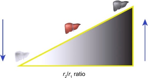
Although nearly all MRI contrast agents affect both T1 and T2, the effects of contrast agents are usually more pronounced for either T1 or T2, leading to their categorization as either T1 or T2 contrast agents. Contrast enhancement is measured by the relaxation rate Ri =1/Ti (s−1), where i =1 or 2. The most important parameter for defining the efficiency of a contrast agent is its relaxivity, ri (ri = Ri/cCA(mM−1 s−1), where cCA is the analytical concentration of ion responsible for the contrast). The r2/r1 ratio is also used to indicate the contrast efficiency; the higher the ratio, the greater the efficiency of a T2 contrast agent and vice versa for a T1 contrast agent ().
Figure 1 Influence of the r2/r1 ratio on the efficiency of a contrast agent.
Notes: High values of r2/r1 are characteristic of T2 contrast agents, which produce a hypointense signal in T2-weighted images, and thus organs appear darker in the image. Low values of r2/r1 define T1 contrast agents, and the associated images are clearer and brighter.
The relaxivity for an MRI contrast agent is defined as the increase in the relaxation rate of the solvent (water) induced by 1 mmol·L−1 of the active ion of the contrast agent, and it is calculated according to
The relaxivity is dependent on the magnetic field applied and the temperature, so it should be reported together with both these parameters.
Most T1 contrast agents currently available are paramagnetic complexes, while those classed as T2 contrast agents are mostly superparamagnetic iron oxides.Citation18
T1 contrast agents
The first generation of exogenous T1 contrast agents (also called positive contrast agents) consisted of high-spin paramagnetic metal ions such as manganese (Mn2+), iron (Fe3+), or gadolinium (Gd3+).Citation21 These contrast agents produce hyperintense signals in T1-weighted images. To obtain significant changes in proton relaxation and therefore a good contrast, the paramagnetic ion needs to be in close contact with the protons of the surrounding water molecules. However, owing to the toxicity associated with these cations (eg, transient destruction of professional macrophages, exchange with endogenous calcium ions, etc), they are used after complexation with low-molecular-weight chelating molecules with no explicit core and surface coating.
Gd is the most clinically used metal ion in paramagnetic T1 contrast agents. It possesses an electron spin of 7/2 and hence seven unpaired electrons promoting spin relaxation due to flipping spins and rotational motion. Free Gd ions are cytotoxic and are retained in liver, spleen, and bone.Citation22 To avoid this toxicity, a chelating process is applied to Gd, in which large organic molecules form a stable complex around the Gd. The chelate reduces the chances of toxicity that result from exposure to Gd. The stable complex is predominantly eliminated via the kidneys. Examples of chelating compounds are diethylene-triamine-pentaacetic acid (DTPA), 1,4,7,10-tetraazacyclo-dodecane-1,4,7,10-tetraacetic acid (DOTA), and dipyridoxyl-di-phosphate (DPDP).Citation19 The chemical structures of such T1 agents are typically characterized by neutral or anionic metal complexes of the type [M(H2O)(L)] or [M(H2O) (L)]n−, where M is the paramagnetic Gd3+ or Mn2+/3+ ion, and L a macrocyclic or acyclic polyaminopolycarboxylate.Citation23
Different types of Gd-containing contrast agents have been approved by the European Medicines Agency (EMEA) and the US Food and Drug Administration (FDA) () for use in MRI as a contrast agent to provide improved images of organs and tissues.
Table 1 Gadolinium-based contrast agents approved for use in humans by the EMEA or FDA
However, although Gd is regarded as safe when administered as a chelated compound, the use of some Gd chelates in people with renal disease has been linked to a rare but severe complication; the medical condition referred to as “nephrogenic systemic fibrosis.” For this reason, the World Health Organization (WHO) issued a restriction on the use of Gd contrast agents, informing that these compounds were contraindicated in patients with chronic severe renal insufficiency, in those with acute renal insufficiency of any severity due to hepatorenal syndrome, or in the perioperative liver transplantation period, and in newborn babies up to 4 weeks of age.Citation24
Because of their low molecular weight, conventional Gd-based contrast agents are mostly nonspecific extracellular contrast agents and exhibit rapid extravasation from the vascular space. In this way, after being intravenously injected, these agents rapidly leak from the blood pool into the interstitium with a distribution half-life (t1/2) of about 5 min. They are mainly cleared by the kidneys with an elimination t1/2 of about 80 min.Citation19 This limitation, which is inherent to MRI, is known as the partial volume dilution effect, and involves a loss of apparent activity in small objects or regions because of the limited resolution of the imaging system. The partial volume dilution effect has often led to the failure of targeted contrast in vivo.Citation25 Extracellular agents are typically Gd chelates of linear or macrocyclic polyaminocarboxylate ligands, and constitute the most important class of MRI contrast agents available. Initial attempts to target MRI focused on coupling Gd atoms directly to antibodies or proteins, but these approaches delivered insufficient paramagnetic material to effectively decrease local relaxation times, and provided inadequate MR signal enhancement in T1 images at typical clinical field strengths. Moreover, for certain purposes such as MR angiography (MRA: a special type of MRI used to study blood vessels) the time window for contrast-enhanced images is very narrow, due to rapid extravasation, which limits the acquisition of high-resolution images. For MRA, contrast agents must be blood-pool agents (also known as intravascular contrast agents) and are characterized by their high molecular weight (<20 kDa) and higher relaxivities. Their large size prevents diffusion through the vascular epithelium and leakage into the interstitial space, and so they reside in the vascular system for an extended period of time. Thus, they are eliminated much more slowly from circulation than their extracellular counterparts, providing a larger imaging time window. Examples of blood-pool contrast agents are Gd-based complexes that interact noncovalently with human serum albumin, and Gd chelates complexed to polymers (eg, dextrans, polylysine derivatives, and polyamidoamines [PANAM™, GE Healthcare Institute, Waukesha, WI, USA]).Citation26
To produce targeting agents, macromolecular constructs, such as liposomes, micelles, fluorinated nanoparticles, dendrimers, and polymers, have been prepared.Citation17,Citation25,Citation27 The resulting nanoparticles have greater paramagnetic metal surface payloads that rotate or tumble more slowly than small-molecule organometallic compounds typically used as blood-pool agents.Citation28
Mn2+, with five unpaired electrons, is another cation used as a contrast agent. Mn-based paramagnetic nanoparticles can be classified into two categories: small-molecule agents and nanoparticulate agents. Small-molecule agents are formed by complexing Mn ions with chelates such as DPDP, DTPA, or even porphyrin rings, just as Gd chelates are. The FDA approved, in May 1997, a Mn-based contrast agent, the injectable mangafodipir trisodium (Teslacan®, St Louis MO, USA) to image the liver. However, in 2012, the EMEA was notified by the marketing authorization holder responsible for Telescan the decision to voluntarily withdrawl the marketing authorization in the European Union.Citation29
Mn chelates can be further modified by their incorporation into lipid bilayers. Such nanoparticulate systems are made of manganese oxides such as MnO, MnO2, and Mn3O4. After dissolution in cells due to proteolytic degradation, these particles convert from T2 contrast agents to T1 contrast agents.Citation30 Although Mn2+ is a natural cellular constituent that resembles Ca2+, its toxicity is also known from dust containing Mn at high doses. Moreover, in view of the capacity of Mn2+ to enter cells through calcium channels, Mn complexes, dendritic Mn chelates, and even Mn nanoparticulate systems have potential applications in neuroimaging. However, this also implies that the brain may be vulnerable to Mn exposure.
One of the limitations of the majority of the contrast agents used, but that especially affects paramagnetic chelates, is that their efficiency decreases at higher magnetic fields. For example, Gd complexes are optimal at fields below 1 T; even at the clinical field of 3 T, the T1 relaxivity of Gd-based contrast agents is reduced by as much as one-third compared with its maximum, while at higher magnetic fields, r1 falls to zero.Citation31 Moreover, for in vitro cell labeling experiments or long-term in vivo cell tracking studies, the clearance of the particles needs to be far slower, which impedes the use of Gd-based chelate agents for these purposes. Hence, owing to their short blood circulation times, poor detection sensitivity, and toxicity concerns, MRI research has shifted to T2 contrast agents, especially to superparamagnetic iron oxide nanoparticles.
T2 contrast agents
T2 contrast agents (or negative contrast agents) decrease the MR signal intensity of the regions they are delivered to. Consequently, they produce hypointense signals in T2- and T2*-weighted images,Citation32 and thus the affected regions appear darker. The phenomenon can be said to result from the large heterogeneity of the magnetic field around the nanoparticle through which water molecules diffuse, since diffusion induces dephasing of the proton magnetic moments, resulting in T2 shortening. T2 contrast agents are also called susceptibility agents because of their effect on the magnetic field. T2 shortening is a remote effect, whereas the T1 shortening process requires a close interaction between the water molecules and T1 agents, as mentioned.Citation16 Another difference with T1 contrast agents is that under high magnetic fields, R2, the relaxation rate, tends asymptotically to a positive constant.Citation10
Iron oxide nanoparticles have been used as T2 contrast agents for more than 25 years. These iron oxides can be ferromagnetic or superparamagnetic, depending on the size of the core of the nanoparticle. Two iron oxides are generally considered for biomedical applications: magnetite (Fe3O4) and its oxidized and more stable form of maghemite (γ-Fe2O3). The critical upper size limit for the observation of superparamagnetism is approximately 25 nm for magnetite and 30 nm for maghemite.Citation33 The two compounds fulfill the prerequisites of: 1) chemical stability under physiological conditions, 2) low toxicity, and 3) sufficiently high magnetic moments.Citation34
Since these two iron oxides exhibit superparamagnetic behavior, the loss of their net magnetization in the absence of an external magnetic field limits their tendency for self-aggregation, and this helps to obtain a good biological response.Citation10 Unfortunately, the ubiquitous Van der Waals forces induce natural aggregation of the particles, and to circumvent this problem, a large portfolio of chemical approaches exists that stabilize the particles. These approaches include the modification of the surface of the particles with diverse materials. Polymers are the most widely used stabilizing materials, and can be classified as hydrophilic or amphiphilic, neutral or charged, homopolymers or copolymers. The polymers can be adsorbed into or anchored onto the iron oxide surface via hydrogen bonds, electrostatic forces, or pseudo-covalent bonding. Among the materials used are poly(ethylene glycol) (PEG), dextran and its derivatives, alginate, chitosan, starch, polyvinyl alcohol, albumin, poly(ethylene imine), organic siloxane, sulphonated styrene-divinyl-benzene, and bioactive molecules and structures such as liposomes.Citation35,Citation36
Since the biological distribution of the nanoparticles is directly dependent on their size, they have been classified according to the overall size of the particles as follows: 1) ultra-small superparamagnetic iron oxide nanoparticles (USPIONs) with diameter (d) less than 50 nm, 2) superparamagnetic iron oxide nanoparticles (SPIONs) with size of hundreds of nanometers, and, ultimately, 3) micron-sized particles of iron oxide (MPIO) with a diameter higher than 1 μm.Citation37 While the overall size of the first two classes allows them to be administered intravenously, the larger particles are usually administered orally, limiting their use to the exploration of the gastrointestinal track. There are also other formulations, such as monocrystalline iron oxide particles (MION) and cross-linked iron oxides (CLIO).Citation19
Several SPION formulations for intravenous or oral administration have been approved for clinical use as MRI contrast agents by the EMEA and FDA; however, the majority of the compounds that were approved for intravenous administration have, at present, been taken off the market.Citation38 Only the SPION for oral administration, Gastromark® (AMAG Pharmaceuticals, Waltham, MA, USA; ferumoxsil, silicone-coated SPIONs), is currently on the market for gastrointestinal bowel marking. The most widely applied coatings for FDA-approved SPIONs are dextran and carboxydextran. compares the properties of nanoparticles coated with one or other polymer.
Table 2 Properties of iron oxide nanoparticles and relaxivity values of three nanoparticles coated with hydrophilic polymers
It is important to note that both the type of coating and its thickness affect the value of r2, although the influence is unclear as studies report different effects. For instance, it has been reported that as the coating thickness increases, the ratio r2/r1 decreases.Citation39 This is due to the inner hydrophobic layer excluding water, while the outer hydrophilic PEG layer allows water to diffuse within the coating zone. Increasing the PEG chain length leads to a reduction in r2 values, although another study has shown that when water molecules are not excluded from regions close to the SPION core, r2 relaxivity increases with increased chain length.Citation40
The overall size of the particle governs its pharmacokinetics and biodistribution. Nanoparticles with a size <5.5 nm are cleared by the kidneys.Citation41,Citation42 SPIONs whose overall diameter is larger than 200 nm are quickly taken up by phagocytic cells and accumulate in the monocyte phagocyte system (MPS), specifically in liver and spleen macrophages. When administered intravenously, approximately 80% of the dose is found in liver and 5%–10% in spleen, with it having a plasma half-life of less than 10 min.Citation16 Therefore, such SPIONs decrease the liver and spleen signal within several minutes of administration. Malignant tumors or metastases have a lack of Kupffer cells, and due to the negligible uptake of nanoparticles, they produce a strong contrast between normal and abnormal tissue on T2-weighted images. USPIONs evade MPS uptake and consequently increase their blood half-life (>2 h). This increased blood circulation maximizes the odds of SPIONs reaching their target tissue. SPIONs and USPIONs are metabolized into a soluble and nonsuperparamagnetic form of iron, which is incorporated into the normal iron pool (eg, ferritin, hemosiderin, hemoglobin) within a couple of days.Citation19
As with any nanoparticle, SPIONs can invade small solid tumors and metastatic cells thanks to passive targeting through the enhanced permeation and retention (EPR) effect. The EPR effect aids nanoparticle uptake by way of leaky vasculature, which permits particles of nanometric size (more or less, with a hydrodynamic radius of less than 100 nm) to cross from the vasculature into the interstitium.Citation43 Poor lymphatic drainage then aids the entrapment of particles in solid tumors.
SPIONs are used as negative contrast for liver imaging, whereas the typical application of USPIONs is lymph node imaging. USPIONs have been tested as blood-pool agents because they are readily distributed in the intravascular extracellular space. In this way, USPIONs are used as contrast for lymphographyCitation44,Citation45 and angiography,Citation46,Citation47 as a bone marrow contrast, or as a perfusion agent in brain and kidney.
Iron oxide nanoparticles require specific approaches to target cells other than macrophages. Since biochemical epitopes of interest are often present in nanomolar or picomolar concentrations, particle relaxivities of around 1,000,000 mM−1·s−1 are required to achieve acceptable contrast-to-noise ratios at the typical field strength.Citation48 In this way, contrast agents that target specific tissue can increase the sensitivity by increasing the local SPION concentration. To achieve such sensitivity in the nanomolar range, the surface of the SPIONs may be modified by active targeting strategies, such as the addition of ligands that are recognized by molecular signatures of afflicted cells. Polyethyleneimine (PEI) is one of the more used ligands.Citation49 The same groupCitation50,Citation51 has reported that hyaluronic acid targeted iron oxide nanoparticles are efficient probes for targeted MRI of cancer cells in vitro and xenografted tumor model in vivo. Other types of ligands have been studied for the targeting of such markers including antibodies, small peptides, lectins, aptamers, engineered proteins, and protein fragments.Citation52 For instance, USPIONs of less than 10 nm in hydrodynamic diameter were tested for tumor-specific MRI targeting. In that study, the USPIONs were stabilized by 4-methylcatechol, and a cyclic arginine–glycine–aspartic acid (cRGD) peptide was coupled via the Mannich reactionCitation53 ().
Figure 2 Schematic illustration of the coupling of cRGD peptide to the SPIONs.
Notes: (A) MRI cross-section image of the U87MG tumors implanted in mice; (B) without the nanoparticles; and (C) with the injection of 300 μg of cRGD-SPIONs. Reprinted with permission from Ho D, Sun X, Sun S. Monodisperse magnetic nanoparticles for thera nostic applications. Acc Chem Res. 2011;44:875–882.Citation60 Copyright 2011 American Chemical Society.
Abbreviations: cRGD, cyclic arginine-glycine-aspartic acid; SPIONs, superparamagnetic iron oxide nanoparticles; MRI, magnetic resonance imaging.
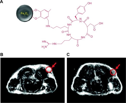
The peptide RGD binds the αVβ3-integrin, a cell adhesion molecule that is overexpressed in tumor vasculature and invasive tumor cells.Citation54 After the administration of the RGD-nanoparticles, the tumor MRI signal intensity decreased by 40%.Citation53
One of the largest drawbacks in using SPIONs is related to the contrast mechanism that they generate. As mentioned, they are negative imaging agents, which produce a signal-decreasing effect. The resulting dark signal could be confused with other pathogenic conditions, and renders images of lower contrast than T1 contrasted images. Moreover, the high susceptibility of T2 contrast agents induces distortion of the magnetic field on neighboring normal tissues. This distortion of the background is called a susceptibility artifact or “blooming effect,” and generates dark images with no background around the lesions.Citation55 This effect prevents their clinical use in low signal body regions, in organs with intrinsic high magnetic susceptibility (eg, lung), or in the presence of hemorrhagic events.Citation56
In addition to nanoparticles whose metal core is fully based on iron oxides, other nanosystems with different magnetic cores have been introduced to improve the signal sensitivity and enhance MRI diagnostics.Citation57 Since the transverse relaxivity r2 depends, apart from size, on the saturation magnetization (Ms), the optimization of Ms is one of the most effective ways to achieve magnetic nanoparticles with high MRI sensitivity. It has been reported that, owing to the higher magnetization, iron nanoparticles have a higher T2 relaxivity than analogous systems containing iron oxides.Citation58 Alloy-based nanomaterials are good candidates for developing T2 contrast agents with higher relaxivities. The substitution of one of the Fe ions in an iron oxide for a different magnetic atom (Mn, Zn, Co, Ni, etc) produces the compounds known as ferrites (Mn-ferrite, Zn-ferrite, etc) characterized by their high Ms, and this enhanced Ms increases the relaxation rate. Yang et alCitation59 have reported the suitability of Mn-ferrites as MRI contrast agents. More pronounced contrast effects are even possible when nonmagnetic ions replace the Fe ions.Citation60 Interestingly, the substitution of Fe2+ with nonmagnetic Zn2+ results in an increase in the net magnetization of the nanoparticles. Ms increases with Zn2+ doping and becomes maximum with a value of x =0.4 in (ZnxM1−x) Fe2O4 (M = Mn2+, Fe2+).For comparison, the Ms of (Zn0.4Mn0.6) Fe2O4 is 175 emu·g−1 magnetic atom, whereas the corresponding value for Fe3O4 is 96 emu·g−1 magnetic atom. The value of r2 reaches 676 mM−1·s−1 for the Zn-doped magnetic nanoparticles, and 98 mM−1·s−1 for Fe3O4.Citation61
Other nanoparticles with potential applications in MRI include gold-iron oxide (Au-Fe3O4) nanoparticles, metallic ion nanoparticles, porous hollow Fe3O4 nanoparticles, and Fe-based alloy nanoparticles, such as iron–cobalt (FeCo) and iron–platinum (FePt) nanoparticles. However, metallic nanoparticles are normally very reactive and are subject to fast oxidation in biological solutions. Once they are coated with a layer of polycrystalline Fe3O4 or a graphitic shell, these metallic nanoparticles are more stable and provide better contrast in MRI. FePt nanoparticles are chemically more stable than Fe and FeCo nanoparticles, and have been shown to have great potential as contrast agents for MRI and CT.Citation28 However, it is worth pointing out that such systems could be used for preclinical experiments, but clinical assessment of their acute and long-term toxicity is required.
Recently, some paramagnetic ions, such as dysprosium (Dy3+), have been proposed as good alternatives to iron oxide T2 contrast agents in high-field MRI, because of their high magnetic moments.Citation62 Dy3+ has been used as a chelate (eg, Dy3+-DTPA) or as nanoparticles (eg, Dy2O3).Citation63,Citation64 One type of Dy3+-based nanoparticles are β-NaDyF4 nanoparticles; their relaxivity has been studied at 3 and 9.4 T in nanoparticles of 5.4, 9.8, and 20.3 nm. It has been reported that their r2 relaxivity is 6–9 times greater at 9.4 T than at 3 T, and that the larger nanoparticles show higher r2 values than the smaller ones, whereas the r1 relaxivities are almost constant for the three sizes at 3 and 9.4 T. At 9.4 T, the r2/r1 ratio is 306 for nanoparticles of 20.3 nm, 230 for those of 9.8 nm, and 106 for those of 5.4 nm.
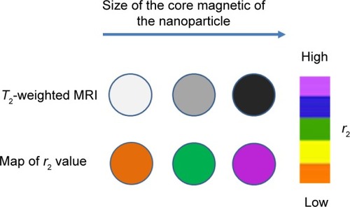
One important result of classical outer-sphere relaxation theoryCitation65 is that the r2/r1 ratio increases with increasing particle size, and thus, smaller particles are much better T1-shortening agents then larger ones ().
Figure 3 T2-weighted contrasts and r2 color maps for iron oxide nanoparticles of different size.
As a consequence of their larger size and magnetic moments, SPIONs were initially developed as T2-agents. However, a new generation of USPIONs, with diameters less than 10 nm, has also been reported to have excellent T1-enhancing properties.Citation28,Citation66 Compared with paramagnetic ions, SPIONs have higher molar relaxivities, and, when used as blood-pool and tissue-specific agents, may offer advantages at low concentrations.Citation67
As indicated above, SPIONs generate dark or negative contrast at the target site with a marked blooming effect from magnetite susceptibility artifacts. Moreover, for MI applications, persistent T2* effects from circulating SPIONs delay MRI by 24–72 h after injection.
To overcome these limitations, specific off-resonance pulse sequences capable of generating a bright contrast in the presence of SPIONs have been proposed.Citation68,Citation69 More recently, another approach, called inversion recovery ON-resonant water suppression (IRON)-MRI, has been developed to obtain a positive contrast.Citation70 Unfortunately, these techniques do not eliminate the signal loss or magnetic susceptibility artifacts, but rather exploit them to generate more easily perceived bright images of the contrast effects, often at the expense of the surrounding anatomical detail. Moreover, the undesirable 24–72 h delay between injection of the agent and the imaging result remains. One approach to resolve the prolonged delay between treatment and imaging has been to consider the use of microparticles of iron oxide (MPIO, size range 0.76–1.63 μm), which rapidly pass into MPS organs.Citation71 However, such large iron oxide particles were prone to aggregation and rapid pulmonary entrapment, and despite the leukocyte mimicking dual homing ligand approach proposed, the potential of the approach will depend on overcoming many challenges.Citation72,Citation73
A more recent approachCitation28 is the preparation of a colloidal iron oxide nanoparticle platform (CION), which is achieved by embedding oleate-coated magnetite particles in a hydrophobic matrix composed of vegetal oil and partially cross-linked phospholipids. Contrary to expectations, this formulation decreased T2 effects, thus allowing positive T1-weighted contrast detection. A CION may be used to detect biosignatures via voxels at very low nanomolar densities.
Dual (T1 and T2) contrast agents
Conventional MRI contrast agents are mostly effective only in a single imaging mode: either T1 or T2. They frequently result in ambiguities in diagnostics, especially when the biological targets are small. The combination of simultaneously strong T1 and T2 contrast effects in a single contrast agent could be a new breakthrough, since it can potentially provide more accurate MRI via self-confirmation with better differentiation of normal and diseased areas. Dual contrast agents would eliminate the possible ambiguity of a single-mode contrast agent when some of the in vivo artifacts are present.Citation57 However, the production of such a contrast has proved extremely challenging.Citation74
USPIONs with a core of less than 10 nm in diameter are capable of producing positive contrast in T1-weighted images when administered in moderate concentrations.Citation75,Citation76 While positive T1 contrast is possible with USPIONs, this benefit is at the expense of their T2 effects.Citation77,Citation78 For this reason, mixing both types of iron oxides, SPIONs and USPIONs, to form a single contrast agent could potentially be a good choice. However, an important problem arises as a consequence of the strong magnetic coupling between the T1 and T2 contrast agents when they are in close proximity: the spin–lattice relaxation process of T1 contrast materials is significantly diminished. One strategy to overcome this phenomenon is the inclusion of a separation layer to modulate the magnetic coupling. To this end, micellar structures incorporating organic block copolymers, inorganic porous materials, and core–shell-type inorganic materials have been considered as possible frameworks.Citation52 For instance, a core–shell-type T1–T2 dual-mode nanoparticle contrast has been described, where the T1 contrast material, Gd2O(CO3)2 of 1.5 nm, is located on the shell so as to come into direct contact with water molecules, for high T1 contrast effects; while the superparamagnetic T2 contrast material, MnFe2O4 of 15 nm, is located at the core, from where it induces a long-range magnetic field for the relaxation of water molecules. The two materials are separated by SiO2. By adjusting the thickness of the SiO2, the magnetic coupling between the T1 and T2 contrast agents is controlled. As the SiO2 becomes thicker, T1 quenching reduces and, concurrently, r1 increases; while the decrease in the T2 effects is relatively weaker. When the SiO2 layer is 16 nm thick, both T1 and T2 contrast effects become larger than the effects of the individual single-mode contrast effects ().Citation79
Figure 4 Schematic image of core–shell-type dual-mode nanoparticle contrast agent [MnFe2O4@SiO2@Gd2(CO3)2].
Notes: The T1 contrast material is positioned on the shell to have direct contact with the water for high T1 contrast effects, and the superparamagnetic T2 contrast material is located at the core, inducing a long-range magnetic field for the relaxation of water.
![Figure 4 Schematic image of core–shell-type dual-mode nanoparticle contrast agent [MnFe2O4@SiO2@Gd2(CO3)2].Notes: The T1 contrast material is positioned on the shell to have direct contact with the water for high T1 contrast effects, and the superparamagnetic T2 contrast material is located at the core, inducing a long-range magnetic field for the relaxation of water.](/cms/asset/a9bcfa19-6c2e-4b3e-a1fd-0233d323128c/dijn_a_76501_f0004_c.jpg)
In other studies, colloidal suspensions of Fe/Fe2O3 nanoparticles capable of providing both T1- and T2-weighted images were synthesized.Citation80 Similarly, an iron core (with its subsequent oxidation giving a ferrite shell) with added nickel ions to form nickel ferrite shell nanoparticle has been studied; its surface treated with dopamine-PEG to make it dispersible, and it acts as a dual-mode T1 and T2 contrast agent.Citation81
Another approach has been the addition of a Gd chelate to the polymer coating of SPIONs.Citation82,Citation83 This dual contrast agent efficiently reduces both T1 and T2 relaxation times and achieved a good contrast in mice for both T1- and T2-weighted images. This unique combination allows for the acquisition of both highly detailed T1- and T2-weighted images with a single imaging nanoprobe. In addition, such a contrast probe could provide enhanced T1-weighted imaging for brain tumors. Common clinical Gd chelates such as Gd–DTPA cannot traverse the blood–brain barrier (BBB) without the use of invasive techniques,Citation84 which limits their application in brain tumor imaging. A significant advantage of SPIONs is their relatively large surface area, which allows for the efficient addition of biologically active moieties such as BBB-penetrating peptides for noninvasive brain tumor imaging.
Paramagnetic/superparamagnetic liposomes: versatile MRI probes
For several years, liposomes have been the center of interest with regard to MRI probes because of their multiple advantages.Citation85 One is their capability to encapsulate hydrophilic substances in their aqueous inner core. However, liposomes can, furthermore, encapsulate hydrophobic compounds within a bilayer. Another advantage is their biocompatibility, understood as the quality of having no toxic or injurious effects on biological systems. Both properties enable liposomes to be utilized as carriers, either for therapeutics or diagnostics in vivo. In this way, liposomes have been used to carry either Gd-based contrast agents (paramagnetic liposomes) or Fe-based contrast agents (superparamagnetic liposomes).
As mentioned above in the “T1 contrast agents” section, when administered in vivo, Gd chelates rapidly diffuse into tissue and interstitial space and result in decreased lesion/vessel signal intensity and a concomitantly enhanced signal from surrounding tissues. As a result, contrast between the lesion/vessel and surrounding tissue is reduced, especially in areas where the vasculature is compromised.Citation85 Furthermore, due to the latent toxicity of Gd chelates, an efficient renal clearance is highly desirable.Citation19 However, for in vitro cell labeling experiments or long-term in vivo cell tracking studies, the clearance of the particles is required to be far lower, which impedes the use of Gd chelates for these purposes, since the rapid reduction in blood concentrations of these agents limits the amount of imaging time available after injection. In this case, active targeting or cell labeling contrast agents must be used. For this, contrast agents must be able to recognize specific molecular sites (eg, cell-specific receptors) at the cellular membrane and to accumulate at those sites. The development of approaches that use probes capable of recognizing and imaging a specific molecular marker of a given pathological process makes diagnosis and therapy much easier. However, the main problem with approaches that use Gd-based contrast agents is the low sensitivity of the resultant MRI, so that to reach 50% contrast enhancement it is necessary to have a local concentration of contrast agent of the order of 0.5 mM. To increase the payload of binding groups delivered to the target site, it is possible to use a single carrier that can bear many ligands to bind to molecular markers. Liposomes and other colloidal structures can be such carriers.
Liposomes can be rendered T1 MRI active by the incorporation into the bilayer of Gd conjugated with lipid moieties (for instance, Gd–DTPA–cholesterol) or by encapsulation of Gd within the aqueous space (for instance, in the form of Gd–DTPA).Citation86 As a high payload of Gd-containing amphiphilic lipid can be incorporated to the bilayer, the relaxivity per particle increases spectacularly, countering the sensitivity problem.Citation87 Nevertheless, the addition of large amounts of Gd alone does not guarantee a low detection limit, since negative effects on the sensitivity may result from a disproportionate increase of r2 with respect to r1, leading to a reduction of the r1/r2 ratio. Moreover, relaxivity rates are a function of the exchange rate of the inner and outer sphere water molecules. As liposome rigidity limits the flux of water between the liposome aqueous cavity and outer bulk water, the physical state of the liposome and its content of cholesterol can modify the overall impact of the contrast agents on local tissue water relaxivity. Moreover, water diffusion at the interface with the phospholipid surface is strongly influenced by the excluded volume provided by the dynamic lipid molecules and head groups.Citation88 These facts mean that the lipid composition of liposomes influences the relaxivity produced by the contrast agents encapsulated or incorporated within them.
The encapsulation of a Gd chelate in conventional liposomes results in better circulation properties than those of the free Gd chelate, and encapsulated Gd stays in the circulating blood for longer than free Gd chelate does. However, recognition by serum proteins may compromise the stability of the encapsulated Gd and may thus produce leakage of the Gd chelate in circulation. The leaked Gd chelate can diffuse into the extravascular space and diminish the quality of the image. Moreover, conventional liposomes are prone to agglomerate when they are obtained at a high lipid concentration, as is usual in these cases. If they were prepared with a low lipid concentration, the dose of liposomes required to obtain a significant signal in vivo would involve an excessively high volume of liposomal suspension. The incorporation of PEG into the liposomal membrane by means of a covalent bound creates sterically stabilized liposomes (Stealth® liposomes, SEQUUS Pharmaceuticals, Menlo Park, CA, USA) that have longer blood persistence times. An example of paramagnetic liposomes are PEGylated liposomes encapsulating Gd–DTPA.Citation89,Citation90 The relaxation characteristics of Gd PEGylated liposomes differ from those of free Gd-DTPA. As expected, the encapsulation significantly lowers the r1 value to 1.04 mM−1·s−1 compared with 4.0 mM−1·s−1 for free Gd-DTPA. The low relaxivity of liposomal Gd is due to the limited access of the Gd atoms to the bulk water molecules, because of the liposomal bilayer, which lowers the rate of water exchange between the bulk and the interior of the liposome. Furthermore, if the lipid contains cholesterol, this reduces the permeability coefficient of the water molecules, leading to a greater reduction in the observed relaxivity.Citation91 Size is another factor that strongly affects relaxivity. Small liposomes present higher relaxivities than large liposomes. The difference is due either to the high surface-area-to-volume ratio in small liposomes, which facilitates proton transport across the liposomal bilayer, or to the decreased time of residence of water inside smaller vesicles.
As indicated, the encapsulation of a hydrophilic Gd-based contrast agent in liposomes involves a reduction of the relaxivity. This loss of relaxivity is overcome by an increase in the concentration of Gd in the PEGylated liposomes, which results in a significant shortening of the blood relaxation time T1.
Given the relaxivity problems resulting from the encapsulation of Gd inside liposomes, the incorporation of Gd chelates into the liposome bilayer is preferable. This approach is expected to enhance the water contact of the Gd chelate, potentially leading to significantly enhanced relaxivity.Citation86 To recognize and image a specific molecular marker of a given pathological process or state (MI), such as inflammation, atherosclerosis, angiogenesis, apoptosis, or the presence of tumors, the paramagnetic liposomes are required to target specific molecular markers. Targeted MI of sparsely expressed receptors in tissues, where positive contrast is preferred due to intrinsic T1 and T2 relaxation times, requires amplification strategies because of the relatively low relaxivities of T1-reducing ions, including Gd. Therefore, it is advantageous to use liposomes in which a large payload of paramagnetic lipids can be incorporated into the bilayers, resulting in efficient T1- and T2-shortening lipidic nanoparticles. As an example, Gd-DTPA-bis(steraylamide) was incorporated into the lipid bilayer of PEGylated liposomes. Then multiple human recombinant annexin A5 molecules were covalently coupled to introduce specificity for apoptotic cells. The resulting contrast agent increased the relaxation rates of apoptotic cell pellets compared with untreated control cells and apoptotic cells that were treated with nonfunctionalized nanoparticles.Citation92
Although recent research has progressed in the preparation and formulation of applications of paramagnetic liposomes for nanoparticle imaging systems, the use of liposomes as contrast agents is mainly based on superparamagnetic liposomes.
Superparamagnetic liposomes are known as magnetoliposomes (MLPs). MLPs were the first multifunctional hybrid liposome/nanoparticle assembly, and they have received considerable attention since their introduction in 1988.Citation93 If the superparamagnetic particles are SPIONs, the MLPs can be used as T2 contrast agents, whereas if USPIONs are used, the MLPs produce a T1 contrast.Citation75,Citation77,Citation94 The term MLP denotes several types of phospholipid–iron oxide constructs with totally different properties. The original MLPs consisted of iron oxide cores, upon which a single phospholipid bilayer was adsorbed. The size of such MLPs is approximately 20 nm (for a review of the original MLPs, see De CuyperCitation95). Another kind of MLPs is extruded MLPs, which consist of large unilamellar vesicles (with diameters of the order of a few hundred nanometers) encapsulating several small nanometer-sized water-dispersible iron oxide cores in the aqueous cavity.Citation96,Citation97 As an alternative to the extrusion method, encapsulation of magnetic particles can also be achieved by sonication, inverse phase evaporation, or a combination of these techniques.Citation98–Citation100 Finally, a third kind of MLPs are formed via the precipitation of iron oxides in the inner space of the vesicles.Citation101 This method has many drawbacks (for instance, little control over the size and size distribution, and the need for large amounts of starting ferrous and ferric salts, which greatly affects the pH and the peroxidation of unsaturated phospholipids). For this reason, “MLPs” usually denotes either original or extruded liposomes.
It has been shown that endosomal localization of different iron oxide particles results in their degradation and reduced MR contrast, the rate of which is governed mainly by the stability of the coating.Citation102 The encapsulation of the iron cores in PEGylated liposomes affords, as indicated for paramagnetic liposomes, biological stability that improves the contrast agents. Moreover, unlike in the case of SPIONs, liposomes may have some advantages, especially in the field of theranostic agents, eg, nanoscale devices that integrate diagnostic and therapeutic functions. The versatility of lipid types and lipid conjugation permits the combination of the magnetic cores with drugs, fluorescent–lipid conjugates, and ligands to design a single nanosystem that can be detected with multiple imaging techniques.Citation103
An additional advantage of MLPs over liposomes or over naked iron oxide nanoparticles is that they can be successfully targeted to body parts of interest, to tumors, for example, and their progression in the body can be followed by MRI. Such targeting can be achieved in two ways: 1) by attaching antibodies or ligands to the vesicle surface that can be selectively recognized by specific receptors present in the cells (biological targeting);Citation104 2) by applying an external magnet near specific body regions where MLPs can then be accumulated (magnetic targeting).Citation105 Both approaches allow a reduction in the total number of targeted nanoparticles used compared with untargeted nanoparticles.
MLPs have been used to combine T1 and T2 MRI contrast agents in a single system to obtain bilabeled contrast agents.Citation106,Citation107 Gd ions were anchored to the surface of MLPs, with up to 500 Gd ions per magnetic vesicle.
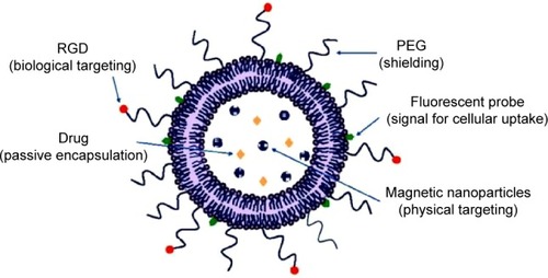
is a scheme of a multimodal particle based on a liposomal structure that allows theranostic applications. This liposome contains several individual cores (or a cluster of cores) of SPIONs, and, moreover, it can enclose a drug. The presence of magnetic nanoparticles makes bioimaging possible or the generation of heat in therapeutic hyperthermia, and also magnetic targeting. Optionally, the liposome can also encapsulate a drug. The shell material is responsible for its surface properties, because of the presence of reactive moieties on the surface. In this way, PEG is attached covalently to the surface of phospholipids in order to prevent aggregation and opsonization. The shell can be tuned to provide binding to molecules; as an example, the peptide RGD is bound at the distal end of some PEG chains for the purpose of targeted drug delivery. Such peptides facilitate the interaction of the liposome with integrins: proteins present on the cellular surface that recognize the peptide RGD. This biological targeting promotes the internalization of liposomes into cells. Moreover, the liposomal bilayer can contain a fluorescent probe, which permits its interaction with cells to be visualized by confocal microscopy.
Figure 5 Scheme of multifunctional liposome for molecular imaging, drug delivery, and therapy.
Abbreviations: RGD, arginine-glycine-aspartic acid; PEG, poly(ethyelene) glycol.
Conclusion
Over the last 25 years, various nanoparticles and complexes have been studied as MRI contrast agents, and several formulations have been approved for clinical use. These contrast agents are formed either of transition and lanthanide metals or of iron oxide nanoparticles and, more recently, ferrite nanoparticles. The transition or lanthanide metals, whose most significant representative is the ion gadolinium (Gd3+), have been extensively used as T1 contrast agents since they increase longitudinal relaxation times. A new generation of T1 contrast agents is formed by Gd complexes immobilized in various nanostructured materials (nanoporous silicas, dendrimers, perfluorocarbon nanoparticles, and nanotubes). Iron oxide nanoparticles with overall diameters greater than 50 nm can also be used as MRI contrast agents owing to their capacity to shorten T2* relaxation times in liver, spleen, and bone marrow by selective uptake and accumulation in MPS cells. Iron oxide nanoparticles with diameters <50 nm have been used for lymph node imaging; moreover, depending on the size of the iron core and their concentration, these small iron oxide nanoparticles can enhance T1 relaxation times. Moreover, iron oxide nanoparticles functionalized with bioactive materials have been used for targeted imaging via the site-specific accumulation.
The presence of dual-mode agents with strong T1–T2 contrast effects in a single construct is very challenging, since such dual agents improve the accuracy of biomedical imaging. Moreover, the development of nanomaterials that can filter the MRI artifacts allows the discrimination between signals coming from contrast agents or artifacts.
MLPs are an example of multifunctional platforms for either multimodal imaging or simultaneous imaging and therapy. MLPs can be carefully manipulated in their composition to incorporate cationic lipids, fluorescent-lipid conjugates, targeting ligands, drugs, and PEG, containing all in a single nanosystem.
The most notable limitations associated with the use of such contrast agents are the current detection limits and the lack of tissue specificity. Current detection limits need to be improved for the successful translation of nanoparticles to in vivo applications. These limitations have been overcome by recent developments in both MRI acquisition methods and postsynthesis modification of nanoparticles.
Acknowledgments
The authors are grateful to the Spanish Ministerio de Economía y Competitividad (MINECO) for financial support of the project MAT2012-36270-C04-03.
Disclosure
The authors report no conflicts of interest in this work.
References
- KherlopianARSongTDuanQA review of imaging techniques for systems biologyBMC Systems Biol2008274
- JamesMLGambhirSSA molecular imaging primer: modalities, imaging agents, and applicationsPhysiol Rev20129289796522535898
- HuangYHeSCaoWCaiKLiangXJBiomedical nanomaterials for imaging-guided cancer therapyNanoscale201246135614922929990
- JokerstJVGambhirSSMolecular imaging with theranostic nanoparticlesAcc Chem Res2011441050106021919457
- MassoudTFGambhirSSMolecular imaging in living subjects: seeing fundamental biological processes in a new lightGenes Dev20031754558012629038
- WeisslederRRossBDRehemtullaAGambhirSSMolecular Imaging: Principles and PracticeShelton, CT, USAPeople’s Medical Publishing House2010
- RudinMWeisslederRMolecular imaging in drug discovery and developmentNature Rev20032123131
- CherrySRMultimodality in vivo imaging systems: twice the power or double the trouble?Annu Rev Biomed Eng20068356216834551
- WeisslederRPitterMImaging in the era of molecular oncologyNature200845258058918385732
- LamTPouliotPAvtiPKSuperparamagnetic iron oxide based nanoprobes for imaging and theranosticsAdv Colloid Interface Sci2013199–20095113
- OpinaACGhaghadaKBZhaoPKieferGAnnapragadaASherryADTmDOTA-tetraglycinate encapsulated liposomes as pH-sensitive LipoCEST agentsPLOS One20116e2737022140438
- AimeSCastelliDDTerrenoEHighly sensitive MRI chemical exchange saturation transfer agents using liposomesAngew Chem Int Ed20054455135515
- WuYSoesbeTCKieferGEZhaoPSherryDA responsive europium (III) chelate that provides a direct readout of pH by MRIJ Am Chem Soc2010132140021400320853833
- HashimZGreenMChungPHGd-containing conjugated polymer nanoparticles: bimodal nanoparticles for fluorescence and MRI imagingNanoscale201468376838624941427
- StephenZRKievitFMZhangMMagnetic nanoparticles for medical MR imagingMater Today201114330338
- MornetSVasseurSGrassetFMagnetic nanoparticle design for medical diagnosis and therapyJ Mater Chem20041421612175
- RümenappCGleichBHaaseAMagnetic nanoparticles in magnetic resonance imaging and diagnosticsPharm Res2012291165117922392330
- LaurentSVander ElstLRochAStructure, synthesis and characterization of contrast agents for magnetic resonance molecular imagingCarretaPLascialfariANMR-MRI, SR and Mössbauer Spectroscopies in Molecular MagnetsMilan, ItalySpringer20077187
- GeraldesCFLaurentSClassification and basic properties contrast agents for magnetic resonance imagingContrast Media Mol Imaging2009412319156706
- LouieAMultimodality imaging probes: design and challengesChem Rev20101103146319520225900
- CaravanPEllisonJJMcMurryTJLaufferRBGadolinium (III) chelates as MRI contrast agents: structure, dynamics, and applicationsChem Rev1999992293235211749483
- WiegersCBWelchMJSharpTLEvaluation of two new gadolinium chelates as contrast agents for MRIMagn Res Imaging199210903911
- LeeGHChangYKimTJBlood-pool and targeting MRI contrast agents: from Gd-chelates to Gd-nanoparticlesEur J Inorg Chem20121019241933
- World Health OrganizationPharmaceuticals: Restrictions in Use and AvailabilityWHO/EMP/QSM/2010.3Geneva: Switzerland201014
- WinterPAtheyPKieferGImproved paramagnetic chelate for molecular imaging with MRIJ Magn Magn Mater2005293540545
- KobayshiHSatoNKawamotoSW. 3D MR angiography of mice using macromolecular MR contrast agents with polyamindoamine dendrimer core with reference to their pharmacokinetic propertiesMagn Reson Med20014545446011241704
- FríasJCWilliamsKJFisherEARecombinant HDL-like nanoparticles: a specific contrast agent for MRI of atherosclerosis plaquesJ Am Chem Soc2004126163161631715600321
- SenpanACaruthersSDRheeIConquering the dark side: colloidal iron oxide nanoparticlesACS Nano200933917392619908850
- European Medicines AgencyEMA/486286/2012LondonUK: European Medicines Agency2012
- PanDCaruthersSDSenpanARevisiting and old friend: manganese-based MRI contrast agentsWiley Interdiscip Rev Nanomed Nanobiotechnol2011316217320860051
- CaravanPStrategies for increasing the sensitivity of gadolinium based MRI contrast agentsChem Soc Rev20063551252316729145
- ParkJYChoiESBaekMJWater-soluble ultra small paramagnetic or superparamagnetic metal oxide nanoparticles for molecular MR ImagingEur J Inorg Chem2009924772481
- KrishnanKBiomedical nanomagnetics: a spin through possibilities in imaging, diagnostics, and therapyIEEE Trans Magn2010462523255820930943
- FilippousiMAngelakerisMSikiniMSurfactant effects on the structural and magnetic properties of iron oxide nanoparticlesJ Phys Chem C20141181620916217
- ShokrollahiHStructure, synthetic methods, magnetic properties and biomedical applications of ferrofluidsMater Sci Eng C Mater Biol Appl2013332476248723623058
- PflipsenCForgeDBenaliSImproved stability and relaxivity of a commercial magnetic ferrofluidJ Phys Chem C20131172091920926
- BulteJWKraitchmanDLIron oxide MR contrast agents for molecular and cellular imagingNMR Biomed20041748449915526347
- BulteJWIn vivo MRI cell tracking: clinical studiesAm J Roentgenol200919331432519620426
- LaConteLENitinNZurkiyaOCoating thickness of magnetic iron oxide nanoparticles affects R2 relaxivityJ Magn Reson Imaging2007261634164117968941
- HuFJoshiHMDravidVPHigh-performance nanostructured MR contrast probesNanoscale201021884189120694208
- ChoiHSLiuWMisraPRenal clearance of nanoparticlesNat Biotechnol2007251165117017891134
- LongmireMChoykePLKobayashiHClearance properties of nano-sized particles and molecules as imaging agents: considerations and caveatsNanomedicine2008370371718817471
- MaedaHWuJSawaTTumor vascular permeability and the EPR effect in macromolecular therapeutics: a reviewJ Control Release20006527128410699287
- HammBIron-oxide-enhanced MR lymphography: just a new toy or a breakthrough?Eur Radiol20021295795811976839
- LindKKresseMDebusNPMullerRHA novel formulation for superparamagnetic iron oxide (SPIO) particles enhancing MR lymphography: comparison of physicochemical properties and the in vivo behaviorJ Drug Target20021022123012075823
- MaesRMLewinJSDuerkJLA new type of susceptibility-artefact-based magnetic resonance angiography: intra-arterial injection of superparamagnetic iron oxide particles (SPIO) A Resovist in combination with TrueFisp imaging: a feasibility studyContrast Media Mol Imaging2006118919517193696
- SchmitzSAAlbrechtTWolfKJMR angiography with superparamagnetic iron oxide: feasibility studyRadiology199921360360710551249
- MorawskiAWinterPCrowderKTargeted nanoparticles for quantitative imaging of sparse molecular epitopes with MRIMagn Reson Med20045148048615004788
- CaiHAnXCuiJFacile hydrothermal synthesis and surface functionalization of polyethyleneimine-coated iron oxide nanoparticles for biomedical applicationsACS Appl Mater Interfaces201351722173123388099
- LiJHeYSunWHyaluronic acid-modified hydrothermally synthesized iron oxide nanoparticles for targeted tumor MR imagingBiomaterials2014353666367724462358
- ZhangHLiJSunWHyaluronic acid-modified magnetic iron oxide nanoparticles for MR imaging of surgically induced endometriosis model in ratsPLOS One20149e9471824722347
- GuptaAKGuptaMSynthesis and surface engineering of iron oxide nanoparticles for biomedical applicationsBiomaterials2005263995402115626447
- XieJChenKLeeHYUltrasmall c(RGDyK)-coated Fe3O4 nanoparticles and their specific targeting to integrin alpha(v)-beta(3)-rich tumor cellsJ Am Chem Soc20081261145811459
- DesgrosellierJSChereshDAIntegrins in cancer: biological implications and therapeutic opportunitiesNat Rev Cancer20101092220029421
- Bin NaHChan SongIHyeonTInorganic nanoparticles for MRI contrast agentsAdv Mater20092121332148
- TerrenoECastelliDDVialeAChallenges for molecular magnetic resonance imagingChem Rev20101103019304220415475
- YooDLeeJHShinTHTheranostic magnetic nanoparticlesAcc Chem Res20114486387421823593
- HadjipanayisCJBonderMJBalakrishnanSMetallic iron nanoparticles for MRI contrast enhancement and local hyperthermiaSmall200841925192918752211
- YangHZhangCShiXWater-soluble superparamagnetic manganese ferrite nanoparticles for magnetic resonance imagingBiomaterials2010313667367320144480
- HoDSunXSunSMonodisperse magnetic nanoparticles for theranostic applicationsAcc Chem Res20114487588221661754
- JangJTNahHLeeJHCritical enhancements of MRI contrast and hyperthermic effects by dopant-controlled magnetic nanoparticlesAngew Chem Int Ed20094812341238
- DasGKJohnsonNJCramenJNaDyF4 nanoparticles as T2 contrast agents for ultrahigh field magnetic resonance imagingJ Phys Chem Lett20123524529
- BulteJWWuCBrechbielMWDysprosium-DOTA-PANAM dendrimers as macromolecular T2 contrast agents: preparation and relaxometryInvest Radiol1998338418459818319
- DasGKZhangYSilvaLDSingle-phase Dy2O3:Tb3+ nanocrystals as dual-modal contrast agent for field high magnetic resonance and optical imagingChem Mater20112324392446
- GueronMNuclear relaxation in macromolecules by paramagnetic ions: a novel mechanismJ Magn Reson1975195866
- TaboadaERodríguezERoigARelaxometric and magnetic characterization of ultrasmall iron oxide nanoparticles with high magnetization. Evaluation as potential T1 magnetic resonance imaging contrast agents for molecular imagingLangmuir2007234583458717355158
- ShubayevVIPisanicTRIIJinSMagnetic nanoparticles for theragnosticsAdv Drug Deliver Rev200961467477
- CunninghamCHAraiTYangPCPositive contrast magnetic resonance imaging of cells labeled with magnetic nanoparticlesMagn Reson Med200553999100515844142
- HuangJZhongXWangLImproving the magnetic resonance imaging contrast and detection methods with engineered magnetic nanoparticlesTheranostics201228610222272222
- StuberMGilsonWDSchärMPositive contrast visualization of iron oxide-labeled stem cells using inversion-recovery with ON-resonant water suppression (IRON)Magn Reson Med2007581072107717969120
- McAteerMASibsonNRvon zur MuhlenCIn vivo magnetic resonance imaging of acute brain inflammation using microparticles of iron oxideNature Med2007101253125817891147
- McAteerMASchneiderJEAliZAMagnetic resonance imaging of endothelial adhesion molecules in mouse atherosclerosis using dual targeted microparticles of iron oxideArterioscler Thromb Vasc Biol200828778317962629
- MuhlenVon zurvon ElverfeldtDMoellerJAMagnetic resonance imaging contrast agent targeted toward activated platelets allows in vivo detection of thrombosis and monitoring of thrombolysisCirculation200811825826718574047
- CorotCRobertPIdèeJMRecent Advances in iron oxide nanocrystal technology for medical imagingAdv Drug Deliv Rev2006581471150417116343
- ParkJYDakshaPLeeGHHighly dispersible PEG surface modified ultra-small superparamagnetic iron oxide nanoparticles useful for target-specific biomedical applicationsNanotechnology20081936560321828874
- Di MarcoMSadunCPortMPhysicochemical characterization of ultrasmall superparamagnetic iron oxide particles (USPIO) for biomedical application as MRI contrast agentsInt J Nanomedicine2007260962218203428
- GossuinYGillisAHocqAMagnetic resonance relaxation properties of superparamagnetic particlesWiley Interdiscip Rev Nanomed Nanobiotechnol2009129931020049798
- JunYWHuhYMChoiJSNanoscale size effect of magnetic nanocrystals and their utilization for cancer diagnosis via magnetic resonance imagingJ Am Chem Soc20051275732573315839639
- ChoiJSLeeJHShinTHSelf-confirming “AND” logic nanoparticles for fault-free MRIJ Am Chem Soc2010132110151101720698661
- BonmatíMOGossuinYMoralesMComparative analysis of the 1H NMR relaxation enhancement produced by iron oxide and core-shell iron–iron oxide nanoparticlesMagn Reson Imaging2007251437144117566686
- ShultzMDCalvinSFatourosPPEnhanced ferrite nanoparticles as MRI contrast agentsJ Magn Magn Mater2007311464468
- BaeKYKimYBLeeYBioinspired synthesis and characterization of gadolinium-labeled magnetite nanoparticles for dual contrast T1- and T2-weighted magnetic resonance imagingBioconjugate Chem201021505512
- YangHZhuangYSunYTargeted dual contrast T1- and T2-weighted magnetic resonance imaging of tumors using multifunctional gadolinium-labeled superparamagnetic iron oxide nanoparticlesBiomaterials2011324584459321458063
- DingDKanalyCWCummingsTJLong-term safety of combined intracerebral delivery of free gadolinium and targeted chemotherapeutic agent PRX321Neurol Res20103281081520021739
- TorchilinVPLiposomes as delivery agents for medical imagingMol Med Today199622422498796897
- KamalyNMillerADParamagnetic liposome nanoparticles for cellular and tumour imagingInt J Mol Sci2010111759177620480040
- StrijkersGJMulderWJvan HeeswijkRBRelaxivity of liposomal paramagnetic MRI contrast agentsMAGMA20051818619216155762
- VictorKGKorbJPBryantRGTranslational dynamics of water at the phospholipid interfaceJ Phys Chem B2013117124751247824059874
- AyyagariLAZhangXGhaghadaKBAnnapragadaALong-circulating liposomal contrast agents for magnetic resonance imagingMagn Reson Med2006551023102916586449
- TilcockCMacDougallPUngerEThe effect of lipid composition on the relaxivity of Gd-DTPA entrapped in lipid vesicles of defined sizeBiochim Biophys Acta199010221811862306454
- UngerECMacDougallPCullisPLiposomal Gd-DTPA: effect of encapsulation on enhancement of hepatoma model by MRIMagn Reson Imaging198974174232811620
- Van TilborgGAMulderWJDeckersNAnnexin A5-functionalized bimodal lipid-based contrast agents for the detection of apoptosisBioconjugate Chem200617741749
- De CuyperMJoniauMMagnetoliposomes: formation and structural characterizationEur Biophys J1988153113193366097
- SoenenSJHodeniusMDe CuyperMMagnetoliposomes: versatile innovative nanocolloids for use in biotechnology and biomedicineNanomedicine (Lond)2009417719119193184
- De CuyperMThe original magnetoliposomes: from the physicochemical basics to theranostic nanomedicineCallejas-FernándezJEstelrichJQuesada-PérezMForcadaJSoft Nanoparticles for Biomedical ApplicationsCambridge, UKRoyal Society of Chemistry2014
- MartinaMSFortinJPMenagerCGeneration of superparamagnetic liposomes revealed as highly efficient MRI contrast agents for in vivo imagingJ Am Chem Soc2005127106761068516045355
- SabatéRBarnadas-RodríguezRCallejas-FernándezJPreparation and characterization of extruded magnetoliposomesInt J Pharm200834715616217692483
- ChenYBoseABothunGDControlled release from bilayer-decorated magnetoliposomes via electromagnetic heatingACS Nano201043215322120507153
- García-JimenoSEscribanoEQueraltJMagnetoliposomes prepared by reverse-phase followed by sequential extrusion: characterization and possibilities in the treatment of inflammationInt J Pharm201140518118721129463
- FrascioneDDiwokyCAlmerGUltrasmall superparamagnetic iron oxide (USPIO)-based liposomes as magnetic resonance imaging probesInt J Nanomedicine201272349235922661890
- MannSHanningtonJPFormation of iron oxides in unilamellar vesiclesJ Colloid Interface Sci1988122326335
- SoenenSJHimmelreichUNuyttenNIntracellular nanoparticle coating stability determines nanoparticle diagnostics efficacy and cell functionalitySmall201062136214520818621
- FattahiHLaurentSLiuFMagnetoliposomes as multimodal contrast agents for molecular imaging and cancer nanotheragnosticsNanomedicine2011652954421542690
- CasalsEVerdaguerATondaRAtomic force microscopy of liposomes bearing fibrinogenBioconjugate Chem200314593600
- García-JimenoSEscribanoEQueraltJExternal magnetic field-induced selective biodistribution of magnetoliposomes in miceNanoscale Res Lett2012745222883385
- De CuyperMSoenenSJCoenegrachtsKSurface functionalization of magnetoliposomes in view of improving iron oxide-based magnetic resonance imaging contrast agents: anchoring of gadolinium ions to a lipophilic chelateAnal Biochem200736726627317568553
- SoenenSJDesenderLDe CuyperMComplexation of gadolinium (III) ions on top of nanometer-sized magnetoliposomesInt J Environ Anal Chem200787783796