
 ?Mathematical formulae have been encoded as MathML and are displayed in this HTML version using MathJax in order to improve their display. Uncheck the box to turn MathJax off. This feature requires Javascript. Click on a formula to zoom.
Abstract
Selenium is an important component of human diet and a number of studies have declared its chemopreventive and therapeutic properties against cancer. However, very limited studies have been conducted about the properties of selenium nanostructured materials in comparison to other well-studied selenospecies. Here, we have shown that the anticancer property of biostabilized selenium nanorods (SeNrs) synthesized by applying a novel strain Ess_amA-1 of Streptomyces bikiniensis. The strain was grown aerobically with selenium dioxide and produced stable SeNrs with average particle size of 17 nm. The optical, structural, morphological, elemental, and functional characterizations of the SeNrs were carried out using techniques such as UV-vis spectrophotometry, transmission electron microscopy, energy dispersive X-ray spectrometry, and Fourier transform infrared spectrophotometry, respectively. The MTT (3-(4,5-dimethylthiazol-2-yl)-2,5-diphenyltetrazolium bromide) assay revealed that the biosynthesized SeNrs induces cell death of Hep-G2 and MCF-7 human cancer cells. The lethal dose (LD50%) of SeNrs on Hep-G2 and MCF-7 cells was recorded at 75.96 μg/mL and 61.86 μg/mL, respectively. It can be concluded that S. bikiniensis strain Ess_amA-1 could be used as renewable bioresources of biosynthesis of anticancer SeNrs. A hypothetical mechanism for anticancer activity of SeNrs is also proposed.
Introduction
Selenium (Se) is an important trace element that plays a crucial role in human health and regulates many crucial cellular functions mediated through its incorporation into selenoproteins.Citation1,Citation2 This inorganic element also occurs in trigonal and monoclinic phases of crystalline microstructures. The monoclinic phase is less stable and occurs in α, β, and γ forms, which differ only in the way that the crystals are packed.Citation3 The antioxidant function of Se is conferred by some of these selenoproteins that directly or indirectly protect against oxidative stress. The extensive experimental evidence indicates based on the in vitro, animal, geographic, and prospective studies that Se supplementation reduces the incidence of various types of cancers. Since as early as the 1960s, geographical studies have proved a consistent trend for populations with low Se compounds intake to have higher cancer mortality rates.Citation2,Citation4 However, at elevated doses, Se compounds usually turn into a prooxidant with well-established cell growth inhibiting/killing properties.Citation1 Thus, the uses of Se compounds for anticancer therapy have been greatly explored during the last decade and results of studies have shown that Se compounds reduce the risk of various types of cancers, such as mammary, prostate, lung, colon, and liver cancers.Citation1,Citation4–Citation7 The research findings also suggest that the concentration, chemical species, and redox potential of Se compounds are critical for their anticancer activity.Citation1,Citation5 These Se compounds have more promising anticancer activity at high dosage; however, high doses of Se compounds give rise to greater concerns about its toxicity.Citation1,Citation5 In this regard, selenium nanostructured materials (SNMs) could reduce the risk of Se toxicity and be widely used in cancer biology due to its promising anticancer activity and less toxicity, compared to Se compounds (inorganic and organic).Citation6–Citation9 SNMs also exhibit unique physical, chemical, and biological properties compared to that of Se compounds.Citation10 Various types of SNMs, which are stabilized and modified with different kinds of biological macromolecules, are reported to possess excellent anticancer activities.Citation1,Citation11–Citation13 Based on this, some researchers suggest that biological macromolecules stabilized and modified may have potential applications as anticancer agents for the killing of human cancer cells. For these reasons, the study of SNMs has gained considerable importance in recent years and therefore, various types SNMs have been obtained by employing physico-chemical methods, ie, amorphous,Citation3,Citation11 trigonal, nanorods,Citation12,Citation13 nanoribbons,Citation14 hexagonal prism,Citation13 nanoplates,Citation15 nanotubes,Citation16 and spheres.Citation13 Therefore, SNMs are being widely used in basic and applied areas of chemistry, physics, environmental science, material science, and biomedicine.Citation7,Citation13,Citation17,Citation18 However, concern is now growing regarding the environmental impact of the SNM synthesis process based on physico-chemical methods that require high pressures, temperatures, and toxic chemicals. These methods have some drawbacks: (i) production of stable SNMs dispersions only at lower concentrations and unsuitability for large-scale production; (ii) requirement of additional stabilizing agents; (iii) production of hazardous by-products, and (iv) increased pollution in the environment.
Consequently, significant efforts are ongoing toward the development of novel nontoxic methods for the synthesis and surface modification/stabilization of SNMs.Citation19–Citation21 Biogenic methods are a renewable, clean, nontoxic, and environmentally friendly procedure for the synthesis of these types of nanomaterials.Citation7,Citation22,Citation23 Recently, biogenic methods have been utilized for the synthesis of a variety of SNMs at ambient conditions.Citation6,Citation19,Citation24–Citation26 It is well established that biosynthesized SNMs have several important characteristics, including more stability and higher biological activity, due to the surface functionalization by biological macromolecules secreted by fungi and bacteria.Citation7,Citation9,Citation17,Citation23,Citation27 Among the different microorganisms, Actinomycetes are less explored for the synthesis of SNMs. Actinomycetes are a diverse group of filamentous true bacteria found in a variety of habitats in terrestrial and aquatic environments.Citation28 Hence, there are reports that have shown that actinomycetes are efficient bioagents for the intracellular and extracellular synthesis of metal nanostructured materials (MNMs).Citation22 Most of the studies have been done on species of Streptomyces genus due to its inherent capability for the production of redox active macromolecules/secondary metabolites.Citation29,Citation30 The capability of Streptomyces genus for the biosynthesis of MNMs has previously been reported.Citation18,Citation22,Citation31–Citation34 However, the synthesis of biostabilized selenium nanorods (SeNrs) using any strain of actinomycetes has not been reported yet. In the present study, we reported: (i) a simple, environmentally friendly, and renewable biogenic method for synthesizing disperse and stable SeNrs by Streptomyces bikiniensis strain Ess_amA-1 and (ii) presented evidence that biosynthesized SeNrs have the anticancer activity. To the best of our knowledge, this is the first report on the synthesis of SeNrs by S. bikiniensis as a novel renewable bioresource and opens up the possibility of commercially viable biogenic production of SeNrs for novel anticancer nanostructured materials.
Materials and methods
Isolation of S. bikiniensis
An insect Tapinoma simrothi was collected from Eldrieh, Riyadh, Saudi Arabia (24.7 N latitude and 46.7 E longitude), and used for the isolation of S. bikiniensis. The suspension of T. simrothi was prepared in normal saline solution (NSS) for the isolation of saccharolytic actinomycetes and an appropriate dilution was spread on the starch casein agar (SCA) medium (pH 7.2±0.2), supplemented with antibiotics (cycloheximide [40 mg/L], nystatin [30 mg/L], and nalidixic acid [10 mg/L]).Citation35,Citation36 The inoculated Petri plates were incubated aerobically at 30°C until the appearance of powdery texture colonies with branching filaments and aerial mycelia. The selected colonies were subcultured and further purified by streaking and among them the strain Ess_amA-1was selected and maintained on International Streptomyces Project 2 (ISP-2) agar medium by periodical subculturing.
Morphological and physiological characterization of S. bikiniensis strain Ess_amA-1
The color of aerial mycelium was determined from mature and sporulating aerial mycelia of the actinomycete colonies on different media such as ISP-2, ISP-4, ISP-6, ISP-7, Czapek Dox, and SCA. The color was determined using color names lists.Citation37 The color of the soluble pigments was determined visually by observing the color changes in the medium due to the diffusing pigments produced by strain Ess_amA-1.Citation38 Carbohydrates and physiological tests were performed using the specific media and methods.Citation39–Citation41 All the cultures were incubated at 30°C for 7 days. The assay for enzymatic activity was performed according to Bibb et al.Citation42
Genomic DNA extraction and purification
Total genomic DNA of strain Ess_amA-1 was isolated from the mycelium biomass (0.1 g), which was harvested from the freshly grown culture in ISP-2 medium as described. Briefly, the collected mycelium biomass was crushed with liquid nitrogen and the powder was mixed with 500 μL lysis buffer (containing 50 mM Tris-HCl, pH 8.0; 5 mM EDTA, pH 8.0; 50 mM NaCl; and 20 μL lysozyme, 10 mg/mL). The cells were lysed by vigorous vortexing and lysate was incubated at 37°C for 30 minutes. Subsequently, 20 μL SDS (10% w/v) and 20 μL of proteinase K (10 mg/mL) were added into the Eppendorf tube and incubated at 55°C for 30 minutes. The cell lysate was cooled down and extracted once with an equal volume of phenol and chloroform (1:1 v/v). The aqueous phase was collected by centrifugation at 10,000 rpm for 5 minutes. Total genomic DNA was precipitated from the obtained aqueous phase by the addition of two volumes of chilled isopropanol. The precipitated genomic DNA was palletized by centrifugation at 13,000 rpm for 30 minutes and the pellet was washed with 70% ethanol. The washed pellet was air dried under laminar flow and dissolved in 50 μL TE buffer (containing 50 mM Tris-HCl and 1 mM EDTA; pH 7.2).
Polymerase chain reaction amplification of 16S ribosomal RNA (16S rRNA) gene and nucleotide sequencing
For molecular characterization, 16S ribosomal RNA (16S rRNA) gene was amplified using primer set Star-F (5′-GAGTTTGATCMTGGCTCAG-3′) and 1387-R (5′-CGGGCGGTGTGTACAAGG-3′). Briefly, ~50 ng genomic DNA was used as the template in 25 μL of polymerase chain reaction (PCR) mixture containing 0.20 mM dNTPs (Deoxyribonucleotide triphosphates), 25 pmole of each primer, 5× Taq polymerase buffer (5 μL), 2.0 mM MgCl2, and 0.2 U Taq DNA polymerase. The PCR parameters consisted of 35 cycles of denaturation at 94°C for 1 minute, annealing at 51°C for 1 minute, extension at 72°C for 1 minute with an initial denaturation, and the final extension was performed for 3 minutes and 7 minutes at 94°C and 72°C, respectively. Amplification was performed on the PCR machine (Sens Quest co., D-37085 Goettingen, Germany). The amplicon was analyzed and sequenced by GATC Biotech, European Custom Sequencing Centre (Cologne, Germany).
Multiple sequence alignments and phylogenetic analysis
The obtained 16S rRNA gene sequence was compared with the homologous sequences retrieved from GenBank using the Blastn tool.Citation43 Multiple sequence analysis with the sequences of different actinomycetes groups was performed using CLUSTALW with default parameters.Citation44 A phylogenetic tree was constructed by the neighbor-joining method with nucleotide pair-wise genetic distances corrected by the Kimura two-parameter methodCitation45 using the TreeCon tool. The reliability of the tree topology was subjected to a bootstrap test and numbers at the nodes indicate bootstrap support values as a percentage of 1,000 replications. All branches with <50% bootstrap support were judged as inconclusive and were collapsed and branch lengths for all trees were normalized to 0.02% divergence. Based on biochemical and molecular characterization, the characterized strain was designated as S. bikiniensis strain Ess_amA-1.
Biosynthesis of SeNrs
Briefly, sterile 100 mL ISP-2 medium containing 1 mM selenium dioxide (SeO2) was inoculated with 1 mL of the fresh inoculums (OD600, 0.5) of strain Ess_amA-1 and incubated in an orbital shaker incubator (150 rpm) at 30°C for 48 hours. A control flask containing ISP-2 without SeO2 was inoculated with a test strain and incubated under the same conditions. The reduction of SeO2 into the elemental selenium (S0) and the nucleation/growth of SeNrs were monitored by sampling an aliquot of the medium at different time periods of incubation (6 hours, 12 hours, and 48 hours). The cells were then removed by filtration and the resulting cell-free filtrate was then centrifuged at 14,000 rpm for 15 minutes to obtain the biosynthesized SeNrs.
Characterization of biosynthesized SeNrs
The optical, structural, morphological, elemental, and functional characterizations of the SeNrs were carried out using UV–Vis spectrophotometer, transmission electron microscope (TEM), energy dispersive X-ray (EDAX) spectrometer, and Fourier transform infrared (FTIR) spectrophotometer, respectively. In order to ascertain the optical characteristics of synthesized SNTs, the absorption spectrum was recorded by Lambda 35 double beam UV–Vis spectrophotometer (Hitachi, Japan) in the wavelength range of A200–A800 nm using a quartz cuvette. The size and structure of the biosynthesized SeNrs were analyzed by JEM-1010 TEM (JEOL, Tokyo, Japan) at an accelerating voltage of 80 kV. For this analysis, the sample was prepared by placing drops of SeNrs aqueous solution on carbon coated copper grids and air dried under dark conditions. The elemental analysis of SeNrs was done using the energy dispersive X-ray spectroscopy (EDS) equipped with JSM-6380 LA scanning electron microscope (SEM) (JEOL, Tokyo, Japan). For functional characterization of SeNrs, the FTIR spectrum was recorded in the range of 400–4,000 wave number (cm−1) on Nicolet 6700 FTIR spectrometer in the transmittance mode at a 4 cm−1 resolution.
3-(4,5-dimethylthiazol-2-yl)-2,5-diphenyltetrazolium bromide assay
The anticancer activity of biosynthesized SeNrs was tested on human breast adenocarcinoma cell line (MCF-7) and human liver carcinoma cell line (Hep-G2) cells (ATCC, Manassas, VA, USA) using the 3-(4,5-dimethylthiazol-2-yl)- 2,5-diphenyltetrazolium bromide (MTT) dye reduction assay. This assay is based on the reduction of MTT dye to a blue colored formazan product by mitochondrial dehydrogenase. The cells were cultured in a humid environment at 37°C and 5% CO2 in a cell culture minimum essential medium (Thermo Fisher Scientific, Waltham, MA, USA) supplemented with 15% fetal bovine serum and 1% penicillin/streptomycin (Thermo Fisher Scientific). At 85%–90% confluence, cells were harvested using 0.25% trypsin/EDTA solution and subcultured into a 96-well plate. The MTT colorimetric assay developed by MosmannCitation46 with modification was used to screen the cytotoxic activity of SeNrs. Briefly, the MCF-7 and Hep-G2 cells (1×10⁴ cells/well) were grown overnight in 96-well flat bottom culture plates, and then exposed to seven different concentrations (1.0, 2.0, 5.0, 10, 25, 50, and 100 μg/mL) of SeNrs for 24 hours. In addition, negative/vehicle control and positive control (doxorubicin) were also used for comparison. After the completion of the desired treatment, 10 μL of MTT reagent (Thermo Fisher Scientific) was added to each well and further incubated for 3 hours at 37°C. Finally, the medium with MTT solution was removed, and 100 μL of DMSO (Sigma-Aldrich Co., St Louis, MO, USA) was added to each well and further incubated for 20 minutes. The optical density (OD) of each well was measured at 570 nm using a microplate reader (Synergy; BioTek, Winooski, VT, USA). The percentage of cytotoxicity compared to the untreated cells was determined. Triplicates were maintained for each treatment. Lethal concentration (LC50) was determined by calculating the cell viability:
Statistical analysis
The triplicate sets of data for the various parameters evaluated were subjected to analysis of variance in accordance with the experimental design (completely randomized design) using SAS statistical packages (Cary, version 6.12; SAS Institute Inc., NC, USA) to quantify and evaluate LSD. The values were calculated at P level of 0.05%.
Results and discussion
Characterization of strain Ess_amA-1
In the present study, the actinomycete strain Ess_amA-1 was isolated from an insect T. simrothi with the aim of exploiting its SeNrs synthesizing potential for anticancer therapy. The presumptive taxonomic identification of the strain was done by its morphological and biochemical characteristics. For the identification of morphological characteristics, a specimen of the strain was examined under the bright-field microscope and analysis revealed that the strain Ess_amA-1 produces a light brown and gray substrate (). These morphological characteristics of the strain were precisely confirmed by the SEM. The image showed that the smooth-surfaced spores are held in straight chains (rectiflexibiles) ().Citation47,Citation48 The morphological characteristics of the strain Ess_amA-1 were further substantiated by physiological and biochemical tests. The results of the physiological and biochemical characteristics are summarized in and . The strain showed fast growth behavior on media (ISP-2, 4, 6, and 7) and moderate growth behavior on ISP-5 and Czapek-Dox agar medium; melanin pigment production was determined on ISP-6 medium. Thus, based on the morphological and biochemical characteristics of strain Ess_amA-1 was identified as a member of Streptomyces genus.Citation38 For the authentic taxonomic characterization of the strain, 16S rRNA gene sequencing was performed and obtained data were analyzed carefully. The strain was identified as a member of S. bikiniensis by 16S rRNA gene sequencing and in silico analysis that has been deposited in NCBI GenBank (Accession Number: KF588366). The total length of the 16S rRNA gene sequenced in the present study has 1,206 base pairs, and NCBI Blastn analysis revealed ~99% sequence homology with the strain of S. bikiniensis clone ZD Hu (GenBank, Accession No: AY946043.1). The related sequence data sets were retrieved from the Blastn result and phylogenetic analysis indicated a close genetic relatedness of strain Ess_amA-1 with S. bikiniensis and we therefore designated the strain as S. bikiniensis strain Ess_amA-1 (). The members of the Streptomyces genus have been largely exploited for the production of bioactive secondary metabolites (ie, antimicrobials, antitumorals, antihypertensives, and immunosuppressants) with wide uses in medicine and agriculture.Citation30,Citation49,Citation50 Thus, the species of the genus Streptomyces are well established bioresources for the production of valuable nanostructured materials.Citation18,Citation22,Citation31–Citation34,Citation51–Citation57
Table 1 Morphological characteristics and growth behavior of Streptomyces bikiniensis strain Ess_amA-1 on different culture media
Table 2 Physiological and biochemical characteristics of Streptomyces bikiniensis strain Ess_amA-1
Figure 1 Isolation and morphological characterizations of Streptomyces bikiniensis strain Ess_amA-1.
Notes: (A) Growth behavior of the isolated strain Ess_amA-1on ISP-2 solid medium Petri plate after 48 hours incubation at 30°C. The strain shows the production of light brown and gray substrate and aerial mycelium on solid medium; (B) scanning electron microscope image of strain Ess_amA-1 shows the morphological characteristics of mycelium and spores at 2 μM scale.
Abbreviation: ISP-2, International Streptomyces Project 2.
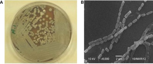
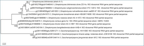
Figure 2 Molecular characterization of Streptomyces bikiniensis strain Ess_amA-1.
Notes: Phylogenetic tree showing the relationship of strain Ess_amA-1with other strains of Streptomyces species based on 16S rRNA gene sequences retrieved from NCBI GenBank. Arrow represents the status of strain am-1 from this study in the phylogenetic tree.
Biosynthesis of SeNrs
The bioreductive capability of the strain was utilized for the synthesis of SeNrs. The strain when challenged with 1 mM SeO2 exhibited a time-dependent change in the color of the ISP-2 liquid culture medium from light gray to red after 6 hours incubation period. The intensity of the red color of culture medium increased upon further incubation up to 48 hours (). The emergence of a red-brick color in the culture medium after incubation of 48 hours was a clear indication that the strain biogenically easily reduces selenite ions to insoluble elemental Se (Se0) form.Citation6,Citation8,Citation25,Citation27,Citation58 The yield of SeNrs was determined and was approximately 7.74 mg/100 mL. The reaction mixture of SeNrs biosynthesis can be optimized to increase the yield and purity by altering the physico-chemical and cultural conditions in the used medium: i) precursor salt, ii) carbon and nitrogen source, iii) pH and oxygen supply, and iv) addition of electron donor, etc.Citation59
Figure 3 Biosynthesis of SeNrs using strain Ess_amA-1.
Notes: (A) ISP-2 medium containing 1 mM selenium dioxide (SeO2) was inoculated with 1 mL of the fresh inoculums of strain Ess_amA-1 and incubated in an orbital shaker incubator at 30°C for 48 hours. The in situ reduction of SeO2 into the SeNrs (brick red color development) is shown by strain Ess_amA-1; (B) a control flask containing ISP-2 without SeO2 was inoculated with Ess_amA-1 and incubated under the same conditions.
Abbreviations: SeNrs, selenium nanorods; ISP-2, International Streptomyces Project 2.

Characterizations (optical, morphological, elemental, and functional) of SeNrs
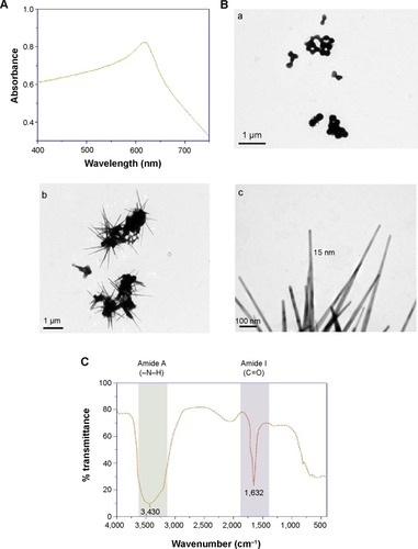
The synthesis of SeNrs in liquid culture medium was monitored by the UV–Vis spectroscopy that showed a strong and broad surface plasmon resonance (SPR) peak at ~620 nm, which is a characteristic of SeNrs (). However, no absorption peak corresponding to the SeNrs in the control flask (without SeO2) was observed. It is well known that due to Mie scattering, SeNrs exhibit absorption at the wavelength ~620 nm. As evident from previous reports, the presence of a single SPR peak evokes biogenic synthesis of SeNrs by S. bikiniensis strain Ess_amA-1, and this was further confirmed by TEM and EDS techniques.Citation58 The time-dependent intensity of red color increase of the culture medium indicated the gradual growth in the size and shape of SeNrs during the incubation period. Hence, the SeNrs were analyzed at three different incubation times, 6 hours, 24 hours, and 48 hours, employing the TEM. The time-dependent change in the shape and morphology of SeNrs was noticed. During the incubation, the shape of reduced elemental selenium (Se0) gradually changed from the spherical structure to a rod-like structure. shows the spherical shape of Se0 irregular nanospheres, which possess an average diameter of 50–100 nm after 6 hours of incubation. However, these spherical shape structures start to lose their integrity as relatively low aspect-ratio anisotropic structures (rods). After 12 hours, aggregates of higher aspect-ratio rods emerging from a few growth centers were observed (). After 48 hours, the length of the structures increased in one dimension and they were converted into the rod-like structures (). The SeNrs with an average length of 600 nm, average diameter of 17 nm, and aspect ratio of 35:1 were observed. Nevertheless, most of the biosynthesis methods have reported the production of spherical shape selenium nanoparticles (SeNPs).Citation24,Citation26,Citation27,Citation60 However, few recent studies reported the biosynthesis of Se nanorods via spherical SeNPs that acted as seeds for growth.Citation6,Citation17 The high free energy of SeNPs evoked the Ostwald ripening process, which may be responsible for the growth of spherical SeNPs into SeNrs.Citation17,Citation61 The TEM data also revealed the nucleation/growth of the SeNrs (6 hours and 12 hours incubation) that is probably due to the presence of aromatic amino acids produced by the strain Ess_amA-1, thus indicating possible adhesion of biological macromolecules on the surface of SeNrs. These data are consistent with the previously documented occurrence of biological macromolecules associated to SNMs of microbial origin.Citation7,Citation9,Citation22 The EDX analysis revealed the presence of Se peak at 1.37 keV, confirming that the SeNrs was successfully synthesized.Citation23 However, the peaks of carbon (C) and oxygen (O) were believed to be derived from biological macromolecules present on the surface of SeNrs (). These biological macromolecules may be responsible for the reduction, growth, and stabilization of biosynthesized SeNrs.Citation25 FTIR spectroscopy was performed to identify the functional groups of the biological macromolecules responsible for the reduction of SeO2 into the elemental selenium (S0) and the nucleation/growth of SeNrs. The FTIR spectrum shows the characteristic stretching vibration bands of proteins on the surface of biosynthesized SeNrs (). The band positions at 3,430 cm−1 were assigned to the stretching vibrations of –N–H and C=O from the amide A and amide I bands of proteins/enzymes, respectively.Citation62 The free amine or cysteine groups of proteins have a strong ability to bind to metal NPs.Citation63,Citation64 FTIR data suggest that the proteins/enzymes produced by the strain Ess_amA-1 are primarily responsible for the synthesis of the SeNrs. The proteins/enzymes present on the surface of SeNrs were acting as natural capping agents, preventing agglomeration and promising anticancer activity. Taken together, the data obtained from EDX and FTIR analyses revealed the purity of SeNrs on the basis of absence of signature peaks of other species of SNMs such as SeO2 NPs.
Table 3 Elemental composition of biosynthesized SeNrs using Streptomyces bikiniensis strain Ess_amA-1
Figure 4 Characterization of biosynthesized SeNrs.
Notes: (A) UV–Vis spectrum of SeNrs shows characteristic absorption at ~A620 nm wavelength due to the localized surface plasmon resonance property; (B) the representative TEM micrographs of in situ SeNrs nucleation and growth at different time periods of growth of strain Ess_amA-1 (6 hours, 12 hours, and 48 hours) was carried out on JEM-1010 TEM (JEOL, Tokyo, Japan) at an accelerating voltage of 80 kV; (Ba) shows spherical shape of Se0 irregular nanospheres, after a 6-hour growth period; (b) aggregates of higher aspect-ratio rods emerging from a few growth centers from Se0 irregular nanospheres, after a 12-hour growth period; (c) the length of the structure increased in one dimension and converted into the nanorod-like structure (SeNrs), after 48-hour growth period; (C) FTIR spectrum of biosynthesized SeNrs suggests the presence of proteins/enzymes produced by the strain Ess_amA-1 that are primarily responsible for the nucleation/growth of the SeNrs.
Abbreviations: SeNrs, selenium nanorods; TEM, transmission electron microscope.
Assessment of anticancer activity of SeNrs
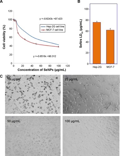
Nanostructured selenium materials have attracted substantial attention due to its excellent biological activity and low toxicity.Citation17 With promising applications in cancer nanotechnology, SNMs are being touted as the new anticancer and chemopreventive agents.Citation65 Very recently, the cytotoxic effect of the inorganic and organic Se compounds on the MCF-7 cell line was assessed.Citation1,Citation9 Various types of SNMs stabilized and surface modified with different types of biological macromolecules/functional groups are reported to possess excellent anticancer activities via the including induction of reactive oxygen species (ROS) production, cell cycle arrest, mitochondrial dysfunction, DNA fragmentation, and cell apoptosis.Citation1,Citation66 These SNMs have also been shown to augment the anticancer properties of chemotherapeutic drugs like adriamycin and doxorubicin.Citation67,Citation68 Therefore, the SeNrs were evaluated for their anticancer properties against Hep-G2 and MCF-7 cell lines. The biosynthesized SeNrs showed growth inhibition of Hep-G2 and MCF-7 cells in a dose-dependent manner (). The inhibitory effect of the SeNrs was significantly higher on the MCF-7 cells than on the Hep-G2 cells. For instance, SeNrs at 10 μg/mL, 25 μg/mL, 50 μg/mL, and 100 μg/mL reduced MCF-7 cell viability to 69.1%, 54.4%, 44.3%, and 37.5%, respectively. All of those values were significantly lower than those of the Hep-G2 cells: 86.9%, 72.5%, 56.4%, and 42.3%, respectively, at LSD (0.05) =5.7% (). The lethal dose (ID50%) of SeNrs on Hep-G2 and MCF-7 cells was obtained at 75.96 μg/mL and 61.86 μg/mL, respectively, as shown in . The data indicated that the effect of SeNrs on MCF-7 cells was significantly more than on Hep-G2 cells. Moreover, microscopic cell morphological observation after MTT staining showed that Hep-G2 and MCF-7 cells treated with SeNrs showed a dose-dependent reduction in cell numbers, loss of cell-to-cell contact, cell shrinkage, and formation of apoptotic bodies (; MCF-7 cells data not shown). These results collectively suggested that biosynthesized SeNrs have the anticancer property against cancer cells and can serve as potential anticancer agents. However, a few recent studies have reported lower toxicity and selectivity of SNMs toward normal cells.Citation1,Citation20 The mechanism involved in the selectivity of the SNMs remains unexplained. Therefore, we tried to explain the plausible mechanism of SNMs for selective killing of cancer cells.
Table 4 Analysis of variance of the effect of different concentrations of biosynthesized SeNrs on the linear growth of Hep-G2 and MCF-7 cell lines
Figure 5 Anticancer activity of biosynthesized SeNrs.
Notes: (A) Effect of SeNrs on the viability of Hep-G2 and MCF-7 human cancer cells under in vitro condition. Human cancer cells were treated with various concentrations (10 μg/mL, 25 μg/mL, 50 μg/mL, and 100 μg/mL) of SeNrs and the MTT assay revealed that the biosynthesized SeNrs induces cell death of Hep-G2 and MCF-7 human cancer cells; (B) determination of lD50% of SeNrs for the Hep-G2 and MCF-7 human cancer cells. The ID50% of SeNrs on Hep-G2 and MCF-7 cells was recorded at 75.96 μg/mL and 61.86 μg/mL, respectively. (C) Microscopic cell morphological observation of Hep-G2 cells after MTT staining showed that cells treated with SeNrs exhibited some important killing features, including reduction in cell viability, loss of cell-to-cell contact, cell shrinkage, and formation of apoptotic bodies.
Abbreviations: SeNrs, selenium nanorods; MTT, 3-(4,5-dimethylthiazol-2-yl)-2,5-diphenyltetrazolium bromide; ID50%, lethal dose.
Plausible anticancer mechanism of SeNrs
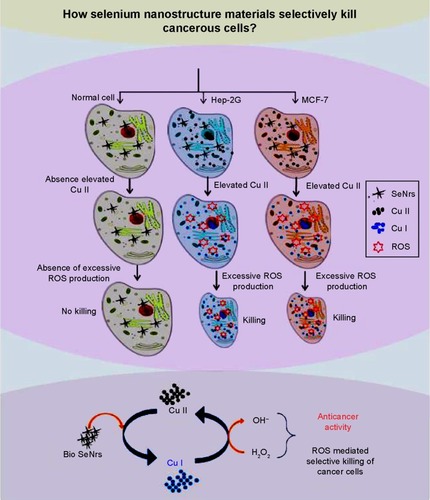
The inorganic and organic selenium compounds play an essential role in human life and a number of them are considered to possess chemopreventive and therapeutic properties against cancer.Citation66 In situ surface functionalized SNMs via the biosynthesis procedure have recently gained much attention as potential anticancer agents due to their excellent anticancer activity, biocompatibility, and low toxicity, when compared to inorganic and organic Se compounds.Citation1,Citation9,Citation69 Conjugation with functional ligands/groups, indeed, can not only prevent the aggregation of SNMs via plus-to-minus charge interactions, but also enhance the anticancer efficacy.Citation1 These SNMs are established as promising antioxidants (redox modulating) but can also act as prooxidants and thereby exhibit potential anticancer properties in the presence of transition metal ions (Cu). It is well established that tissue, cellular, and serum copper levels are considerably elevated in various malignancies.Citation70,Citation71 These SNMs are able to bind cell chromatin materials (both DNA and Cu[II]) forming a ternary complex. A redox reaction of the Se compound and Cu(II) in the ternary complex may occur, leading to the reduction of Cu(II) to Cu(I), whose reoxidation generates a variety of ROS. Therefore, cancer cells may be more subject to electron shuttle between copper ions and SeNrs to generate ROS, thereby exhibiting the killing effect on Hep-G2 and MCF-7 cells (). Thus, our hypothesis is that the anticancer mechanism of SeNrs involves the mobilization of endogenous copper, possibly chromatin-bound copper, and the consequent prooxidant action.
Figure 6 Plausible anticancer activity of biosynthesized SeNrs.
Notes: The anticancer mechanism of SeNrs involves the mobilization of endogenous copper (Cu), possibly chromatin-bound copper, and the consequent prooxidant action. Cancer cells may be more subject to electron shuttle between copper ions and SeNrs to generate ROS, thereby exert the killing effect on Hep-G2 and MCF-7 cells.
Abbreviations: SeNrs, selenium nanorods; ROS, reactive oxygen species.
Conclusion
The isolated S. bikiniensis strain Ess_amA-1 has the inherent potential to produce more stabilized, bioefficacious, and ecofriendly SeNrs than physico-chemically synthesized SNMs, and can be exploited for mass-scale production. The biosynthesized SeNrs showed anticancer activity against Hep-G2 and MCF-7 cells under in vitro conditions. SNMs are potent anticancer agents, with a modest effect on normal cells. The exact mechanism by which this anticancer activity is mediated remains unclear to the scientific community. In this paper, we suggest a hypothesis that the anticancer mechanism of SeNrs involves mobilization of elevated endogenous copper of cancer cells and consequent prooxidant action. Nevertheless, in-depth studies should be conducted to investigate the anticancer action mechanism of SNMs.
Acknowledgments
This work was supported by King Saud University, Deanship of Scientific Research, College of Science, Research Center.
Disclosure
The authors report no conflicts of interest in this work.
References
- Aboul-FadlTSelenium derivatives as cancer preventive agentsCurrent Medicinal Chemistry – Anti-Cancer Agents20055663765216305485
- RaymanMPSelenium and human healthLancet201237998221256126822381456
- ShikuoLYuhuaSAnjianXRapid, room-temperature synthesis of amorphous selenium/protein composites using Capsicum annuum L extractNanotechnology20071840405101
- RaymanMPSelenium in cancer prevention: a review of the evidence and mechanism of actionProc Nutr Soc200564452754216313696
- GaoFYuanQGaoLCytotoxicity and therapeutic effect of irinotecan combined with selenium nanoparticlesBiomaterials201435318854886625064805
- SrivastavaNMukhopadhyayMBiosynthesis and structural characterization of selenium nanoparticles mediated by Zooglea ramigeraPowder Technol20132442629
- BeheshtiNSoflaeiSShakibaieMEfficacy of biogenic selenium nanoparticles against Leishmania major: in vitro and in vivo studiesJ Trace Elem Med Biol201327320320723219368
- TorresSKCamposVLLeónCGBiosynthesis of selenium nanoparticles by Pantoea agglomerans and their antioxidant activityJ Nanopart Res201214111236
- ForootanfarHAdeli-SardouMNikkhooMAntioxidant and cytotoxic effect of biologically synthesized selenium nanoparticles in comparison to selenium dioxideJ Trace Elem Med Biol2014281757924074651
- LampisSZonaroEBertoliniCBernardiPButlerCSValliniGDelayed formation of zero-valent selenium nanoparticles by Bacillus mycoides SeITE01 as a consequence of selenite reduction under aerobic conditionsMicrob Cell Fact20141313524606965
- JohnsonJASaboungiMLThiyagarajanPCsencsitsRMeiselDSelenium nanoparticles: a small-angle neutron scattering studyJ Phys Chem B199910315963
- ZhangBYeXDaiWHouWZuoFXieYBiomolecule-assisted synthesis of single-crystalline selenium nanowires and nanoribbons via a novel flake-cracking mechanismNanotechnology2006172385390
- KumarASevonkaevIGoiaDVSynthesis of selenium particles with various morphologiesJ Colloid Interface Sci2014416011912324370410
- CaoXXieYZhangSLiFUltra-thin trigonal selenium nanoribbons developed from series-wound beadsAdv Mater2004167649653578
- YinHXuZBaoHBaiJZhengYSingle crystal trigonal selenium nanoplates converted from selenium nanoparticlesChem Lett2005341122123
- ZhangHYangDJiYMaXXuJQueDSelenium nanotubes synthesized by a novel solution phase approachJ Phys Chem B2004108411791182
- SrivastavaPBragancaJMKowshikMIn vivo synthesis of selenium nanoparticles by Halococcus salifodinae BK18 and their anti-proliferative properties against HeLa cell lineBiotechnol Prog20143061480148725219897
- SubbaiyaRSelvamMSynthesis and characterization of silver nanoparticles from Streptomyces olivaceus sp-1392 and its anticancerous activity against non-small cell lung carcinoma cell line (NCI-H460)Curr Nanosci2014102243249
- SarkarJSahaSDeyPAcharyaKProduction of selenium nanorods by phytopathogen, Alternaria alternataAdv Sci Lett2012101111114
- FengYSuJZhaoZDifferential effects of amino acid surface decoration on the anticancer efficacy of selenium nanoparticlesDalton Trans20144341854186124257441
- YangFTangQZhongXSurface decoration by Spirulina polysaccharide enhances the cellular uptake and anticancer efficacy of selenium nanoparticlesInt J Nanomedicine2012783584422359460
- GolinskaPWypijMIngleAPGuptaIDahmHRaiMBiogenic synthesis of metal nanoparticles from actinomycetes: biomedical applications and cytotoxicityAppl Microbiol Biotechnol201498198083809725158833
- ShakibaieMForootanfarHGolkariYMohammadi-KhorsandTShakibaieMRAnti-biofilm activity of biogenic selenium nanoparticles and selenium dioxide against clinical isolates of Staphylococcus aureus, Pseudomonas aeruginosa, and Proteus mirabilisJ Trace Elem Med Biol201529023524125175509
- MishraRRPrajapatiSDasJDangarTKDasNThatoiHReduction of selenite to red elemental selenium by moderately halotolerant Bacillus megaterium strains isolated from Bhitarkanika mangrove soil and characterization of reduced productChemosphere20118491231123721664643
- SrivastavaNMukhopadhyayMBiosynthesis and structural characterization of selenium nanoparticles using Gliocladium roseumJ Cluster Sci2015
- OremlandRSHerbelMJBlumJSStructural and spectral features of selenium nanospheres produced by Se-Respiring bacteriaAppl Environ Microbiol2004701526014711625
- MahmoudvandHFasihi HarandiMShakibaieMScolicidal effects of biogenic selenium nanoparticles against protoscolices of hydatid cystsInt J Surg201412539940324686032
- TiwariKGuptaRKDiversity and isolation of rare actinomycetes: an overviewCrit Rev Microbiol201339325629422901022
- Dela CruzRGaoYPenumetchaSSheplockRWengKChanderMExpression of the Streptomyces coelicolor SoxR regulon is intimately linked with Actinorhodin productionJ Bacteriol2010192246428643820952574
- HwangKSKimHUCharusantiPPalssonBTLeeSYSystems biology and biotechnology of Streptomyces species for the production of secondary metabolitesBiotechnol Adv201432225526824189093
- ZonoozNFSaloutiMBiological synthesis of gold nanoparticles by cell free supernatant of Streptomyces sp. ERI-3Asian J Chem2012241046474649
- ThenmozhiMKannabiranKKumarRGopiesh KhannaVAntifungal activity of Streptomyces sp. VITSTK7 and its synthesized Ag2O/Ag nanoparticles against medically important Aspergillus pathogensJ Mycol Med20132329710323706303
- SubashiniJGopiesh KhannaVKannabiranKAnti-ESBL activity of silver nanoparticles biosynthesized using soil Streptomyces speciesBioprocess Biosyst Eng2014376999100624122217
- SadhasivamSShanmugamPYunKBiosynthesis of silver nanoparticles by Streptomyces hygroscopicus and antimicrobial activity against medically important pathogenic microorganismsColloids Surf B Biointerfaces201081135836220705438
- SrinivasanMCLaxmanRSDeshpandeMVPhysiology and nutritional aspects of actinomycetes: an overviewWorld J Microbiol Biotechnol19917217118424424929
- CochraneVWPhysiology of actinomycetesAnnu Rev Microbiol1961151124
- PridhamTGColor and Streptomycetes: report of an international workshop on determination of color of streptomycetesAppl Microbiol1965131436114264847
- ShirlingEBGottliebDMethods for characterization of Streptomyces speciesInt J Syst Bacteriol1966163313340
- PridhamTGGottliebDThe utilization of carbon compounds by some Actinomycetales as an aid for species determinationJ Bacteriol194856110711416561537
- GorajanaAVenkatesanMVinjamuriSResistoflavine, cytotoxic compound from a marine actinomycete, Streptomyces chibaensis AUBN1/7Microbiol Res2007162432232716580188
- LuedemannGMBrodskyBCTaxonomy of gentamicin-producing micromonosporaAntimicrob Agents Chemother (Bethesda)196316111612414274877
- BibbMFreemanRHopwoodDPhysical and genetical characterisation of a second sex factor, SCP2, for Streptomyces coelicolor A3(2)Mol Gen Genet19771542155166
- AltschulSFMaddenTLSchäfferAAGapped BLAST and PSI-BLAST: a new generation of protein database search programsNucleic Acids Res19972517338934029254694
- ThompsonJDHigginsDGGibsonTJCLUSTAL W: improving the sensitivity of progressive multiple sequence alignment through sequence weighting, position-specific gap penalties and weight matrix choiceNucleic Acids Res19942222467346807984417
- KimuraMA simple method for estimating evolutionary rates of base substitutions through comparative studies of nucleotide sequencesJ Mol Evol19801621111207463489
- MosmannTRapid colorimetric assay for cellular growth and survival: application to proliferation and cytotoxicity assaysJ Immunol Methods1983651–255636606682
- OhnishiYIshikawaJHaraHGenome sequence of the streptomycin-producing microorganism Streptomyces griseus IFO 13350J Bacteriol2008190114050406018375553
- de Lima ProcópioREda SilvaIRMartinsMKde AzevedoJLde AraújoJMAntibiotics produced by StreptomycesBraz J Infect Dis201216546647122975171
- AigleBLautruSSpitellerDGenome mining of Streptomyces ambofaciensJ Ind Microbiol Biotechnol201441225126324258629
- Van WezelGPMcDowallKJThe regulation of the secondary metabolism of Streptomyces: new links and experimental advancesNat Prod Rep20112871311133321611665
- ManivasaganPVenkatesanJKangKHSivakumarKParkSJKimSKProduction of α-amylase for the biosynthesis of gold nanoparticles using Streptomyces sp. MBRC-82Int J Biol Macromol201572717825128097
- SoniNPrakashSAntimicrobial and mosquitocidal activity of microbial synthesized silver nanoparticlesParasitol Res201411431023103025544704
- ManikprabhuDLingappaKSynthesis of silver nanoparticles using the Streptomyces coelicolor klmp33 pigment: an antimicrobial agent against extended-spectrum beta-lactamase (ESBL) producing Escherichia coliMater Sci Eng C Mater Biol Appl20144543443725491848
- SanjenbamPGopalJVKannabiranKAnticandidal activity of silver nanoparticles synthesized using Streptomycessp.VITPK1J Mycol Med2014
- KarthikLKumarGKirthiAVRahumanAABhaskara RaoKVStreptomyces sp. LK3 mediated synthesis of silver nanoparticles and its biomedical applicationBioprocess Biosyst Eng201437226126723771163
- El-NaggarNEAAbdelwahedNADarweshOMFabrication of biogenic antimicrobial silver nanoparticles by Streptomyces aegyptia NEAE 102 as eco-friendly nanofactoryJ Microbiol Biotechnol201424445346424375417
- SivalingamPAntonyJJSivaDAchiramanSAnbarasuKMangrove Streptomyces sp. BDUKAS10 as nanofactory for fabrication of bactericidal silver nanoparticlesColloids Surf B Biointerfaces201298121722652354
- Mary JacobJBalakrishnanRMKumarUBBiosynthesis of lead selenide quantum rods in marine Aspergillus terreusMater Lett20141240279281
- KorbekandiHAshariZIravaniSAbbasiSOptimization of biological synthesis of silver nanoparticles using Fusarium oxysporumIran J Pharm Res201312328929824250635
- DhanjalSCameotraSSAerobic biogenesis of selenium nanospheres by Bacillus cereus isolated from coalmine soilMicrob Cell Fact201095220602763
- JeongUXiaYSynthesis and crystallization of monodisperse spherical colloids of amorphous seleniumAdv Mater Deerfield2005171102106
- BarthAInfrared spectroscopy of proteinsBiochim Biophys Acta2007176791073110117692815
- Saravana KumarPBalachandranCDuraipandiyanVRamasamyDIgnacimuthuSAl-DhabiNExtracellular biosynthesis of silver nanoparticle using Streptomyces sp. 09 PBT 005 and its antibacterial and cytotoxic propertiesAppl Nanosci201552169180
- MandalSPhadtareSSastryMInterfacing biology with nanoparticlesCurr Appl Phys200552118127
- EstevezHGarcia-LidonJCLuque-GarciaJLCamaraCEffects of chitosan-stabilized selenium nanoparticles on cell proliferation, apoptosis and cell cycle pattern in HepG2 cells: comparison with other selenospeciesColloids Surf B Biointerfaces201412218419325038448
- PiJYangFJinHSelenium nanoparticles induced membrane bio-mechanical property changes in MCF-7 cells by disturbing membrane molecules and F-actinBioorg Med Chem Lett201323236296630324140445
- TanLJiaXJiangXIn vitro study on the individual and synergistic cytotoxicity of adriamycin and selenium nanoparticles against Bel7402 cells with a quartz crystal microbalanceBiosens Bioelectron20092472268227219101136
- RamamurthyCHSampathKSArunkumarPGreen synthesis and characterization of selenium nanoparticles and its augmented cytotoxicity with doxorubicin on cancer cellsBioprocess Biosyst Eng20133681131113923446776
- PrasadKSSelvarajKBiogenic synthesis of selenium nanoparticles and their effect on As(III)-induced toxicity on human lymphocytesBiol Trace Elem Res2014157327528324469678
- HadiSMBhatSHAzmiASHanifSShamimUUllahMFOxidative breakage of cellular DNA by plant polyphenols: a putative mechanism for anticancer propertiesSemin Cancer Biol200717537037617572102
- AzmiASBhatSHHanifSHadiSMPlant polyphenols mobilize endogenous copper in human peripheral lymphocytes leading to oxidative DNA breakage: a putative mechanism for anticancer propertiesFEBS Lett2006580253353816412432