Abstract
Objective
To evaluate the cytotoxicity of iron nanoparticles on cardiac cells and to determine whether they can modulate the biological activity of 7-ketocholesterol (7KC) involved in the development of cardiovascular diseases. Nanoparticles of iron labeled with Texas Red are introduced in cultures of nonbeating mouse cardiac cells (HL1-NB) with or without 7-ketocholesterol 7KC, and their ability to induce cell death, pro-inflammatory and oxidative effects are analyzed simultaneously.
Study design
Flow cytometry (FCM), confocal laser scanning microscopy (CLSM), and subsequent factor analysis image processing (FAMIS) are used to characterize the action of iron nanoparticles and to define their cytotoxicity which is evaluated by enhanced permeability to SYTOX Green, and release of lactate deshydrogenase (LDH). Pro-inflammatory effects are estimated by ELISA in order to quantify IL-8 and MCP-1 secretions. Pro-oxidative effects are measured with hydroethydine (HE).
Results
Iron Texas Red nanoparticles accumulate at the cytoplasmic membrane level. They induce a slight LDH release, and have no inflammatory or oxidative effects. However, they enhance the cytotoxic, pro-inflammatory and oxidative effects of 7KC. The accumulation dynamics of SYTOX Green in cells is measured by CLSM to characterize the toxicity of nanoparticles. The emission spectra of SYTOX Green and nanoparticles are differentiated, and corresponding factor images specify the possible capture and cellular localization of nanoparticles in cells.
Conclusion
The designed protocol makes it possible to show how Iron Texas Red nanoparticles are captured by cardiomyocytes. Interestingly, whereas these fluorescent iron nanoparticles have no cytotoxic, pro-inflammatory or oxidative activities, they enhance the side effects of 7KC.
Introduction
Currently, nanomaterials such as nanotubes, nanowires, fullerene derivatives and quantum dots have received enormous attention to create new types of analytical tools for biotechnology and life sciences.Citation1–Citation3 Thus, nanomaterials have been used to create unique devices, possessing novel physical and chemical functional properties.Citation4 Although there are potentially numerous applications of nanomaterials in modern technologies applied to biology and medicine (imaging, delivery of contrast agents, drugs, genes/oligonucleotides and protein/peptides, allowing the monitoring of biodistribution and therapeutic activity, simultaneously),Citation5,Citation6 there is a serious lack of information concerning their effects on human health especially in patients with cardiovascular risks characterized by elevated plasmatic levels of oxysterols such as 7-ketocholesterol.Citation7 The main toxicological concern is the fact that some nanoparticles such as iron nanoparticles, are redox active,Citation4,Citation8,Citation9 and consequently can have some cytotoxic effects on various cellular models,Citation10,Citation11 and can induce important side effects on small animals.Citation12,Citation13 Moreover, as some animal models have shown that nanoparticles can access the vascular domain as well as major organs such as brain, kidneys, and liver,Citation14–Citation16 we asked whether nanoparticles can interact with cardiac cells and induce some cytotoxic effects (induction of cell death, inflammation, and oxidation) on cardiomyocytes to favor cardiac dysfunctions. We also asked whether nanoparticles increase the cytotoxic, pro-inflammatory, and pro-oxidative effects of 7-ketocholesterol (7KC) known to trigger a wide number of side effects on numerous cell types.Citation17
Indeed, oxysterols are biologically active molecules resulting from the oxidation of cholesterol. As they penetrate cell membranes, their concentrations can reach harmful levels in various cell types.Citation7,Citation17 New findings suggest that the effects of oxysterols on cardiomyocytes can lead to cell hypertrophy and death. The pathological actions of oxysterols on smooth muscle cells and cardiomyocytes were shown to depend on dysfunctional Ca2+ signalling.Citation7,Citation17,Citation18 This makes oxysterols one of the major factors precipitating morbidity in cardiac diseases and inflammation-induced heart complications.
Since little information is available on nanomaterial toxicity, simple in vitro toxicity models are of major importance especially for iron nanoparticles, which are frequently used to perform medical imaging.Citation19
So, the aim of the present in vitro study performed on non-beating murine cardiac cells (HL1-NB cells)Citation20,Citation21 which have been cultured in the absence or in the presence of fluorescent iron nanoparticles labeled with Texas Red associated or not with 7KC, was: 1) to obtain cytological and biochemical information (toxicity, cellular localization) on the effect of these nanoparticles on cardiac cells, and 2) to determine whether they can modulate the biological activity of 7KC itself which is known to contribute to the development of cardiovascular diseases. The cellular interactions able to induce cell death, and the pro-oxidative and pro-inflammatory effects of fluorescent iron nanoparticles associated or not with 7KC after different times of treatment were determined by biochemical techniques, flow cytometry (FCM), and/or confocal laser scanning microscopy (CLSM) coupled with factor analysis of medical image sequences (FAMIS). FAMIS provides factor images corresponding to each fluorescent compound.Citation22–Citation24 This method uses physical properties of fluorochromes,Citation25 and enables to isolate and visualize fluorochromes by means of their spectral pattern,Citation26 as well as their velocity.Citation27 In the present study, the toxicity was measured with SYTOX Green (0.5 μM; 5-min incubation), which stains dead cells,Citation28 and by the quantification of lactate dehydrogenase (LDH) activity in the culture medium.Citation29 Pro-inflammatory effects were evaluated by ELISA via the secretion levels of IL-8 and MCP-1 in the culture medium.Citation30 Pro-oxidative effects were quantified by flow cytometry with hydroethidine (HE).Citation31 The kinetics of capture of nanoparticles and SYTOX Green were memorized simultaneously using CLSM during a 10-min period of time. Sequences of images were processed according to a FAMIS based method providing dynamic or spectral components. Sequences of images were obtained according to a protocol requiring either the memorization of an image every 3 or 10 s inside a spectral window, or the scanning along the emission spectrum (525–715 nm). Using these image sequences, the aim of the work was to 1) characterize the incorporation and exit dynamics of nanoparticles, 2) differentiate the emission spectra of SYTOX Green and of nanoparticles. Computed dynamic and spectral curves (factors) and corresponding factor images generated by FAMIS are used to visualize the capture and final localization of nanoparticles in HL1-NB cells.
Our data support that the iron nanoparticles have very slight cytotoxic effects on HL1-NB cells (no increase of SYTOX Green associated fluorescence, slight increase of LDH release), that they are captured by cells, and that they do not stimulate IL-8 and MCP-1 secretion nor reactive oxygen species (ROS) production. However, when associated with 7KC, iron nanoparticles enhance the cytotoxicity as well as the pro-inflammatory and pro-oxidative effects of this compound. Our approach, which can provide a valuable tool to differentiate the biological activities of various nanoparticles associated or not with other compounds in living cells, underlines that iron nanoparticles can reinforce the side effects of potential cardiovascular risk factors such as 7KC.
Materials and methods
Cells, cell culture, and cell treatments
The HL-1 cell line derives from tumoral atrial cardiac myocytes from transgenic mice, and was a gift from Dr WC Claycomb (Louisiana State University Medical Center).Citation20 HL1-NB cells obtained as described beforeCitation21 were cultured in Claycomb medium (JRH Biosciences Ltd.) supplemented with 10% fetal bovine serum, 4 mM L-glutamine, 100 U/mL penicillin, 100 mg/mL streptomycin, 0.3 mM ascorbic acid and 10 mM norepinephrine, at 37 °C in a humid atmosphere of 5% CO2/95% air. F25 flasks (for cell production), 12 mm glass cover plates (in 35 mm boxes, for microscopy), and 6 wells cell culture plates (for flow cytometry) were coated with a mixture of gelatin (0.02%) and fibronectin (12.5 mg/L). The Claycomb medium was renewed daily. Cells were split at a mean density of 15,000/cm2 and were allowed to reach confluence before treatments with iron nanoparticles and/or 7-ketocholesterol (7KC).
7KC was provided by Sigma (L’Isle d’Abeau Chesnes, France). Stock solution of 7KC was prepared at 800 μg/mL: 800 μg of 7KC were dissolved in 50 μL of absolute ethanol, 950 μl of culture medium were added, and the solution was sonicated. To obtain a 40 μg/mL final 7KC concentration (100 μM), 50 μL of the initial solution was added per ml of culture medium on confluent cells. In our culture conditions, confluence was reached at 48 h of culture, and To corresponds to the introduction of 7KC associated or not with MACS iron nanoparticles (Miltenyi Biotec, Germany) in the culture medium.
Iron nanoparticles used at 10 μg/mL final concentration were prepared as follows. MACS nanoparticles (diameters: 20–50 nm), which are goat anti-rabbit IgG microbeads (Miltenyi Biotec, Ref: 130-048-602) were incubated for 30 min at 21 °C with Texas Red conjugated rabbit IgG (Rockland, PA, USA; Ref: 011-0902) in order to obtain fluorescent nanoparticles. Fluorescent nanoparticles were collected with a magnet (STEMCELL Technologies, Grenoble, France), resuspended in distilled water (concentration adjusted at 1 mg/mL), and stored at 4 °C for a period of time not exceeding 6 months. While some iron nanoparticles with a size comprised between 250–300 nm were observed by fluorescence microscopy, the presence of smaller nanoparticles (20–250 nm) is also theoretically highly probable.
Evaluation of cell death by quantification of LDH and by nuclear staining with SYTOX green
HL1-NB confluent cells were cultured with or without fluorescent iron nanoparticles associated or not with 7KC (40 μg/mL) for 30 h, and the release of LDH in the culture supernatants, which increases with cell death, was measured by using a Vitros 950 (Ortho Clinical Diagnostics, Rochester, NY, USA). Moreover, the percentages of SYTOX Green positive cells (corresponding to dead cells) were also simultaneously determined.Citation32 SYTOX Green (Molecular Probes/Invitrogen, Cergy Pontoise, France) was used at a final concentration of 0.5 μM. After 10 min of incubation at room temperature, the samples were analyzed, and the fluorescence of the dye was collected with a 520/10 nm band pass filter. Data were collected on a logarithmic scale on a GALAXY/PAS flow cytometer (Partec Gmbh, Münster, Germany), and further analyzed with FlowMax software (Partec).
Measurement of IL-8 and MCP-1 secretion by ELISA
To measure interleukin-8 (IL-8) and monocyte chemotactic protein-1 (MCP-1) secretion by ELISA, confluent HL1-NB cells were incubated for 30 h with or without fluorescent iron nanoparticles associated or not with 7KC (40 μg/mL). At the end of the incubation time, the culture medium was collected by centrifugation, and stored at −80 °C. Samples were defrosted just before ELISA was performed in accordance with the procedures of the manufacturers (IL-8: Bender MedSystemsTM; Vienna, Austria; MCP-1: PeproTech EC, London, UK).
Flow cytometric measurement of the production of reactive oxygen species
The production of reactive oxygen species (ROS) was determined on HL1-NB confluent cells cultured for 30 h with or without iron nanoparticles in the absence or presence of 7KC (40 μg/mL). At the end of the treatment, cells were detached by trypsinization and resuspended in the culture medium at 106 cells/mL. To measure the production of ROS, cells were incubated with HE, which allows the identification of superoxide anions. Indeed, HE is a nonfluorescent compound which can diffuse through cell membranes, and which is rapidly oxidized in ethidium under the action of superoxide anions.Citation33,Citation34 HE (Molecular Probes/Invitrogen, Cergy Pontoise, Paris) was initially prepared at a concentration of 10 mM in DMSO, and was used at a 4 μM final concentration on cell samples of 106 cells per ml of culture medium. After 15 min of incubation at 37 °C, 10,000 cells were analyzed by flow cytometry with a GALAXY/PAS flow cytometer (Partec) at excitation and emission wavelengths of 488 nm and 590/10 nm, respectively. Data were collected on a logarithmic scale, and further analyzed with FlowMax software (Partec).
Observation by conventional fluorescence microscopy and fluorescence analysis by confocal laser scanning microscopy
The emission of untreated or of 7KC-treated-HL1-NB cell cultures incubated or injected with MACS Texas Red and stained with SYTOX Green was analyzed by means of confocal laser scanning microscopy (CLSM) in three-dimensional (3D) sequences of images obtained by spectral analysis. Sequences were then analyzed by Factor Analysis of Medical Image Sequences (FAMIS) algorithm available via Pixies.Citation22,Citation23 Therefore, emissions of MACS Texas Red and SYTOX Green in untreated HL1-NB cells screened by CLSM and available excitation sources can be differentiated. As a consequence, image analysis performed on spectral sequences of images leads to specific co-localized images of MACS Texas Red and SYTOX Green. Indeed, sequences of images can be investigated by FAMIS and provide factor images corresponding to each fluorescent compound. This method uses physical properties of fluorochromes, and permits the isolation of fluorochromes by means of their spectral pattern, as well as their velocity.
Image analysis
Following general factor analysis techniques, FAMIS was developed to process biomedical image sequences.Citation22,Citation23 The FAMIS process has to cope with mixtures of components characterized by their physical behavior as well as linearity and component positivity basic hypothesis.
Sequences of images were further processed by FAMIS, available at Apteryx under the name of Pixies (www.apteryx.fr). FAMIS decomposes image sequences into a smaller number of images called factor images, and curves, called factor curves.Citation35,Citation36 Factor curves estimate individual spectral or temporal behavior in the sequence of images. Factor images correspond to spatial distribution components. The basic idea of FAMIS is to process the curves that represent the evolution of fluorescence intensity of each pixel in the image spectral or temporal sequence (). FAMIS assumes that each pixel is a mixture of different patterns and aims to unmix them. Factors are estimated in a two-step procedure from the image sequence.
Figure 1 Basic presentation of FAMIS. Factors are estimated in a two-step procedure from the image sequence: 1) correspondence analysis and 2) oblique analysis are performed to obtain positive factor curves and images. Factor images are recomputed back to the original sampling by oblique projection on the factor curves.
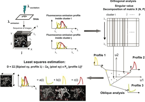
Pixels of the images are combined into 4 × 4 clusters. Correspondence analysisCitation37,Citation38 is first performed on the intensity evolution of each cluster. Then oblique analysis is performed on the results of correspondence analysis,Citation22 requiring positive factor curves and images. Factor images are recomputed in the original sampling by oblique projection, on the factor curves and the estimation is performed using the least-squares method. Here, the factor curves correspond to the emission spectra and the capture or staining kinetics of the fluorochromes. Factor images provide images of stained fluorescent structures. Superimposition in true color of these factor images provides a supplementary tool for interpretation.
Statistical analysis
Statistical analyses were performed on at least three independent experiments with SigmaStat 2.03 software (Systat Software Inc) with the Student t test. Data were considered statistically different at a P value of 0.05 or less.
Results
Confocal laser scanning microscopic analysis of iron nanoparticles in untreated or 7KC-treated HL1-NB cells counterstained with SYTOX Green
Action of MACS Texas Red iron nanoparticles on cells
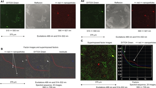
In the case of spectral observations of MACS Texas Red treated cells (1: 6–10 h; 2: 20–24 h) in which emissions are collected through band-pass filters then processed before interpretation, the excitation at 488 nm and 514–532 nm was performed and emission was collected in the regular mode (515–550 nm, 590–621 nm) (; ), and the spectral mode in 10 nm filters from blue to red (555 ≥ 705 nm). The resulting spectral sequences were investigated by means of FAMIS. A green emission corresponding to SYTOX Green is visualized in the first factor image and a red emission (610 nm) is visualized in the second factor image (). Superimposition in true color of these factor images () provides a supplementary tool for interpretation.
Figure 2 Case of spectral observations of iron nanoparticles conjugated with Texas Red incubated in untreated murine cardiac HL1-NB cells and counterstained with SYTOX Green (1: 6–10 h, 2: 20–24 h) in which emissions are collected through band-pass filters. A1–A2) Regular mode through band-pass filters. B) Spectral mode through 10 nm band-pass filters to obtain sequences of images and process by means of FAMIS. A green emission (535 nm) corresponding to SYTOX Green is visualized in the first factor image and a red emission (610 nm) is visualized in the second factor image. C) Superimposition in true color of these factor images.
Injection of MACS Texas Red iron nanoparticles on cells
In the case of temporal and spectral observations of MACS Texas Red nanoparticles injected in a culture of untreated cells in which emissions are collected through band-pass filters then processed before interpretation, the excitation at 488 nm and 514–532 nm was performed and emission was collected in the regular mode (515–550 nm, 590–621 nm) after injection (). The emission was then collected in the spectral mode in 10 nm filters from blue to red (525 ≥ 715 nm) and subsequently in the temporal mode in a long pass filter (>525 nm). The resulting spectral and temporal sequences were investigated by means of FAMIS.
Figure 3 Case of temporal and spectral observations of iron nanoparticles conjugated with Texas Red injected in a culture of untreated murine cardiac HL1-NB cells counterstained with SYTOX Green in which emissions are collected through band-pass filters. A) Regular mode through band-pass filters after injection. B) The emission is then collected in the temporal mode in a long-pass-filter and processed by means of FAMIS. A stable emission corresponding to SYTOX Green and an emission uptake corresponding to red nanoparticles are visualized to localize nanoparticles in cell compartments. C) Superimposition in true color of the factor image. In some cells, the presence of a high signal emphasizes the fact that nanoparticles accumulate inside cytoplasm. D) Spectral mode through 10 nm band-pass filters. Investigation by means of FAMIS. A green emission (535 nm) corresponding to SYTOX Green in the first factor image and a red emission (610 nm) corresponding to red nanoparticles are visualized in the second factor image. Nanoparticles are either captured or not by the cells. E) Superimposition in true color of these factor images is performed to localize nanoparticles in cell compartments.
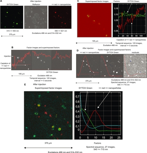
Temporal sequence: a stable emission corresponding to SYTOX Green is visualized in the second and third factor images, and an emission uptake corresponding to Texas Red is visualized in the first factor image (). Superimposition in true color of these factor images () provides a supplementary tool for interpretation.
Spectral sequence: a green emission (535 nm) corresponding to SYTOX Green is visualized in the first factor image and red emission (610 nm) is visualized in the second factor image (). Superimposition in true color of these factor images () provides a supplementary tool for interpretation.
Action of 7KC with MACS Texas Red iron nanoparticles on cells
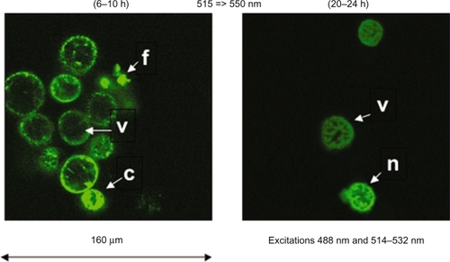
Spectral observations of 7KC treated cells (6–10 h; 20–24 h) were performed in which emissions were collected through band-pass filters and processed before interpretation, to visualize the possible toxicity of products by means of SYTOX Green. The excitation at 488 nm and 514–532 nm was performed and emission was collected in the regular mode (515–550 nm, 590–621 nm). Evidence of toxicity of 7KC at 20–24 h is thus obtained together with the nontoxicity of 7KC at 6–10 h (). When 7KC is combined with MACS Texas Red iron nanoparticles to process cells, no higher toxicity could be visualized at 6–10 h. As for a comparison, evidence of higher toxicity on cells of decane (6 h) and europium chloride (1 h) at similar concentrations was demonstrated (data not shown), by using the same protocol. In these cases most of the cells did accumulate SYTOX Green.
Figure 4 Case of spectral observation of the action of 7-ketocholesterol on murine cardiomyocytes HL1-NB cells counterstained with SYTOX Green. Spectral observations of 7-ketocholesterol-treated cells (6–10 h; 20–24 h) show the possible toxicity of products by means of SYTOX Green. At 6–10 h, most visible cells are viable (v), but typical figures of apoptosis are also observed (cells with condensed (c) or fragmented (f) nuclei), probably corresponding to spontaneous apoptosis. At 20–24 h, the structure of the nuclei suggests necrosis (n), which can either correspond to primary or secondary necrosis.Citation7,Citation17
Quantification of the cytotoxic, pro-inflammatory and pro-oxidative effects of iron nanoparticles associated or not with 7-ketocholesterol on HL1-NB cells
The ability of iron nanoparticles to induce cell death, and to trigger pro-inflammatory, and pro-oxidative effects on HL1-NB cells was evaluated comparatively to untreated cells (control), 7KC-, and (7KC + iron nanoparticles)-treated cells by using flow cytometric, biochemical and microscopic methods at 30 h of culture (). Compared to untreated cells, slight cytotoxic effects of iron nanoparticles were found in the presence of iron nanoparticles. These cytotoxic effects revealed by a significant increase of LDH release in the culture medium were not associated with an enhanced percentage of SYTOX Green positive cells (corresponding to dead cells). Moreover, no effects of iron nanoparticles were found on the levels of IL-8 and MCP-1. Indeed, similar levels of IL-8 and MCP-1 were found () in the culture medium of untreated cells and of cells cultured in the presence of iron nanoparticles. It should be noted that in agreement with data obtained on promonocytic U937 cells,Citation31 significant cytotoxic and pro-oxidative effects were found on 7KC-treated HL1-NB cells whereas no pro-inflammatory activities were observed (). Interestingly, the ability of 7KC to induce cell death, overproduction of ROS, and IL-8 secretion was enhanced in the presence of iron nanoparticles ().
Table 1 Evaluation of the cytotoxic, pro-inflammatory, and pro-oxidative effects of iron nanobeads associated or not with 7-ketocholesterol on murine cardiomyocytes HL1-NB
Discussion
The present study performed on living murine cardiac HL1-NB cells was carried out to identify the biological activities of fluorescent iron nanoparticles, and to evaluate whether they are able to induce cell death and stimulate the production of ROS and/or the secretion of cytokines (IL-8, MCP-1) capable of favoring the recruitment of monocytes, granulocytes, and T-cells, and therefore to promote inflammatory processes.Citation31,Citation39 As it has been reported that exposure to ambient air nanoparticles can increase peripheral thrombosis, and atherosclerotic lesion formation,Citation40 additional investigations are important to evaluate the contribution of nanoparticles to specific aspects of cardiovascular diseases, and to determine whether nanoparticles can influence major cardiovascular risk factors, such as diabetes, hypertension, cholesterol as well as some of its main oxidized derivatives such as 7KC which plays a major part in cardiovascular diseases.Citation41,Citation42 Therefore, the present study was undertaken on living cardiomyocytes (HL1-NB cells) to better understand the response of cardiac cells when they are exposed to iron nanoparticles in the absence or in the presence of 7KC.
To this end, flow cytometric and confocal microscopic methods of investigation were used. Cell viability, analyzed by means of staining with SYTOX Green, was also evaluated by measuring the release of lactate deshydrogenase (LDH) in the culture medium.Citation30,Citation43 Pro-inflammatory effects were evaluated by measuring IL-8 and MCP-1 secretion in the culture medium,Citation43 and overproduction of ROS was quantified by flow cytometry after staining with HE.Citation33,Citation34 Confocal observations were made more effective and specific by using the sequences of images that can be obtained by spectral or temporal modes, and were then processed by image analysis. Indeed, factor analysis (FAMIS) makes it possible to differentiate spectral or temporal emissions of fluorochromes in a multistaining system and to provide the images corresponding to each fluorochrome.Citation44 The use of the FAMIS image processing technique also allows the differentiation of mixed fluorescent emissions from dyes and nanoparticles,Citation45 and factor images make it possible to obtain an overview of the uptake and effect of nanoparticles at the single cell level.Citation46
Since nanoparticles can interact with various molecules, receptors, and/or macromolecular complexes, iron nanoparticles can contribute to activate, repress, or modulate some metabolic pathways. As the early biological activities of 7KC on the cytoplasmic membrane involves the PDK1/Akt signalling pathway,Citation47 which subsequently activates various signalling pathways (especially those triggering cell death),Citation17 it is important to determine whether iron nanoparticles interacting with the cytoplasmic membrane are able to modulate some biological activities of 7KC strongly suspected to play important roles in cardiovascular diseases. Noteworthy, our data show that these iron nanoparticles have slight cytotoxic activities, and that they do not trigger the release of major chemokines, such as IL-8 and MC-P1, known to favor the recruitment of inflammatory cells into the vascular wall contributing therefore to the development of vascular lesions.Citation43,Citation30 Moreover, iron nanoparticles do not stimulate the production of ROS. However, interaction of iron nanoparticles with murine cardiac HL1-NB cells increases 7KC induced cell death, inflammation, and oxidation. Indeed, an increased proportion of SYTOX Green positive cells and higher levels of LDH release were observed, and IL-8 secretion, and ROS production were significantly enhanced. Thus, whereas additional in vivo experiments will be required to establish that lipid disorders can influence the biological activities of nanoparticles, our data highlight that iron nanoparticles are able to increase the side effects of a major cholesterol oxide derivative: 7KC. As iron nanoparticles are widely used for in vivo imaging,Citation48,Citation49 our data suggest that they could be used with caution in atherosclerotic patients, who often have high risk of cardiovascular diseases.
Considering the results obtained from the evaluation of the ability of nanoparticles to promote cell death, pro-inflammatory and/or pro-oxidative processes, cellular deposits were used to perform CLSM analysis in order to link cell death (evaluated by means of SYTOX Green staining) with the accumulation of iron nanoparticles of untreated HL1-NB cells. In our conditions, we specifically screened the injection of iron nanoparticles in the culture medium of untreated and 7KC-treated cells. It resulted in temporal sequences of images, which were investigated by means of FAMIS. A green emission (535 nm) corresponding to SYTOX Green was observed inside cells together with the red emission (610 nm) of nanoparticles. This analysis provides information on the localization of the nanoparticles, and shows that they accumulate in cells. This combined spectral and temporal analysis of SYTOX Green and fluorescent nanoparticles makes the characterization of the interaction of nanoparticles on cells more specific. In processed confocal sequences of images, the repartition dynamics of nanoparticles is also determined, and the emission spectra of SYTOX Green and nanoparticles are differentiated. Computed dynamic and emission curves (factors) and corresponding factor images also show that the nanoparticles are localized at the periphery and inside the cardiac HL1-NB cells. As the biological activity of 7KC is associated with its accumulation in the cytoplasmic membrane, especially in lipid rafts,Citation50 the interaction of iron nanoparticles with the cytoplasmic membrane can explain their ability to modulate the effects of 7KC. The results obtained in the present investigation also indicate that the different methods tested in this study can be applied to characterize other iron nanoparticles such as ultra small iron oxide particles (USPIOs) frequently used to perform magnetic resonance imaging (MRI) of humans and animals.Citation51 Though we can not exclude that the labeling procedure can influence the cellular interactions and the biological activities of the iron nanoparticles that we tested, our approach can constitute a useful tool to early evaluate the possible side effects carried by nanoparticles such as USPIOs (Endorem, Sinerem [Guerbet, France]), which are used to improve MRI clinical investigations.
In conclusion, the combined use of FCM associated with appropriate biochemical and immunological methods, together with CLSM and subsequent spectral methods of analysis such as FAMIS, provide an efficient tool to analyze the interaction and the capture of nanoparticles by living cells, and to determine their abilities to promote cell death, and pro-inflammatory activities. This approach can be generalized and be used as an in vitro model to characterize side effects of various types of nanoparticles,Citation52 and has some applications in human toxicology. In addition, our data underline that the simultaneous use of CLSM with FAMIS is an efficient tool to differentiate colors in a designed multistaining system involving SYTOX Green and nanoparticles. Therefore, the proposed approach opens the way to numerous basic investigations with nanoparticles, allows the visualization of the cellular and the tissue distribution of various molecules associated with nanoparticles, and provides a valuable approach to evaluate the cytotoxic activities of various fluorescent nanoparticles on living cells and on small animal models.
Acknowledgements
This work was supported by the Institut National de la Santé et de la Recherche Médicale (INSERM), the Fondation de France, the University Hospital (CHU Dijon, France), and the Université de Bourgogne (Dijon, France).
Disclosure
The authors declare no conflicts of interest.
References
- BruchezMMoronneMGinPWeissSAlivisatosAPSemiconductor nanocrystals as fluorescent biological labelsScience1998281201320169748157
- TatonTMirkinCLetsingerRScanometric DNA array detection with nanoparticle probesScience20002891757176010976070
- CuiYWeiQParkHLieberCNanowire nanosensors for highly sensitive and selective detection of biological and chemical speciesScience20012931289129211509722
- ColvinVThe potential environmental impacts of engineered nanomaterialsNature Biotechnology20032111661170
- MurthySKNanoparticles in modern medicine: state of the art and future challengesInt J Nanomedicine2007212914117722542
- WeberWACzerninJPhelpsMEHerschmanHRTechnology Insight: novel imaging of molecular targets is an emerging area crucial to the development of targeted drugsNat Clin Pract Oncol20085445418097456
- VejuxALizardGCytotoxic effects of oxysterols associated with human diseases: Induction of cell death (apoptosis and/or oncosis), oxidative and inflammatory activities, and phospholipidosisMol Aspects Med20093015317019248805
- StrohAZimmerCGutzeitCIron oxide particles for molecular magnetic resonance imaging cause transient oxidative stress in rat macrophagesFree Radic Biol Med20043697698415059638
- PetersKUngerREGattiAMSabbioniETsarykRKirkpatrickCJMetallic nanoparticles exhibit paradoxical effects on oxidative stress and pro-inflammatory response in endothelial cells in vitroInt J Immunopathol Pharmacol20072068569518179741
- HussainSMHessKLGearhartJMGeissKTSchlagerJJIn vitro toxicity of nanoparticles in BRL 3A rat liver cellsToxicol In Vitro20051997598316125895
- TsujiJSMaynardADHowardPCResearch strategies for safety evaluation of nanomaterials, part IV: risk assessment of nanoparticlesToxicol Sci200689425016177233
- OberdörsterEManufactured nanomaterials (Fullerenes, C60) induce oxidative stress in the brain of juvenile largemouth bassEnvironmental Health Perspectives20041121058106215238277
- KolosnjajJSzwarcHMoussaFToxicity studies of fullerenes and derivativesAdv Exp Med Biol200762016818018217343
- OberdörsterGOberdörsterEOberdörsterJNanotoxicology: an emerging discipline evolving from studies of ultrafine particlesEnviron Health Perspect200511382383916002369
- KumarVFarellGYuSCell biology of pathologic renal calcification: contribution of crystal transcytosis, cell mediated calcification, and nanoparticlesJ Investig Med200654412424
- TetleyTDHealth effects of nanomaterialsBiochem Soc Trans200735Pt 352753117511644
- VejuxAMalvitteLLizardGSide effects of oxysterols: cytotoxicity, oxidation, inflammation, and phospholipidosisBraz J Med Biol Res20084154555618719735
- LukyanenkoVLukyanenkoYOxysterols in heart failureFuture Cardiol2009534335419656059
- CorotCRobertPIdéeJMPortMRecent advances in iron oxide nanocrystal technology for medical imagingAdv Drug Deliv Rev2006581471150417116343
- ClaycombWCLansonNAStallworthBSHL-1 cells: a cardiac muscle cell line that contracts and retains phenotypic characteristics of the adult cardiomyocytesProc Natl Acad Sci U S A199895297929849501201
- PellouxSRobillardJFerreraRNon-beating HL-1 cells for confocal microscopy: application to mitochondrial functions during cardiac preconditioningProg Biophys Mol Biol20069027029816140363
- Di PaolaRBazinJPAubryFHandling of dynamic sequences in nuclear medecineIEEE Trans Nucl Sci19822913101321
- FrouinFCinottiLBenaliHExtraction of functional volumes from medical dynamic volumetric datasetsComp Med Imaging Graph199317397404
- VejuxALizardGTourneurYRiedingerJMFrouinFKahnEEffects of caspase inhibitors (z-VAD-fmk, z-VDVAD-fmk) on Nile Red fluorescence pattern in 7-ketocholesterol-treated cells: investigation by flow cytometry and spectral imaging microscopyCytometry A20077155056217458884
- KahnEConfocal microscopy: characterization of fluorescent tracers by image processing of optical sectionsPathol Biol20014919419811367552
- KawataSSasakiKMinamiSComponent analysis of spatial and spectral patterns in multispectral images I BasisJOSA A1987421012106
- Lansing TaylorDWaggonerASMurphyRFLanniFBirgeRRApplications of Fluorescence in the Biomedical SciencesNew YorkAlan R Liss Inc1986129140
- de la MonteSMNeelyTRCannonJWandsJREthanol impairs insulin-stimulated mitochondrial function in cerebellar granule neuronsCell Mol Life Sci2001581950196011766890
- CirelliNLebrunPGueuningCSecretory characteristics and viability of human term placental tissue after overnight cold preservationHum Reprod20001575676110739815
- PrunetCMontangeTVéjuxAMultiplexed flow cytometric analyses of pro- and anti-inflammatory cytokines in the culture media of oxysterol-treated human monocytic cells and in the sera of atherosclerotic patientsCytometry A20066935937316604541
- Lemaire-EwingSPrunetCMontangeTComparison of the cytotoxic, pro-oxidant and pro-inflammatory characteristics of different oxysterolsCell Biol Toxicol2005919711416142584
- MoldrichRXBeartPMPascoeCJCheungNSLow-affinity kainate receptor agonists induce insult-dependent apoptosis and necrosis in cultured murine cortical neuronsJ Neurosci Res20005978879610700016
- RotheGValetGFlow cytometric analysis of respiratory burst activity in phagocytes with hydroethidine and 2’,7’-dichlorofluorescinJ Leukoc Biol1990474404482159514
- RotheGValetGFlow cytometric assays of oxidative burst activity in phagocytesMethods Enzymol19942335395488015489
- KahnEFrouinFSouchierCConfocal multilaser focusing and single-laser characterization of UV excitable stains of cellular preparationsCytometry200040424910754516
- KahnELizardGPélégriniMFour-dimensional factor analysis of confocal images sequences (4D-FAMIS) to detect and characterize low numbers of human papillomavirus DNA by FISH in HeLa and SiHa cellsJ Microscopy1999193227243
- HarmanHHModern Factor AnalysisChicagoUniversity of Chicago Press1960
- BenzecriJPL’analyse des Données, Tome 2: L’analyse des CorrespondancesParisDunod1973
- LizardGGueldrySSordetOGluthatione is implied in the control of 7-ketocholesterol-induced apoptosis, which is associated with radical oxygen species productionFASEB J199812165116639837855
- BhatnagarAEnvironmental cardiology. Studying mechanistic links between pollution and heart diseaseCirc Res20069969270517008598
- BrownAJJessupWOxysterols and atherosclerosisAtherosclerosis19991421289920502
- Van ReykDMBrownAJHult’enLMDeanRTJessupWOxysterols in biological systems: sources, metabolism and pathophysiological relevanceRedox Rep20061125526217207307
- LemaireSLizardGMonierSDifferent patterns of IL-1beta secretion, adhesion molecule expression and apoptosis induction in human endothelial cells treated with 7alpha-, 7beta-hydroxycholesterol, or 7-ketocholesterolFEBS Lett19984404344399872417
- KahnEHotmarJFrouinFSpectral and dynamic confocal fluorescence characterization of cytogenetic preparations by factor analysisAnal Cell Pathol19961245568933908
- KahnEReceveurACoullinPMultiple excitation confocal analysis of targets in nuclei of cytogenetic preparationsAnal Quant Cytol Histol2004261715032074
- KahnEVejuxAMenetrierFAnalysis of CD36 expression on human monocytic cells and atherosclerotic tissue sections with quantum dots. Investigation by flow cytometry and spectral imaging microscopyAnal Quant Cytol Histol200628142616566276
- VejuxAGuyotSMontangeTRiedingerJMKahnELizardGPhospholipidosis and down-regulation of the PI3-K/PDK-1/Akt signalling pathway are vitamin E inhibitable events associated with 7-ketocholesterol-induced apoptosisJ Nutr Biochem200920456118495460
- MichaletXPinaudFFBentolilaLAQuantum dots for live cells, in vivo imaging, and diagnosticsScience200530753854415681376
- GaoXDaveSRQuantum dots for cancer molecular imagingAdv Exp Med Biol2007620577318217335
- RoyerMCLemaire-EwingSDesrumauxC7-ketocholesterol incorporation into sphingolipid/cholesterol-enriched (lipid raft) domains is impaired by vitamin E: a specific role for alpha-tocopherol with consequences on cell deathJ Biol Chem2009284158261583419351882
- Di MarcoMSadunCPortMGuilbertICouvreurPDubernetCPhysicochemical characterization of ultrasmall superparamagnetic iron oxide particles (USPIO) for biomedical application as MRI contrast agentsInt J Nanomedicine2007260962218203428
- AuffanMRoseJBotteroJYLowryGVJolivetJPWiesnerMRTowards a definition of inorganic nanoparticles from an environmental, health and safety perspectiveNat Nanotechnol2009463464119809453