
Abstract
In this study, fluorescent dye-conjugated magnetic resonance (MR) imaging agents were investigated in T mode. Gadolinium-conjugated silica nanoparticles were successfully synthesized for both MR imaging and fluorescence diagnostics. Polyamine and polycarboxyl functional groups were modified chemically on the surface of the silica nanoparticles for efficient conjugation of gadolinium ions. The derived gadolinium-conjugated silica nanoparticles were investigated by zeta potential analysis, transmission electron microscopy, inductively coupled plasma mass spectrometry, and energy dispersive x-ray spectroscopy. MR equipment was used to investigate their use as contrast-enhancing agents in T1 mode under a 9.4 T magnetic field. In addition, we tracked the distribution of the gadolinium-conjugated nanoparticles in both lung cancer cells and organs in mice.
Introduction
Generated molecular images can reflect the molecular and metabolic pathways within cells. In particular, tracking biological progress in a physiological environment and detecting possible malfunctions has been advocated as a means of diagnosing disease in its early stages. One commercially important magnetic resonance (MR) enhancing material is tetra-azacyclododecane-tetraacetic acid (TTA) containing gadolinium (Gd).Citation1 Moieties of TTA become paramagnetic by the interaction between the d-orbital of Gd and lone pairs of electrons in nitrogen. The carboxylic ions attached to a macrocyclic ligand can also interact with the Gd ions. It is important that these molecular imaging probes can selectively target specific cells or organs for accurate diagnosis and treatment. Surface-modified silica nanoparticles afford the feasibility of combining various functionalities onto the silica surface.Citation2–Citation21 Moreover, they have good water wettability and low cytotoxicity for in vivo application.Citation22 Cellular imaging agents must produce adequate real-time diagnostic images.Citation23 Real-time dual mode analysis and cell tracking protocols have been studied.Citation24–Citation30 Ligand-decorated silica particles have been widely used as analytical and biosensing tools.Citation31–Citation40 Timely multi-functional MR imaging contrast-enhancing nanoparticles have been studied in biomedical applications.Citation41–Citation52
With the above in mind, we designed silica nanoparticles to have a dual imaging mode, ie, (MR) imaging and fluorescent optical imaging. Poly(ethylene glycol) (PEG) and PEG-containing block copolymers, with very flexible and hydrophilic properties, were conjugated onto the silica nanoparticles to enable them to escape uptake by the mononuclear phagocyte system. Qianjun et al reported that PEGylated nanoparticles reduce nonspecific binding of serum proteins and cellular responses.Citation53 The silica nanoparticles were decorated further with Gd ions and fluorescent dye. The resulting functionalized silica (hereafter referred to as dual imaging silica) nanoparticles were characterized by transmission electron microscopy (TEM), energy-dispersive spectroscopy, dynamic light scattering (DLS), and optical imaging analysis. The particles were further investigated as potential MR imaging agents when studying T1 relaxivity and image-enhancing efficiency. Transfection of lung cells was implemented using lung cancer cells and the corresponding organ cell has been imaged under the multi-modal imaging such as fluorescence and magnetic resonance imaging. The distribution of these dual imaging silica nanoparticles to the organs after intraperitoneal injection was studied in a mouse model by elemental analysis using inductively coupled plasma mass spectrometry (ICPMS).
Materials and methods
Materials
Most of the materials used in this study were obtained from commercial sources. Trimethoxy(3-[oxiran-2-ylmethoxy] propyl)silane (TPS), branched polyethylenimine (PEI, molecular weight 25,000), 4′,6-diamidino-2-phenylindole (DAPI), NaHCO3, silica (LUDOX AS-40) nanoparticles, dichloromethane (DCM), GdCl3·6H2O, dimethyl sulfoxide, sodium sulfate (Na2SO4), sulfonic acid sodium salt 97%, ethyl acetate, rhodamine isothiocyanate (RITC), and hexane were obtained from Sigma-Aldrich. Mono-N-hydroxysuccinate (NHS)-PEG (molecular weight 5,000) was purchased from Sunbio Chem (Korea).
Fabrication of dual imaging silica nanoparticles
Branching of mono-NHS-PEG and PEI
A solution of mono-NHS-PEG (50 mg, 10 mmol) in anhydrous DCM (80 mL) was added to a solution of PEI (250 mg, 10 mmol) in anhydrous DCM (80 mL) with cooling in an ice bath. The mixture was stirred at room temperature and the temperature was allowed to increase naturally from 0°C for 12 hours. Thin layer chromatography with ethyl acetate and hexane at a ratio of 1:1 (v/v) was used to check the progress of the reaction. The solvent was evaporated by speed-vacuum and freeze-drying and the crude compound was kept in a freezer at −20°C. The crude solid material obtained after evaporation of the solvent was then purified by gel permeation chromatography (Sephadex LH20, CH2Cl2).
Branching of PEG-PEI and TPS
A solution of PEG-PEI (100 mg, 3.3 mmol) in DCM (80 mL) was added to a solution of TPS (0.80 mL, 4.6 mmol) in the same solvent (20 mL) with cooling in an ice bath. The mixture was stirred at room temperature; thereafter, the temperature was allowed to increase naturally from 0°C for 12 hours.
Synthesis of PEI-PEG silica nanoparticles
PEI-PEG (53 mg, 0.13 mmol) and TPS were placed in a round bottomed flask and dissolved with 1.25 mL of ethanol, 1.25 mL of water, and 1.25 mL of acetic acid. An aqueous suspension (125 µL) of silica (LUDOX AS-40) nanoparticles (Sigma-Aldrich) was added. The reaction mixture was warmed to 80°C under stirring for 24 hours. The ethanol was evaporated under reduced pressure, and solid NaHCO3 was added to the suspension to reach a pH 7–8. The precipitate was filtered and washed with a borate buffer solution (pH 9.5, 5×2 mL) and with water (5×2 mL). The solid was dried under vacuum and then redissolved in 100 mL of DCM. The organic solution was washed with water (100 mL) and dried over Na2SO4.
Synthesis of RITC-labeled PEI-PEG silica nanoparticles
A colloidal dispersion of PEI-PEG silica nanoparticles (100 µg/mL) was placed in a scintillation tube, and 5 mmol RITC in dimethyl sulfoxide was added (pH 7.7±0.3). The mixture was agitated for 12 hours at room temperature, and then cleaned in DCM. Finally, dialysis was carried out using a dialysis tube for 48 hours (molecular weight cut-off, 50 kDa).
Synthesis of Gd (III)-PEI-PEG-RITC silica nanoparticles
RITC-labeled PEI-PEG silica nanoparticles (5 mg) were placed in a round bottomed flask and deionized water was added to create a neutral pH condition. Additionally, a GdCl3 (10 mM) solution was provided and vortexed for 12 hours at 90°C. Arsenazo (III) solution was used with a colorimetric method to investigate binding between the Gd (III) ions and the PEI-PEG-RITC silica nanoparticles. The color of Arsenazo (III) solution changes to green when free Gd (III) ion are present in solution; further, the bright pink color of Arsenazo III solution changes to dark purple when Gd (III) ion become conjugated with certain ligands.
Quantitative analysis of Gd (III) ions by ICPMS
To verify the concentration of Gd in the purified complexes, ICPMS was performed using a computer-controlled Agilent 7500 device. Samples were prepared by digestion in nitric acid (ratio of nitric acid to sample, 9:1) in a 70°C water bath. The samples were diluted in 15 mL conical vials with a final concentration of 3% (v/v) nitric acid. Gd (III) standards were purchased from AccuStandard (New Haven, CT, USA) and diluted to 0.1, 1, 5, 10, and 50 ng/mL of Gd (III). The samples were treated with 60% HNO3 (1 mL) and 35% HCl (3 mL), and then pre-processed and analyzed with ICPMS (300 W power, 130 psi pressure, and 70°C temperature).
Dynamic light scattering
The complexes were investigated by DLS for particle size and zeta potential using a Nano ZS instrument at 25°C. An He-Ne laser producing vertically polarized light (λ0 approximately 632.8 nm) was used as the light source. Each sample was filtered using a 0.2 µm cellulose acetate membrane.
Cell culture and transfection of PEI-PEG silica nanoparticles and RNA
A549 (human alveolar basal epithelial) cells were sourced from the Korea Cell Line Bank (Seoul, Republic of Korea) and cultured in RPMI 1640 medium supplemented with 10% heat-inactivated fetal bovine serum and 1% antibiotics (Invitrogen, Grand Island, NY, USA) at 37°C in a humidified atmosphere of 5% CO2. The cells were seeded in a 12-well culture plate at a density of 2×10⁵ cells per well and allowed to attach overnight. The incubation medium was then replaced by fresh medium. Dual imaging silica nanoparticles of different concentrations (0.1–1 mg/mL) were transferred into the wells by dropwise. After incubation for 4 hours, the old medium was replaced with fresh medium, and the nanoparticle-treated cells were incubated for 24 hours. The nucleus was stained with DAPI (blue) and the complex was represented with red for successful transfection of Gd(III)-PEI-PEG-RITC-silica nanoparticles. Since A549 cell, human derived neuroblastoma cell, was commercially available, ethics was not sought.
Animal experiments and animal care
Normal SD mouse (20 g weight) was used and animal number was two (Central Lab Animal Inc, Seoul, Korea). First, 100 µL of the dual imaging silica nanoparticle solution was injected slowly using a 1 mL syringe into the tail vein. Concentration of the dual imaging silica nanoparticle colloidal solution 100 µL (2 mg/mL) was injected into the mice. The dual imaging silica nanoparticles were administered by intraperitoneal injection. The mice were euthanized using CO2 gas in the euthanasia chamber supplied by compressed gas cylinders. One hour later, the internal organs, including kidney, spleen, pancreas, heart, lungs, and liver, were removed from each animal. The organs were then fixed in 10% formalin solution and investigated for distribution of Gd by ICPMS.
All animals were housed and maintained at 20°C–24°C on a 50:50 light-dark cycle. They received humane care in compliance with the Guide for the Care and Use of Laboratory Animals prepared by the Institute of Laboratory Animal Resources and published by the US National Institutes of Health and in accordance with the animal experiment guidelines of Samsung Biomedical Research Institute (SBRI). The study was reviewed and approved by the Institutional Animal Care and Use Committee of the SBRI. SBRI is a facility accredited by the Association for Assessment and Accreditation of Laboratory Animal Care International and abides by the Institute of Laboratory Animal Resources guidelines.
Results and discussion
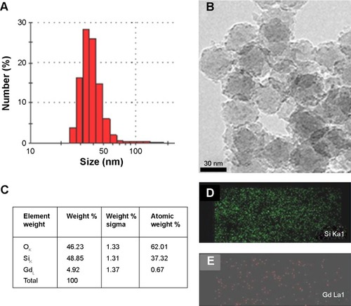
The entire synthesis procedure is summarized in . Branched PEI was PEGylated using amine and NHS conjugation chemistry. PEGylated PEI was conjugated onto silica nanoparticles via TPS. The PEI-PEG silica nanoparticles were investigated by solid-state nuclear magnetic resonance (NMR) (). The particles were reacted further with RITC dye, and Gd was conjugated onto the particles via the polyamine group on the PEI (). The morphological characteristics of the dual imaging silica nanoparticles were investigated by both DLS and TEM. The average particle size was 40±7 nm by DLS () and TEM (). Moreover, elemental analysis by energy-dispersive spectroscopy (interfaced with TEM) showed that weight portion of 64Gd was approximately 4.92% and that of 29Si and 8O was 48.85% and 46.23%, respectively ().
Figure 1 Synthesis of dual imaging silica nanoparticles.
Abbreviations: Gd, gadolinium; NHS, N-hydroxysuccinate; PEG, poly(ethylene glycol); PEI, polyethylenimine; TPS, trimethoxy(3-[oxiran-2-ylmethoxy]propyl)silane.
![Figure 1 Synthesis of dual imaging silica nanoparticles.Abbreviations: Gd, gadolinium; NHS, N-hydroxysuccinate; PEG, poly(ethylene glycol); PEI, polyethylenimine; TPS, trimethoxy(3-[oxiran-2-ylmethoxy]propyl)silane.](/cms/asset/58c240b7-533a-4e7b-8fe3-0cad9329a3cf/dijn_a_88311_f0001_c.jpg)
Figure 2 (A) Particle size distribution (by dynamic light scattering) and (B) morphological analysis (by transmission electron microscopy) of Gd silica nanoparticles, (C) energy-dispersive spectroscopic elemental analysis, and the corresponding elemental mapping of Si (D) and Gd (E).
Abbreviations: Si, silica; Gd, gadolinium.
The hydrophilic properties of silica nanoparticles and the multiple amine functionality grafts on the PEGylated PEI create an environment appropriate for enhancing MR imaging in T1 mode.Citation54 The hydrophobic surface moiety on the dual imaging silica could lead to it being captured and anchored in the liver, while the hydrophilic polar coating by which surface chargesCitation55 can prolong its retention in the circulation.Citation56–Citation59 Further, the surface amine groups on the dual imaging silica can chelate Gd ions and interact in an aqueous environment to produce T1-weighted MR images, with hydration on the dual imaging silica nanoparticles affecting the image contrast. It has been reported that relaxivity is dependent on the hydration environment of Gd conjugate’s its number.Citation59
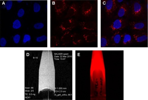
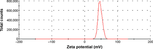
shows that the dual imaging silica nanoparticles upon transfer into A549 (human lung cancer) cells. The nanoparticles were prepared at different concentrations (0.1–1 mg/mL) and transfected into the wells dropwise. After incubation for 4 hours, the old medium was replaced by fresh medium, and the nanoparticle-treated cells were incubated for 24 hours. The nucleus was stained with DAPI (blue), and the dual imaging silica nanoparticles showed red fluorescence (). shows the T1-weighted MR imaging for the colloidal silica solution produced by a microimaging analyzer (9.4 T). In addition, the dual imaging silica nanoparticles had red fluorescence functionality (). Atul et al have reported that the surface charge on nanoparticles can affect cellular localization, and shown that positively charged nanoparticles demonstrate significant uptake in A549 lung cancer cells when compared with negatively charged nanoparticles.Citation60 Our dual imaging silica nanoparticles had a strong positive charge (+50 mV, ). In agreement with Atul et al our positively charged dual imaging silica nanoparticles were easily transfected into A549 lung cancer cells.
Figure 3 (A–C) Feasibility of cell transfection in A549 lung cancer cells in fluorescent imaging mode using gadolinium-conjugated silica nanoparticles (0.2 mg/mL).
Notes: (A) Nuclei stained by 4,6-diamidino-2-phenylindole (blue), (B) dual imaging silica nanoparticles represents red fluorescence (rhodamine isothiocyanate dye). (C) Merged fluorescence microscopic image of dual imaging silica nanoparticles transfected in A549 lung cancer cells. Gadolinium-conjugated silica nanoparticles in colloidal solution using T1-weighted magnetic resonance imaging (D) and fluorescent imaging mode (E).
The longitudinal (r1) MR relaxivity for dual imaging silica nanoparticles was measured (). In addition, each T1 relaxation time is shown in . High-resolution MR imaging was carried out using a Bruker vertical spectrometer (wide-bore, 9.4 T, 400.2 MHz). Gradient echo sequences were used to acquire the MR images (effective spectral bandwidth 101010.1 Hz, echo time 3.000 milliseconds, echo position 50%, repetition time 100.000 milliseconds) in order to compare all the samples (with a fixed receiver gain value of 370.0). A 128×128 acquisition matrix was used for a field of view. The total scan time measured was 125,800 milliseconds in one accumulation experiment. For the T1 measurements, the echo time was regulated at 79.225, 125, 250, 500, 750, 1,000, 2,000, and 3,000 milliseconds. The echo time was 3.640 milliseconds. The field of view was the same as on the two-dimensional image. In the estimated total scan time, it has taken 0 hour 16 minutes 26 seconds 140 milliseconds. The dwell time was 0.004950 milliseconds and the minimum repetition time was 16.685 milliseconds. Relaxivity measurements were acquired by taking the slope of a plot of 1/T1 (s−1) versus concentration (mM). The longitudinal water proton relaxation times (T1) were determined using 400 MHz solid-state NMR with a Bruker micro-imaging probe analyzer operating at 400 MHz and 25°C.
Figure 4 (A, B) Relaxation (r1) rates with different concentrations of dual imaging silica nanoparticles and (C, D) T1-weighted images using a 9.4 T micro-imaging analyzer instrument. Colloidal solution of dual imaging silica nanoparticles in water (C) and a pure water sample (D).
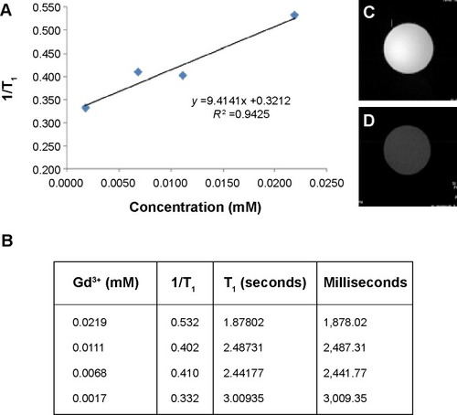
Interestingly, the relaxivity was higher (r1=9.41±0.32 mM−1 s−1) when compared with a previous report for a T1 Gd agent (r1=3 mM−1 s−1).Citation61 We used ICPMS to determine our Gd ion concentration, and T1 relaxivity was measured and compared using four different concentrations. Two-dimensional images were obtained using dual imaging silica nanoparticle dispersion () and pure deionized water () for comparison. Significant signal enhancement in the T1-weighted image was observed for the dual imaging silica nanoparticle dispersion. We believe that the highly hydrophilic environment of the dual imaging silica nanoparticles allows for easy access of water molecules to the Gd (III) ion for water proton relaxation.Citation62–Citation65 Kim et al reported on the use of Gd-TSPETE-silica nanoparticle-[Ru(bpy)3]2+ (r1 =9 mM−1 s−1).Citation64 Therefore, the r1 relaxivity value for our Gd-PEI-PEG silica nanoparticles is similar to that of other silica nanoparticle-based MR imaging agents.
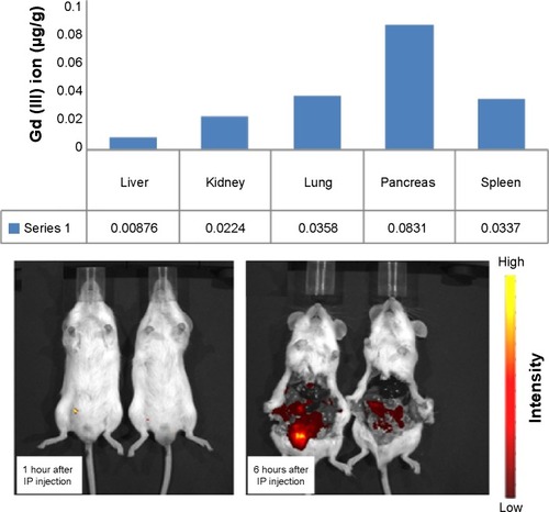
Information on the in vivo distribution of dual mode nanoparticles is useful for bioimaging purposes. compares the biodistribution imaging results at 1 hour and 6 hours after intraperitoneal injection of the nanoparticles. One hour after injection, rich storage was not observed in the pancreas; however, 6 hours after injection, Gd ions were accumulated primarily in the pancreas rather than in other organs such as liver, kidney, lung, pancreas, and spleen. Although Gd enrichment was observed in pancreas, broad distribution in all organs was clear. The circulation time and activity of nanoparticles in blood vessels can be enhanced by surface PEGylation. Our dual imaging silica nanoparticles utilized a PEGylation strategy and consequently showed broad biodistribution in all organs in a mouse model. We believe that the enhanced hydrophilicity of the PEGylated surface on the dual imaging silica nanoparticles increased the particle circulation time. Consequently, broad distribution of Gd was achieved in vivo.Citation55–Citation58
Figure 5 Distribution of gadolinium to the organs in an in vivo mouse model. The upper panel shows organ-dependent inductively coupled plasma mass spectrometry data 6 hours after injection. The lower panel shows the corresponding data at 1 hour and 6 hours after injection.
Abbreviations: IP, Intraperitoneal; Gd, gadolinium.
Conclusion
In summary, we used covalent bonding to construct nano-particles with both MR imaging and fluorescence function-alities. Our Gd-decorated hybrid silica nanoparticles had highly enhanced MR relaxivity. In addition, optical imaging by fluorescence was achieved due to the surface-layered dyes. The polyamine and polycarboxyl groups increased the chelation efficiency of these Gd-silica nanoparticles. We believe that this protocol can be used to develop other useful bioimaging and theranostic silica hybrids for in vivo and in vitro biomedical applications. In the near future, we will report on our in vivo imaging research efforts to enhance the targeting ability of these conjugated hybrid silica nanoparticles.
Acknowledgments
This work was supported by the 2015 Myongji University Research Fund.
Disclosure
The authors report no conflicts of interest in this work.
Supplementary materials
In order to provide more detailed characterization, solid-state nuclear magnetic resonance (NMR) analyses were performed, 600 MHz 1H-NMR spectrum and 400 MHz 29Si, 13C CP MAS NMR spectrum. (A) 29Si CP MAS NMR spectrum of aminopropyl-functionalized silica nanoparticles and PEI 25,000-PEG 5,000 graft silica nanoparticles were compared with silica nanoparticles. (B) 13C CP MAS NMR spectrum of aminopropyl-functionalized silica nanoparticles and PEI 25,000-PEG 5,000 graft silica nanoparticles were also compared with silica nanoparticles. (C) 600 MHz 1H-NMR NMR spectra are shown for PEI 25,000, PEG 5,000, and PEI 25,000-PEG 5,000 copolymer.
Solid-state 29Si CP-MAS NMR analyses were performed using a Bruker Avance 400 MHz spectrometer operating at 79.54 5 MHz. The samples were located in 4 mm ZrO2 rotors. 7 kHz. The spectrum width was 23.8 kHz, and the delay or repetition time was 5.0 seconds. Contact time was performed for 5 milliseconds. The 29Si spectrum was calibrated with the signal obtained using the external standard DSS [3-(trimethylsilyl)-1-propane sulfonic acid, sodium salt 97%, Sigma Chemicals] at 0.00 ppm.
The 13C solid-state NMR experiments were performed with the same NMR instrument, ie, 29Si CP-MAS NMR operating with 100.690 MHz. ZrO2 rotors (4 mm) were used, and magic angle spinning was carried out at a spinning rate of 7 kHz, a 30.3 kHz spectrum width, a 5.0 seconds delay or repetition time, and a 2 milliseconds contact time.
Figure S1 Solid-state NMR analysis of 400 MHz 29Si, 13C CP MAS NMR spectrum. 29Si CP MAS NMR spectrum and 600 MHz 1H-NMR spectrum.
Notes: 29Si CP MAS NMR spectrum of aminopropyl functionalized silica nanoparticles and PEI 25-PEG 5k graft silica nanoparticles as well as silica nanoparticles (A), 13C CP MAS NMR spectrum of aminopropyl functionalized silica nanoparticles and PEI 25-PEG 5k graft silica nanoparticles as well as silica nanoparticles (B), 600 MHz 1H-NMR NMR spectrum of PEI 25k, PEG5k as well as PEI 25k-PEG 5k copolymer (C).
Abbreviations: APTES, (3-aminopropyl)triethoxysilane; NP, nanoparticle; PEG, poly(ethylene glycol); PEI, polyethylenimine; ppm, parts per million, NMR, nuclear magnetic resonance.
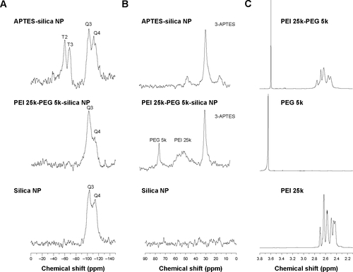
Figure S2 Zeta potential of the dual imaging silica nanoparticles.
References
- WienmannHJBraschRCPressWRWesbeyGECharacteristics of gadolinium-DTPA complex: a potential NMR contrast agentAJR Am J Roentgenol19841426196246607655
- UlbrichWLamprechtATargeted drug-delivery approaches by nanoparticulate carriers in the therapy of inflammatory diseasesJ R Soc Interface200975566
- KeJHLinJJCareyJRChenJSChenCYWangLFA specific tumor-targeting magnetofluorescent nanoprobe for dual–modality molecular imagingBiomaterials2010311707171519969347
- AlloucheJBoissiereMHelaryCLivageJCoradinTBiomimetic core-shell gelatin/silica nanoparticles: a new example of biopolymer-based nanocompositesJ Mater Chem20063031203125
- HuangCCHuangWSuCHFengCNKuoWSYehCSA general approach to silicate nanoshells: gadolinium silicate and gadolinium silicate: europium nanoshells for dual-modality optical and MR imagingChem Commun20092333603362
- YuYYChenCYChenWCSynthesis and characterization of organic-inorganic hybrid thin films from poly(acrylic) and monodispersed colloidal silicaPolymer200344593601
- BorregoTAndradeMPintoMLPhysicochemical characterization of silylated functionalized materialsJ Colloid Interface Sci201034460361020129614
- SalonMCGerbaudGAbdelmoulehMBruzzeseCBoufiSBelgacemMNStudies of interactions between silane coupling agents and cellulose fibers with liquid and solid-state NMRMagn Reson Chem20074547348317431857
- KleemannENeuMJekelNNano-carriers for DNA delivery to the lung based upon a TAT-derived peptide covalently coupled to PEG-PEIJ Control Release2005109209316
- FullerJEZugatesGTFerreiraLSIntracellular delivery of core-shell fluorescent silica nanoparticlesBiomaterials2008291526153218096220
- MijatovicJBinderWHGruberHCharacterization of surface modified silica nanoparticles by 29Si solid state NMR spectroscopyMikrochim Acta2000133175181
- PeerDKarpJMHongSFarokhzadOCMargalitRLangerRNano-carriers as an emerging platform for cancer therapyNat Nanotechnol2007275176018654426
- LipskiAMPinoCJHaseltonFRChenIWShastriVPThe effect of silica nanoparticle-modified surfaces on cell morphology cytoskeletal organization and functionBiomaterials2008293836384618606447
- SantraSZhangPWangKTapecRTanWConjugation of biomolecules with luminophore-doped silica nanoparticles for photostable biomarkersAnal Chem2001734988499311681477
- FengLWangYWangNMaYPreparation of poly(ethylene glycol)-grafted silica nanoparticles using a facile esterification condensation methodPolym Bull200963313327
- JoubertMDelaiteCBourgeat-LamiEDumasPHairy PEO-silica nanoparticles through surface-initiated polymerization of ethylene oxideMacromol Rapid Commun200526602607
- EstevezMCO’DonghueMBChenXTHighly fluorescent dye-doped silica nanoparticles increase flow cytometry sensitivity for cancer cell monitoringNano Res20092448461
- GrafCVossenDLJImhofABlaaderenAVA general method to coat colloidal particles with silicaLangmuir20031966936700
- KimJSRieterWJTaylorKMAnHLinWLinWSelf-assembled hybrid nanoparticles for cancer-specific multimodal imagingJ Am Chem Soc20071298962896317602632
- MishraRSuWPohmannRCell-penetrating peptides and peptide nucleic acid-coupled MRI contrast agents: evaluation of cellular delivery and target bindingBioconjug Chem2009201860186819788302
- ShinJAdnisurRMKoMKImGHLeeJHLeeISHollow manganese oxide nanoparticles as multifunctional agents for magnetic resonance imaging and drug deliveryAngew Chem Int Ed200948321324
- YiDKLeeSSPapaefthymiouGCYingJYNanoparticle architectures template by SiO2/Fe2O3 nanocompositesChem Mater200518614619
- UeckerMZhangSVoitDKarausAMerboldtKDFrahmJReal-time MRI at a resolution of 20 msNMR Biomed20102398699420799371
- SmrnovPGazeauFBeloeilJCDoanBTWilhelmCGilletBSingle-cell detection by gradient echo 9.4T MRI: a parametric studyContrast Med Mol Imaging20061165174
- LiongMLuJKovochichMMultifunctional inorganic nanoparticles for imaging, targeting, and drug deliveryACS Nano2008288989619206485
- SelvanSTanTTYiDKJanaNRFunctional and multifunctional nanoparticles for bioimaging and biosensingLangmuir201026116311164119961213
- GindyMEPrud’hommeRKMultifunctional nanoparticles for imaging, delivery and targeting in cancer therapyExpert Opin Drug Deliv2009686587819637974
- MackayJAChenMMcDanielJRLiuWSimnickAJSelf-assembling chimeric polypeptide-doxorubicin conjugate nanoparticles that abolish tumors after a single injectionNat Mater2009899399919898461
- BuddeMDFrankJAMagnetic tagging of therapeutic cells for MRIJ Nucl Med20095017117419164242
- BargeACravottoGGianolioEFedeliFHow to determine free Gd and free ligand in solution of Gd chelates. A technical noteContrast Med Mol Imaging20061184188
- HillaireauHCouvreurPNanocarriers’ entry into the cell: relevance to drug deliveryCell Mol Life Sci2009662873289619499185
- KorzeniowskaBNooneyRWencelDMcDonaghCSilica nanoparticles for cell imaging and intracellular seedingNanotechnology2013242002
- Brannon-PeppasLBlanchetteJONanoparticle and targeted systems for cancer therapyAdv Drug Deliv Rev2004561649165915350294
- Lessard-VigerMRiouxMRainvilleLBoudreauDFRET-enhancement in multilayer core-shell nanoparticlesNano Lett200993066307119603786
- ParkHYangJLeeJHaamSChoiIHYooKHMultifunctional nanoparticles for combined doxorubicin and photothermal treatmentsACS Nano200932919292619772302
- BardhanBRChenWPerez-TorresCNanoshells with targeted simultaneous enhancement of magnetic and optical imaging and photothermal therapeutic responseAdv Funct Mater20091939013909
- LawBTungCHProteolysis: a biological process adapted in drug delivery, therapy, and imagingBioconjug Chem2009201683169519754162
- ChongHSSongHALimSA novel cholic acid-based contrast enhancement agent for targeted MRIBioorg Med Chem Lett2008182505250818337094
- KamalyNKalberTThanouMBellJDMillerADFolate receptor targeted bimodal liposomes for tumor magnetic resonance imagingBioconjug Chem20092064865519368341
- Al-OgaidiIGouHAl-KazazAKA gold@silica core-shell nanoparticle-based surface-enhanced Raman scattering biosensor for label-free glucose detectionAnal Chim Acta2014811768024456597
- RichardCDoanBTBeloeilJCBessodesMTothESchermanDNoncovalent functionalization of carbon nanotubes with amphiphilic Gd3+ chelates: toward powerful T1 and T2 MRI contrast agentsNano Lett2008823223618088153
- HiraiMMinematsuHKondoNOieKIgarashiKYamazakiNAccumulation of liposome with sialyl Lewis X to inflammation and tumor region: application to in vivo bio-imagingBiochem Biophys Res Commun200735355355817189617
- WanglerCMoldenhauerGSaffrichRPAMAM structure-based multifunctional fluorescent conjugates for improved fluorescent labeling of biomacromoleculesChem Eur J2008148116813018752247
- FletcherSJorgensenMRMillerADFacile preparation of an orthogonally protected, pH-sensitive, bioconjugate linker for therapeutic applicationsOrg Lett200464245424815524454
- MajorJLMeadeTJBioresponsive, cell-penetrating, and multimeric MR contrast agentsAcc Chem Res20094289390319537782
- ThianYLRiddellAMKohDMLiver-specific agents for contrast-enhanced MRI: role in oncological imagingCancer Imaging20131356757924434892
- YoonTJYuKNKimESpecific targeting, cell sorting, and bioimaging with smart magnetic silica core-shell nanomaterialsSmall2006220921517193022
- ChengWPingYZhangYChuangKHLiuYMagnetic resonance imaging (MRI) contrast agents for tumor diagnosisJ Healthc Eng20134234523502248
- YiDKSelvanSTLeeSSPapaefthymiouGCKundaliyaDYingJYSilica-coated nanocomposites of magnetic nanoparticles and quantum dotsJ Am Chem Soc200512749914992
- CaiJShapiroEMHamiltonADSelf-assembling DNA quadruplex conjugated to MRI contrast agentsBioconjug Chem20092020520819125646
- TsaiCPHungYChouYHHigh-contrast paramagnetic fluorescent mesoporous silica nanorods as a multifunctional cell-imaging probeSmall2008418619118205156
- LouieAMultimodality imaging probes: design and challengesChem Rev20101103146319520225900
- HeQZhangJShiJaThe effect of PEGylation of mesoporous silica nanoparticles on nonspecific binding of serum proteins and cellular responsesBiomaterials2010311085109219880176
- ChoJCharKHongJDLeeKBFabrication of highly ordered multilayer films using a spin self-assembly methodAdv Mater20011310761078
- DuLChenJQiYPreparation and biomedical application of non-polymer coated superparamagnetic nanoparticlesInt J Nanomedicine2007280581218203447
- TaupitzMSchnorrJMjukANew generation of monomer stabilized very small superparamagnetic iron oxide particles (VSOP) as contrast medium for angiography: preclinical results in rats and rabbitsJ Magn Reson Imaging20001290591111105029
- SchnorrJWagnerSPilgrimmHPreclinical characterization of monomer-stabilized very small superparamagnetic iron oxide particles (VSOP) as a blood pool contrast medium for MR angiographyAcad Radiol20022S307S30912188256
- WagnerSSchnorrJPilgrimmHMonomer-coated very small superparamagnetic iron oxide particles as contrast medium for magnetic resonance imaging: preclinical in vivo characterizationInvest Radiol20023716717711923639
- ChangCABrittainHGTelserJTweedleMFpH dependence of relaxivities and hydration numbers of gadolinium(III) complexes of linear amino carboxylatesInorg Chem19923155975600
- AsatiASantraSKaittanisCSPerezMJSurface-charge-dependent cell localization and cytotoxicity of cerium oxide nanoparticlesACS Nano201045321533120690607
- HaarPJBroaddusWCChenZJFatourosPPGilliesGTCorwinFDGd-DTPA T1 relaxivity in brain tissue obtained by convection-enhanced delivery, magnetic resonance imaging and emission spectroscopyPhys Med Biol2010553451346520508321
- BoiteauRMeadeTMajorJGadolinium-labeled nanoparticles for magnetic resonance imagingNanoscape200853339
- DavisJJHuangWYDaviesGLLocation-tuned relaxivity in Gd-doped mesoporous silica nanoparticlesJ Mater Chem201222228482285026052183
- KimJYPiaoYHyeonTHMultifunctional nanostructured materials for multimodal imaging, and simultaneous imaging and therapyChem Soc Rev20093837239019169455
- WartenbergNFriesPRaccurtOGuillermoAImbertDMazzantiMA gadolinium complex confined in silica nanoparticles as a highly efficient T1/T2 MRI contrast agentChemistry201396980698323606305