
Abstract
CpG oligodeoxynucleotides (ODNs) stimulate innate and adaptive immune responses. Thus, these molecules are promising therapeutic agents and vaccine adjuvants against various diseases. In this study, we developed a novel CpG ODNs delivery system based on polyethyleneimine (PEI)-functionalized boron nitride nanospheres (BNNS). PEI was coated on the surface of BNNS via electrostatic interactions. The prepared BNNS–PEI complexes had positive zeta potential and exhibited enhanced dispersity and stability in aqueous solution. In vitro cytotoxicity assays revealed that the BNNS–PEI complexes with concentrations up to 100 μg/mL exhibited no obvious cytotoxicity. Furthermore, the positively charged surface of the BNNS–PEI complexes greatly improved the loading capacity and cellular uptake efficiency of CpG ODNs. Class B CpG ODNs loaded on the BNNS–PEI complexes enhanced the production of interleukin-6 and tumor necrosis factor-α from peripheral blood mononuclear cells compared with CpG ODNs directly loaded on BNNS. Contrary to the free CpG ODNs or CpG ODNs directly loaded on BNNS, class B CpG ODNs loaded on the BNNS–PEI complexes induced interferon-α simultaneously. PEI coating may have changed the physical form of class B CpG ODNs on BNNS, which further affected their interaction with Toll-like receptor 9 and induced interferon-α. Therefore, BNNS–PEI complexes can be used to enhance the immunostimulatory effect and therapeutic activity of CpG ODNs and the treatment of diseases requiring interleukin-6, tumor necrosis factor-α, and interferon-α.
Introduction
Bacterial DNA exerts immunostimulatory effects by activating the host defense system and inducing innate and adaptive immune responses.Citation1–Citation4 Unmethylated CpG motif is the specific sequence present in bacterial DNA that is responsible for the aforementioned effects.Citation1,Citation5 Similar to bacterial DNA, synthetic oligodeoxynucleotides (ODNs) containing unmethylated CpG motifs exhibit immunostimulatory effects.Citation1,Citation6 CpG ODNs uptaken by immune cells bind to intracellular Toll-like receptor 9 (TLR-9), a pattern-recognition receptor located mainly in the endolysosomes of B cells and plasmacytoid dendritic cells.Citation2,Citation3,Citation7 The binding process initiates an immunostimulatory cascade that induces the maturation, differentiation, and proliferation of multiple immune cells, such as B and T lymphocytes, natural killer cells, and monocytes/macrophages.Citation8,Citation9 This mechanism further triggers cell signaling pathways, such as mitogen-activated protein kinases and NFκB, which induce multiple proinflammatory cytokines and chemokines as well as modulate cellular inflammatory response.Citation10,Citation11 Thus, CpG ODNs are promising immune adjuvants and immunotherapeutic agents against allergy/asthma, cancer, and infectious diseases.Citation11–Citation14 Clinical studies suggest that CpG ODNs are safe and well tolerated when administered to humans; these molecules reportedly improve vaccine-induced immune responses.Citation2,Citation15 However, the biologic activity of CpG ODNs is transient, and the extreme susceptibility to nuclease degradation in serum and poor cellular uptake of natural CpG ODNs severely limit the therapeutic applications of these molecules.Citation16,Citation17 Therefore, developing approaches to optimize the stimulatory activity of CpG ODNs has attracted considerable interest. Various strategies are involved in the chemical modification of the CpG ODNs backbone. For instance, substituting oxygen with sulfur generates phosphorothioate CpG ODNs, which are highly resistant to nuclease degradation and exert strong immunostimulatory effects.Citation17,Citation18 However, modification of the CpG ODNs backbone causes several severe side effects.Citation19,Citation20 Thus, numerous studies have explored delivery systems that would improve the stimulatory activity of CpG ODNs. Increasing evidence indicates that both innate and adaptive immune responses induced by CpG ODNs can be significantly enhanced using various nanoparticles as carriers.Citation17,Citation18 The exact mechanisms have not been defined, but several possible routes have been proposed. These mechanisms include the protection of CpG ODNs from nuclease degradation, prolonged half-life, increased cellular uptake efficiency, and enhanced delivery to intracellular compartments.Citation18 Therefore, using nanoparticles as carriers may facilitate the use of naturally occurring CpG ODNs in clinical applications.
Boron nitride (BN) has attracted considerable attention because of its outstanding properties and structural similarity with carbon.Citation21,Citation22 BN also exhibits easier cellular uptake and lower cytotoxicity than carbon materials.Citation23,Citation24 Therefore, BN has several applications ranging from composite materials to electrical and optical devices.Citation22,Citation25 BN nanomaterials also have biomedical applications. In vitro and in vivo experiments suggested that BN nanotubes possess optimal biocompatibility, indicating their potential biomedical applications.Citation26–Citation28 We previously used boron nitride nanospheres (BNNS) as carriers for the delivery of unmodified CpG ODNs to activate TLR-9.Citation29 BNNS exhibited no cytotoxicity and protected unmodified CpG ODNs from nuclease degradation. When we added BNNS to 293XL-TLR-9 cells, the BNNS localized in the endolysosome after 24 hours. This localization was maintained even after cell division. TLR-9 also localized in the endolysosome; hence, BNNS can deliver CpG ODNs into the endolysosome compartments of immune cells and stimulate a strong immune response compared with free CpG ODNs. However, CpG ODNs and BNNS are both negatively charged. Therefore, the loading capacity and cellular uptake efficiency of CpG ODNs may be limited by electrostatic repulsion and may not induce a robust cytokine response. Furthermore, class B CpG ODNs loaded on BNNS only induced interleukin 6 (IL-6) and tumor necrosis factor-α (TNF-α) but not interferon-α (IFN-α). However, a high amount of IFN-α induction can be observed when class B CpG ODNs are adsorbed electrostatically onto polystyrene nanoparticles.Citation30 We hypothesized that methods for enhancing CpG ODNs internalization by immune cells may potentiate their immunostimulatory effect. Moreover, CpG ODNs may induce IFN-α when class B CpG ODNs are loaded electro-statically onto BNNS.
In this study, we developed a novel CpG ODNs delivery system based on polyethyleneimine (PEI)-functionalized BNNS (). PEI is the “gold standard” cationic polymer for nucleic acid delivery, either used alone or for surface coating of nanoparticles, to bind negatively charged nucleic acid drugs.Citation31,Citation32 PEI exhibits high transfection efficiency because of enhanced cellular uptake and endosomal escape. Compared with high–molecular weight PEIs, low–molecular weight PEIs are less toxic and more effective as a coating material for nanoparticles to enhance the immunostimulatory effect of CpG ODNs.Citation33 We found that PEI can be coated on negatively charged BNNS through electrostatic interactions, forming BNNS–PEI complexes highly enriched in positive charges that allowed the effective loading of CpG ODNs via a similar process. The formed BNNS–PEI complexes showed no cytotoxicity and significantly enhanced the loading capacity and cellular uptake of CpG ODNs, further inducing a robust cytokine response. Contrary to free CpG ODNs or free CpG ODNs directly loaded on BNNS, class B CpG ODNs loaded on the BNNS–PEI complexes can induce IFN-α. This phenomenon suggests that coating of PEI on BNNS increased the cytokines stimulated by CpG ODNs possibly by altering signal transduction through the interaction of CpG ODNs with TLR-9. Further investigation on the mechanism underlying this phenomenon is currently underway. The current work may provide a promising strategy to enhance the delivery efficiency and immunostimulatory effect of CpG ODNs. Class B CpG ODNs loaded on the BNNS–PEI complexes can simultaneously induce IL-6, TNF-α, and IFN-α. Thus, this study may be beneficial for treating diseases (eg, cancer) requiring the three aforementioned cytokines.
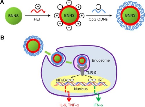
Figure 1 Schematic illustration of the process for preparation and application of the PEI-functionalized BNNS as an efficient CpG ODNs carrier.
Notes: (A) Preparation of the PEI-functionalized BNNS for CpG ODNs loading. (B) Application of the PEI-functionalized BNNS as a carrier for enhancing the immunostimulatory effects of the CpG ODNs.
Abbreviations: BNNS, boron nitride nanospheres; IFN, interferon; IL-6, interleukin-6; IRF, interferon regulatory factor; NFκB, nuclear factor κB; ODN, oligodeoxynucleotide; PEI, polyethyleneimine; TLR-9, Toll-like receptor 9; TNF, tumor necrosis factor.
Materials and methods
Materials
Branched PEI with an average molecular weight of 600 Da was purchased from Wako Pure Chemicals (Osaka, Japan). Phosphodiester-based class B CpG ODNs, referred to as CpG ODNs2006x3-PD,Citation34 were obtained from Fasmac, Inc. (Kanagawa, Japan), diluted in sterile water at 100 μM, and then stored at −20°C. Frozen human peripheral blood mononuclear cells (PBMCs) were purchased from Cellular Technology Limited (Cleveland, OH, USA) and thawed in accordance with the manufacturer’s protocol. Roswell Park Memorial Institute (RPMI)-1640 medium and fetal bovine serum were obtained from Gibco (Grand Island, NY, USA). Cell Counting Kit-8 was purchased from Dojindo (Kumamoto, Japan).
Preparation of BNNS–PEI complexes
High-purity BNNS were synthesized through chemical vapor deposition as previously reported.Citation35 PEI was diluted with Milli-Q water to a concentration of 10% (v/v) prior to use. Working solutions were prepared from the stock by dilution with Milli-Q water. BNNS were functionalized by suspending 1 mg of BNNS in 1 mL of PEI solution, and the resulting solution was stirred at room temperature for 2 hours. The PEI-functionalized BNNS were washed five times with Milli-Q water by centrifugation and redispersed in phosphate-buffered saline (PBS).
Characterizations
Fourier transform infrared (FTIR) spectroscopy was performed using a Spectrum GX spectrophotometer (Perkin Elmer, Boston, MA, USA) at 4 cm−1 resolution with 32 scans. Transmission electron microscopy was conducted using a 3000F, high-resolution, field-emission transmission electron microscope (JEOL, Tokyo, Japan) operated at an acceleration voltage of 300 kV. Dynamic light scattering measurements were obtained using Photal DLS-6000DL (Otsuka, Hirakata, Japan). Zeta potentials were measured using a LEZA-600 electrophoresis zeta potential analyzer (Otsuka, Japan). UV-vis absorption spectra were measured with a U-2900 spectrophotometer (Hitachi, Tokyo, Japan) at room temperature. Thermogravimetric analysis was conducted on an SII TG/DTA 6200 system at a heating rate of 5°C/minute with an open alumina cell.
Cell culture and in vitro cytotoxicity assay
Frozen human PBMCs were thawed in accordance with the manufacturer’s protocol. The cytotoxicities of BNNS and the BNNS–PEI complexes to PBMCs were evaluated using a Cell Counting Kit-8 assay. PBMCs were seeded into a 96-well plate at a density of 5×10⁴ cells per well. After 24 hours of incubation at 37°C with 5% CO2, until the cells adhered to the plate, the cell culture was added with a series of concentrations of BNNS and the BNNS–PEI complexes. After another 24 hours of incubation, each well had 10 μL of Cell Counting Kit-8 solution and then incubated for another 3 hours. Finally, the absorbance at 450 nm was obtained using a microplate reader to determine the relative cell viability. No ethics approval was required from the institutional review board for the use of this cell line.
Preparation of BNNS–PEI/CpG ODN complexes
CpG ODNs solution (25 μL) was added to an equal volume of BNNS–PEI solution (2 mg/mL in PBS at pH 7.4) and then shaken at room temperature for 1 hour. The mixture was then centrifuged at 15,000 revolutions per minute for 15 minutes to collect the BNNS–PEI/CpG ODN complexes. Binding of the CpG ODNs to the BNNS–PEI complexes was further confirmed through gel electrophoresis using 10%–20% gel. BNNS–PEI was incubated with CpG ODNs at various weight ratios (BNNS–PEI/CpG ODNs) from 5 to 100. The mixture was centrifuged, and the supernatant was collected for gel electrophoresis. The CpG ODNs in the gel were visualized by ethidium bromide using a Dolphin-Doc UV transilluminator (Kurabo, Japan). Loading capacity was calculated from the concentration of the unloaded CpG ODNs in the supernatant measured by a NanoDrop 2000 spectrophotometer (Thermo Scientific, Waltham, MA, USA). The BNNS–PEI/CpG ODN complexes were resuspended by adding 50 μL of PBS and used for the following cytokine stimulation.
Uptake of CpG ODNs by human PBMCs
Human PBMCs (8×10⁴) were seeded in a 35 mm Petri dish with a glass bottom and then incubated for 24 hours at 37°C under 5% CO2. BNNS/fluorescein isothiocyanate (FITC)–CpG ODN and BNNS–PEI/FITC–CpG ODN complexes were then added to the dish at a final concentration of 50 μg/mL. Free FITC–CpG ODNs and BNNS were used as controls. After incubation for 24 hours, the cells were washed twice with cold PBS to terminate the uptake and then fixed with 3.7% (v/v) paraformaldehyde. Fluorescence of the fixed cells was visualized under an SP5 confocal laser scanning microscope (Leica, Nussloch, Germany) with a 63× oil immersion objective.
Cytokine assay
Human PBMCs were seeded at a density of 5×10⁶ cells/mL in RPMI-1640 medium supplemented with 10% fetal bovine serum and then immediately stimulated with the BNNS/CpG ODN and BNNS–PEI/CpG ODN complexes. After 48 hours and 8 hours of incubation at 37°C respectively, cell supernatants were collected for further analysis. The concentrations of IL-6, IFN-α, and TNF-α in the medium were determined through enzyme-linked immunosorbent assay using the Ready-SET-Go! human IL-6 kit (eBioscience, San Diego, CA, USA), human TNF-α kit (eBioscience), and human IFN-alpha-Module set (eBioscience) in accordance with the manufacturers’ protocol.
Statistical analysis
Statistical analysis was performed using Student’s t-test. Data are presented as mean ± standard deviation (*P<0.05, **P<0.01).
Results and discussion
Synthesis and characterization of the BNNS–PEI complexes
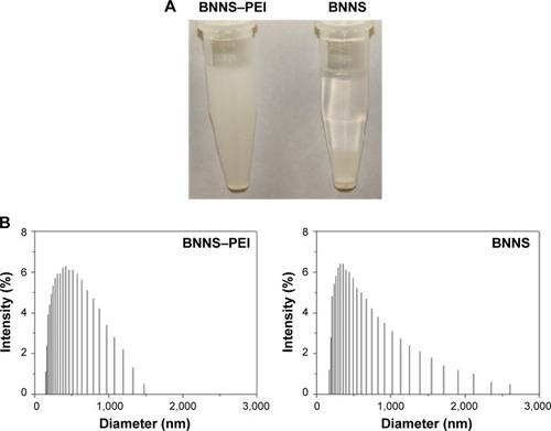
BNNS were synthesized through chemical vapor deposition.Citation35 The as-prepared BNNS showed a uniform spherical shape with an average diameter of approximately 150 nm (). In addition, well-ordered channels were clearly observed from the high-resolution transmission electron micrograph of the BNNS (). We used PEI to functionalize the surface of BNNS and obtain a positively charged surface that facilitates the loading of CpG ODNs onto the BNNS. shows the schematic of the synthesis route. BNNS were first noncovalently functionalized by PEI polymers, forming positively charged BNNS–PEI complexes. Then, the negatively charged CpG ODNs were loaded onto the BNNS–PEI complexes via electrostatic interactions. FTIR results show the PEI coating on BNNS. shows the FTIR spectra of BNNS, PEI, and the BNNS–PEI complexes. Two distinct peaks at 2,700–2,900 cm−1 assigned to the C–H vibrations in PEI were clearly observed in the BNNS–PEI complexes compared with pure BNNS. This result suggests that PEI has been functionalized on the surface of BNNS. Zeta potential measurements further confirmed the successful functionalization of the BNNS with PEI (). The coating of PEI greatly increased the zeta potential of BNNS from −5 mV to +40 mV for the BNNS–PEI complexes. In addition, the surface charge of the BNNS–PEI complexes increased when the initial concentration of the PEI solution was increased during the functionalization of the BNNS. This phenomenon was probably due to the increased amino groups coated on the surface of BNNS. PEI concentrations were optimized to 5% to achieve the highest positive zeta potential and were used in the following studies. Therefore, the positive charge of the BNNS–PEI complexes facilitated the loading of the negatively charged CpG ODNs through electrostatic interactions and enhanced the cellular uptake of the complexes. Thermogravimetric analysis is a convenient technique that reveals the composition and change in the thermal stability of complexes. This process was used to determine the amount of PEI coated on the surface of BNNS. shows that BNNS only exhibited slight mass loss (<5%) because of their superb structural stability and anti-oxidation ability. By contrast, a high weight loss (22%) was observed for the BNNS–PEI complexes. Thus, the weight ratio of PEI in the BNNS–PEI complexes was calculated to be approximately 17%, which possibly facilitated the loading of the CpG ODNs on the BNNS. The dispersity and stability of nanoparticles are crucial for their application as carriers in drug delivery systems. PEI coating enhanced these characteristics of BNNS in physiological solutions () mainly through the interaction of the amino functional group in PEI with BNNS. PEI coating on the surface can trigger electrostatic and steric repulsion among the highly positively charged BNNS–PEI complexes in solutions and stabilize the dispersion. Further measurement of the size distributions showed that PEI coating did not significantly increase the mean particle size of the BNNS–PEI complexes, which exhibited a relatively narrow size distribution in PBS compared with BNNS (). This phenomenon also indicates that the BNNS–PEI complexes may have favorable dispersity in PBS. Thus, the BNNS–PEI complexes can be used as carriers for CpG ODNs delivery.
Figure 2 (A) Typical transmission electron microscopy image of the boron nitride nanosphere. (B) A high-resolution image.
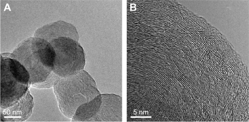
Figure 3 Characterizations of the BNNS and BNNS–PEI complexes.
Notes: (A) FTIR spectra of BNNS, PEI, and BNNS–PEI complexes. (B) Zeta potentials of BNNS and a series of BNNS–PEI complexes at different starting concentrations of PEI. (C) Thermal gravimetric analysis of the BNNS and BNNS–PEI complexes.
Abbreviations: BNNS, boron nitride nanospheres; FTIR, Fourier transform infrared; PEI, polyethyleneimine.
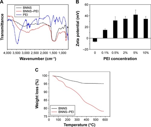
Figure 4 Characterizations of the BNNS and BNNS–PEI complexes.
Notes: (A) Dispersed BNNS and BNNS–PEI complexes in PBS after statically placed for 24 hours. (B) Hydrodynamic diameters distribution of BNNS and BNNS–PEI complexes in PBS.
Abbreviations: BNNS, boron nitride nanospheres; PBS, phosphate-buffered saline; PEI, polyethyleneimine.
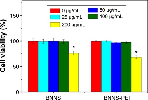
In vitro cytotoxicity
The cytotoxicity of a carrier is critical for its application in drug delivery systems.Citation36 In the present study, the in vitro cytotoxicity of the BNNS–PEI complexes was evaluated using human PBMCs in a water-soluble tetrazolium cell proliferation assay. PEI is a widely used nonviral gene delivery carrier, but it is reportedly toxic to cells. In the present study, both BNNS and BNNS–PEI complexes with concentrations of up to 100 μg/mL showed slight cytotoxicity to human PBMCs (). This phenomenon may be mainly attributed to the low molecular weight (600 Da) of PEI used in our experiment. The cytotoxicity of PEI is closely dependent on its structure and molecular weight. Low–molecular weight PEI always shows much lower cytotoxicity compared with high–molecular weight PEI.Citation37 However, BNNS and the BNNS–PEI complexes at 200 μg/mL decreased cell viability. The concentration of the BNNS–PEI complexes used in the following cell experiments was 50 μg/mL. Therefore, PEI coating did not significantly enhance the cytotoxicity of BNNS to PBMCs, and the BNNS–PEI complexes could be used as carriers for CpG ODNs delivery.
Figure 5 In vitro cytotoxicity assay. Relative cell viability of peripheral blood mononuclear cells incubated with increasing concentrations of BNNS or BNNS–PEI complexes measured by a water-soluble tetrazolium salt assay.
Notes: Data are presented as mean ± standard deviation (n=5). *P<0.05.
Abbreviations: BNNS, boron nitride nanospheres; PEI, polyethyleneimine.
CpG ODNs loading
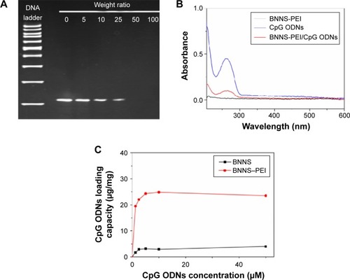
The capability of the BNNS–PEI complexes as carriers for CpG ODNs delivery was investigated by incubating the complexes with CpG ODNs. Loading of CpG ODNs onto the BNNS–PEI complexes was initially confirmed through gel electrophoresis. The band of CpG ODNs from the supernatant became weaker when the BNNS–PEI complexes were mixed with CpG ODNs at a high weight ratio (BNNS–PEI/CpG ODNs) and disappeared at the ratio of 50 (). This result indicates the electrostatic binding of the negatively charged CpG ODNs to the positively charged BNNS–PEI complexes, and all CpG ODNs could be bound by the BNNS–PEI complexes at the weight ratio of 50. The characteristic adsorption peak at 260 nm for CpG ODNs appeared in the UV-vis spectra of the BNNS–PEI/CpG ODN complexes, which also confirmed the binding of CpG ODNs to the BNNS–PEI complexes (). Meanwhile, the loading capacity of the CpG ODNs on BNNS and the BNNS–PEI complexes increased with increasing CpG ODNs concentration and then reached a plateau. The BNNS–PEI complexes possessed eightfold higher capacity to load CpG ODNs (24 μg/mg nanoparticles) than BNNS (). This result can be attributed to the strong positive surface charge of the BNNS–PEI complexes, which enhanced the loading of the CpG ODNs via electrostatic interactions. The release of CpG ODNs from the BNNS–PEI/CpG ODN complexes was further examined under conditions that corresponded to the physiological environment in TLR-9-localized endolysosomes. However, almost no CpG ODNs were released. This result suggests that the BNNS–PEI/CpG ODN complexes are stable and that CpG ODNs cannot be easily released from the BNNS–PEI complexes because of the strong binding.
Figure 6 Loading of CpG ODNs on BNNS–PEI complexes.
Notes: (A) Gel electrophoresis image of the supernatant after interaction of CpG ODNs with BNNS–PEI at increasing weight ratios. (B) UV-vis spectra of CpG ODNs, BNNS–PEI, and BNNS–PEI/CpG ODNs complexes. (C) Loading capacity of CpG ODNs on BNNS and BNNS-PEI complexes with increasing CpG ODNs concentrations, denoted as μg CpG ODNs loaded on 1 mg BNNS. Data are presented as mean ± standard deviation (n=3).
Abbreviations: BNNS, boron nitride nanospheres; ODN, oligodeoxynucleotide; PEI, polyethyleneimine.
Intracellular uptake of CpGODNs
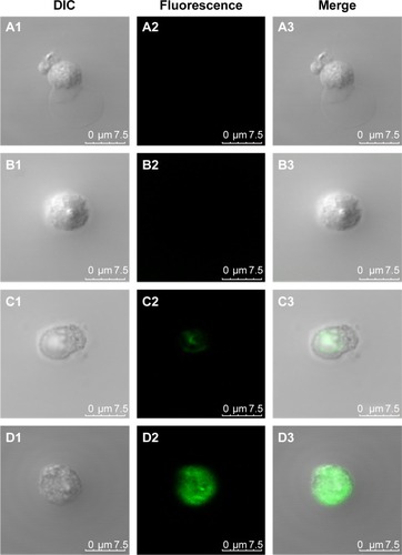
Delivery or transfection efficiency of the carrier is a critical criterion for a drug/gene delivery system. The colloidal characteristics and CpG ODNs loading capacity of the BNNS–PEI complexes may have been improved, but their capability to enhance the cellular uptake of CpG ODNs required further investigation. In the cellular uptake study, FITC-labeled CpG ODNs were used to replace the regular CpG ODNs and loaded on BNNS and the BNNS–PEI complexes. This process produced complexes with green fluorescence when observed via confocal laser scanning microscope. shows that the green fluorescence from the cells incubated with the BNNS–PEI/FITC–CpG ODN complexes was much stronger than that from the cells incubated with the BNNS/FITC–CpG ODN complexes. However, no fluorescence was observed when free FITC-labeled CpG ODNs were applied to the cells, indicating the low cellular uptake and carrier requirement for CpG ODNs delivery. These findings confirmed our hypothesis that the intracellular delivery of CpG ODNs can be enhanced by the BNNS–PEI complexes. After being uptaken by cells, CpG ODNs localize mainly in endosomal compartments and activate TLR-9.Citation2,Citation38,Citation39 This phenomenon induces several cytokines and chemokines, as well as further modulates the immune response.Citation10 Therefore, enhancing the cellular uptake of CpG ODNs by the BNNS–PEI complexes is believed to strengthen the interaction of CpG ODNs with TLR-9 and enhance immune response.
Figure 7 Confocal microscope images of PBMCs after 24 hours of incubation with BNNS (A1-3), free CpG ODNs (B1-3), CpG ODNs loaded on BNNS (C1-3), CpG ODNs loaded on BNNS–PEI complexes (D1-3).
Note: CpG ODNs were labeled with FITC.
Abbreviations: BNNS, boron nitride nanospheres; DIC, differential interference contrast; FITC, fluorescein isothiocyanate; ODN, oligodeoxynucleotide; PBMCs, peripheral blood mononuclear cells; PEI, polyethyleneimine.
Cytokine induction in vitro
The efficacy of the BNNS–PEI complexes as carriers for CpG ODNs delivery was tested by incubating BNNS–PEI/CpG ODNs with PBMCs. The resultant cytokines were determined via enzyme-linked immunosorbent assay. As expected, PBMCs stimulated by the BNNS–PEI/CpG ODN complexes had the highest amounts of IL-6 and TNF-α (), reaching levels approximately six times higher than those in the cells stimulated by BNNS/CpG ODNs. Although PEI can be used alone as a drug delivery carrier, the BNNS–PEI complexes exhibited much higher efficiency, and the BNNS–PEI/CpG ODN complexes induced higher amounts of cytokines (, B). By contrast, free CpG ODNs can only induce a relatively small amount of cytokines. This finding suggests that the high cytokine production of CpG ODNs was due to their high loading capacity and enhanced cellular uptake resulting from the positively charged BNNS–PEI complexes. Similar results were obtained when a BNNS-binding peptide was used as a linker molecule for CpG ODNs delivery; this peptide improved the CpG ODNs internalization by PBMCs.Citation40 However, the simultaneous induction of IL-6 and IFN-α was not achieved with our previous BNNS delivery system.Citation39,Citation40
Figure 8 Cytokine induction from PBMCs stimulated by CpG ODNs loaded on BNNS and BNNS–PEI complexes.
Notes: Loaded BNNS or BNNS–PEI complexes (50 μg/mL). (A) IL-6 production. (B) TNF-α production. (C) IFN-α production. Data are presented as mean ± standard deviation (n=3). *P<0.05, **P<0.01. #not detectable (below detection limit).
Abbreviations: BNNS, boron nitride nanospheres; IFN, interferon; IL-6, interleukin-6; ODN, oligodeoxynucleotide; PBMCs, peripheral blood mononuclear cells; PEI, polyethyleneimine; TNF, tumor necrosis factor.
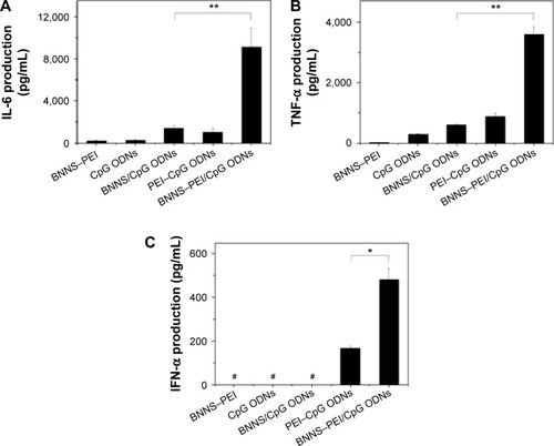
Class B CpG ODNs, which have a linear structure, can stimulate TLR-9 in B cells and induce IL-6 and TNF-α; however, these ODNs cannot induce IFN-α similar to class A CpG ODNs.Citation10,Citation41–Citation43 When we applied the CpG ODNs loaded electrostatically onto PEI-functionalized BNNS to stimulate the PBMCs, class B CpG ODNs acquired the capability to induce IFN-α (). By contrast, free CpG ODNs or the CpG ODNs directly loaded on BNNS cannot stimulate IFN-α secretion. IFN-α induction can also be attributed to the CpG ODNs loaded on the BNNS–PEI complexes but not to the free ones released from the BNNS–PEI/CpG ODN complexes. We did not detect the release of CpG ODNs from the BNNS–PEI/CpG ODN complexes under conditions that corresponded to the physiological environment in the TLR-9–localized endolysosome. A high amount of IFN-α induction was also reported when class B CpG ODNs were adsorbed electrostatically onto polystyrene nanoparticles or silica nanoparticles with cationic polymer-coated surfaces.Citation30 The mechanism by which class B CpG ODNs acquire the potential to induce IFN-α remains unclear, but the physical form of the CpG ODNs may play an important role in this process.Citation18,Citation44,Citation45
The immunostimulatory effects of CpG ODNs depend not only on their base sequence and structure but also on their physical form.Citation44 The greater capability of class A CpG ODNs to induce IFN-α production is due to the formation of a higher order multimeric structure.Citation30 However, such structures are not observed in class B CpG ODNs, which form linear structures.Citation46 An immune response similar to class A CpG ODNs can be obtained by artificially causing class B CpG ODNs to form a multimeric structure using nanoparticles.Citation33,Citation47 This phenomenon suggests that the multimerization of CpG ODNs is crucial for IFN-α induction. Therefore, the class B CpG ODNs loaded onto the positively charged BNNS–PEI complexes through electrostatic interactions formed a higher order multimeric structure similar to class A CpG ODNs and acquired the capability to induce IFN-α. However, similar to free class B CpG ODNs, class B CpG ODNs could not form a multimeric structure when bound directly to negatively charged BNNS because of the weak interactions. Thus, class B CpG ODNs can induce IL-6 and TNF-α but not IFN-α. However, the exact mechanism by which class B CpG ODNs acquire the capability to induce IFN-α remains unclear, and further investigation is currently underway.
Conclusion
PEI-functionalized BNNS were used as nanocarriers for the intracellular delivery of CpG ODNs. PEI was coated on the surface of BNNS through noncovalent electrostatic interactions. The resultant BNNS–PEI complexes possessed strong positive charges and enhanced dispersity and stability in aqueous solution. Furthermore, the BNNS–PEI complexes were nontoxic to human PBMCs up to a concentration of 100 μg/mL. Given their positive surface charge, the BNNS–PEI complexes can effectively enhance the loading capacity and cellular uptake of CpG ODNs. The use of the BNNS–PEI complexes as carriers for CpG ODNs delivery significantly enhanced the production of cytokines, such as IL-6 and TNF-α, from PBMCs compared with the use of BNNS or PEI. Class B CpG ODNs loaded on the BNNS–PEI complexes acquired the capability to induce IFN-α. PEI coating may have changed the physical form of class B CpG ODNs on BNNS, further modulated their interaction with TLR-9, and induced both IL-6 and IFN-α. Our results highlight the potential of the BNNS–PEI complexes as novel nanocarriers for enhancing the immunostimulatory and therapeutic activity of CpG ODNs, especially for treating diseases that require IL-6, TNF-α, and IFN-α simultaneously.
Acknowledgments
This work was supported by Fundamental Research Funds for the Central Universities (No JUSRP11427) and Scientific Research Foundation for the Returned Overseas Chinese Scholars, State Education Ministry.
Disclosure
The author reports no conflicts of interest in this work.
References
- KriegAMYiAKMatsonSCpG motifs in bacterial DNA trigger direct B-cell activationNature199537465225465497700380
- KlinmanDMImmunotherapeutic uses of CpG oligodeoxynucleotidesNat Rev Immunol200444248257
- HemmiHTakeuchiOKawaiTA toll-like receptor recognizes bacterial DNANature2000408681374074511130078
- WagnerHBacterial CpG DNA activates immune cells to signal infectious dangerAdv Immunol19997332936810399010
- AkiraSTakedaKKaishoTToll-like receptors: critical proteins linking innate and acquired immunityNat Immunol20012867568011477402
- KlinmanDMYiAKBeaucageSLConoverJKriegAMCpG motifs present in bacterial DNA rapidly induce lymphocytes to secrete interleukin 6, interleukin 12, and interferon gammaProc Natl Acad Sci U S A1996937287928838610135
- BauerSKirschningCJHäckerHHuman TLR9 confers responsiveness to bacterial DNA via species-specific CpG motif recognitionProc Natl Acad Sci U S A200198169237924211470918
- StaceyKJSweetMJHumeDAMacrophages ingest and are activated by bacterial DNAJ Immunol19961575211621228757335
- SunSQZhangXHToughDFSprentJType I interferon-mediated stimulation of T cells by CgG DNAJ Exp Med199818812233523429858519
- KriegAMCpG motifs in bacterial DNA and their immune effectsAnnu Rev Immunol20022070976011861616
- FonsecaDEKlineJNUse of CpG oligonucleotides in treatment of asthma and allergic diseaseAdv Drug Deliv Rev200961325626219167442
- KlinmanDMKlaschikSSatoTTrossDCpG oligonucleotides as adjuvants for vaccines targeting infectious diseasesAdv Drug Deliv Rev200961324825519272313
- KriegAMTherapeutic potential of toll-like receptor 9 activationNat Rev Drug Discov20065647148416763660
- MuradYMClayTMCpG oligodeoxynucleotides as TLR9 agonists therapeutic applications in cancerBio Drugs2009236361375
- KlinmanDMCurrieDGurselIVerthelyiDUse of CpG oligodeoxynucleotides as immune adjuvantsImmunol Rev2004199120121615233736
- MutwiriGLittel-van Den HurkSVBabiukLAApproaches to enhancing immune responses stimulated by CpG oligodeoxynucleotidesAdv Drug Deliv Rev200961322623219162103
- MutwiriGKNichaniAKBabiukSBabiukLAStrategies for enhancing the immunostimulatory effects of CpG oligodeoxynucleotidesJ Control Release200497111715147800
- HanagataNStructure-dependent immunostimulatory effect of CpG oligodeoxynucleotides and their delivery systemInt J Nanomedicine201272181219522619554
- SheehanJPLanHCPhosphorothioate oligonucleotides inhibit the intrinsic tenase complexBlood1998925161716259716589
- HenrySPBeattieGYehGComplement activation is responsible for acute toxicities in rhesus monkeys treated with a phosphorothioate oligodeoxynucleotideInt Immunopharmacol20022121657166612469940
- ChopraNGLuykenRJCherreyKBoron-nitride nanotubesScience1995269522696696717807732
- GolbergDBandoYTangCCZhiCYBoron nitride nanotubesAdv Mater2007191824132432
- ChenXWuPRousseasMBoron nitride nanotubes are non-cytotoxic and can be functionalized for interaction with proteins and cellsJ Am Chem Soc2009131389089119119844
- JonssonESimonsenLKarlssonMLarssonRThe hollow fiber model: a new method for in vivo-evaluation of antitumor effect, toxicity and pharmacokinetics of new anticancer drugsAnn Oncol199894444
- GaoZHZhiCYBandoYGolbergDSerizawaTIsolation of individual boron nitride nanotubes via peptide wrappingJ Am Chem Soc2010132144976497720297821
- CiofaniGRaffaVMenciassiACuschieriABoron nitride nanotubes: an innovative tool for nanomedicineNano Today200941810
- CiofaniGDantiSNittiSMazzolaiBMattoliVGiorgiMBiocompatibility of boron nitride nanotubes: an up-date of in vivo toxicological investigationInt J Pharm20134441–2858823357257
- CiofaniGDantiSGenchiGGPilot in vivo toxicological investigation of boron nitride nanotubesInt J Nanomedicine20127192422275819
- ZhiCYMengWJYamazakiTBN nanospheres as CpG ODN carriers for activation of toll-like receptor 9J Mater Chem2011211452195222
- KerkmannMCostaLTRichterCSpontaneous formation of nucleic acid-based nanoparticles is responsible for high interferon-α induction by CpG-A in plasmacytoid dendritic cellsJ Biol Chem200528098086809315591070
- PutnamDGentryCAPackDWLangerRPolymer-based gene delivery with low cytotoxicity by a unique balance of side-chain terminiProc Natl Acad Sci U S A20019831200120511158617
- ZhuJTangALawLPAmphiphilic core-shell nanoparticles with poly(ethylenimine) shells as potential gene delivery carriersBioconjug Chem200516113914615656585
- ManoharanYJiQYamazakiTEffect of molecular weight of polyethyleneimine on loading of CpG oligodeoxynucleotides onto flake-shell silica nanoparticles for enhanced TLR9-mediated induction of interferon-alphaInt J Nanomedicine201273625363522888224
- MengWJYamazakiTNishidaYHanagataNNuclease-resistant immunostimulatory phosphodiester CpG oligodeoxynucleotides as human toll-like receptor 9 agonistsBMC Biotechnol2011118821943407
- TangCCBandoYHuangYZhiCYGolbergDSynthetic routes and formation mechanisms of spherical boron nitride nanoparticlesAdv Funct Mater2008182236533661
- ZhuYFIkomaTHanagataNKaskelSRattle-type Fe(3)O(4)@ SiO(2) hollow mesoporous spheres as carriers for drug deliverySmall20106347147819943250
- BrunotCPonsonnetLLagneauCFargePPicartCGrosgogeatBCytotoxicity of polyethyleneimine (PEI), precursor base layer of poly-electrolyte multilayer filmsBiomaterials200728463264017049374
- LatzESchoenemeyerAVisintinATLR9 signals after translocating from the ER to CpG DNA in the lysosomeNat Immunol20045219019814716310
- ZhangHChenSZhiCYamazakiTHanagataNChitosan-coated boron nitride nanospheres enhance delivery of CpG oligodeoxynucleotides and induction of cytokinesInt J Nanomedicine201381783179323674892
- ZhangHYamazakiTZhiCHanagataNIdentification of a boron nitride nanosphere-binding peptide for the intracellular delivery of CpG oligodeoxynucleotidesNanoscale20124206343635022941279
- KrugATowarowskiABritschSToll-like receptor expression reveals CpG DNA as a unique microbial stimulus for plasmacytoid dendritic cells which synergizes with CD40 ligand to induce high amounts of IL-12Eur J Immunol200131103026303711592079
- HartmannGWeeratnaRDBallasZKDelineation of a CpG phosphorothioate oligodeoxynucleotide for activating primate immune responses in vitro and in vivoJ Immunol200016431617162410640783
- HartmannGKriegAMMechanism and function of a newly identified CpG DNA moth in human primary B cellsJ Immunol2000164294495210623843
- GuiducciCOttGChanJHProperties regulating the nature of the plasmacytoid dendritic cell response to toll-like receptor 9 activationJ Exp Med200620381999200816864658
- HondaKYanaiHNegishiHIRF-7 is the master regulator of type-I interferon-dependent immune responsesNature2005434703477277715800576
- KleinDCGLatzEEspevikTStokkeBTHigher order structure of short immunostimulatory oligonucleotides studied by atomic force microscopyUltramicroscopy2010110668969320202756
- ChinnathambiSChenSGanesanSHanagataNBinding mode of CpG oligodeoxynucleotides to nanoparticles regulates bifurcated cytokine induction via toll-like receptor 9Sci Rep2012253422837814