
Abstract
Purpose
Nutrients and immunological factors of breast milk are essential for newborn growth and the development of their immune system, but this secretion can contain organic and inorganic toxins such as barium. Colostrum contamination with barium is an important issue to investigate because this naturally occurring element is also associated with human activity and industrial pollution. The study evaluated the administration of barium nanoparticles to colostrum, assessing the viability and functional activity of colostral mononuclear phagocytes.
Methods
Colostrum was collected from 24 clinically healthy women (aged 18–35 years). Cell viability, superoxide release, intracellular Ca2+ release, and phagocyte apoptosis were analyzed in the samples.
Results
Treatment with barium lowered mononuclear phagocyte viability, increased superoxide release, and reduced intracellular calcium release. In addition, barium increased cell death by apoptosis.
Conclusion
These data suggest that nanoparticles of barium in colostrum are toxic to cells, showing the importance of avoiding exposure to this element.
Introduction
Breast milk contains several immunological components, including macrophages, neutrophils, lymphocytes, cytokines, immunoglobulins, complement proteins, chemokines, growth factors, and proteins, that can affect child’s immune maturation.Citation1–Citation5 Both neutrophils and macrophages play a key role in host defense against microorganisms,Citation6 killing bacteria, fungi, and protozoa by phagocytosis.Citation7–Citation11 However, in addition to beneficial cellular components, breast milk can contain environmental pollutants such as organic or inorganic substancesCitation12 that are transferred from the mother to the baby.Citation13 Thus, interest in the biological effects of exposure to toxic metals has increased, with biological fluids such as blood, urine, and breast milk analyzed by biomonitoring.Citation13–Citation15
Metals are the most important category of natural elements that can be extracted from the environment to be used in industrial processes. They can spread in the environment and accumulate in body tissues, possibly causing toxic effects even in small amounts.Citation16 Some studies report the effects of human exposure to barium (Ba) and other metals.Citation13,Citation17–Citation23 Exposure to barium deserves particular attention because this element is commonly found in drinking waterCitation24 and can be released into the environment by natural disaggregation of rocks and mineralsCitation25 or as polluting waste from industry and human activities.Citation25,Citation26
The toxicity of compounds containing barium depends on their solubility,Citation25 that is, soluble salts are more toxic than insoluble salts.Citation27 The action mechanism of barium appears to involve the blocking of K+ and NaK+ channels in the cell membrane and increasing potassium transfer from an extracellular to intracellular medium.Citation25,Citation28 The main effect of barium is the stimulation of all types of muscles,Citation28,Citation29 and the dominant clinical condition of barium poisoning in humans seems to be hypocalcemia, which paralyzes skeletal muscles.Citation28
Although barium is found at low levels in the environment, the health consequences of long-term exposure have yet to be fully explained.Citation30 Considering that barium can contaminate breast milk, thereby affecting components of the still-developing baby’s immune system, the aim of this study was to evaluate the effects of barium nanoparticles on the viability and functional activity of mononuclear (MN) cells of colostrum.
Materials and methods
Subjects
Approximately 15 mL of colostrum was collected at the Health System Program of Barra do Garças, Mato Grosso, Brazil, from clinically healthy women (18–35 years old; N=24) who had no diagnosed diseases, such as hypertension and diabetes, and who reported not consuming alcoholic beverages or tobacco. All the mothers had given birth to healthy term babies through surgical delivery. This study was approved by the institutional Research Ethics Committee of the Federal University of Mato Grosso (protocol number: 354/CEP-HUJM/07), and all the mothers signed informed consent.
Colostrum sampling and separation of colostral cells
Colostrum was collected in sterile plastic tubes between 48 hours and 72 hours postpartum. The samples were centrifuged (160× g, 4°C) for 10 minutes. The upper fat layer was discarded, and the aqueous supernatant was stored at −80°C for later analyses. Cells were separated by a Ficoll-Paque gradient (Pharmacia, Upsala, Sweden), producing preparations with 98% of pure MN cells at concentration with the average of 4.2±0.7, analyzed by light microscopy. Purified MN cells were resuspended independently in serum-free 199 medium at a final concentration of 2×10⁶ cells/mL. The cells were used for assays of viability, superoxide release, calcium release, and apoptosis.
Treatment of MN colostral phagocytes with barium chloride
To assess the effect of barium chloride on cellular viability, MN phagocytes (2×10⁶ cells/mL) were incubated with 10 μL of barium chloride (Sigma-Aldrich Co., St Louis, MO, USA) at the concentrations of 1 g/L, 1 μg/L, 10 ng/L, and 1 ng/L for 0, 30, 60, 90, 120, and 180 minutes at 37°C.
To investigate the effect of barium chloride on superoxide anion release, intracellular Ca2+ release, and apoptosis, MN phagocytes (2×10⁶ cells/mL) were incubated with 10 μL of barium chloride at concentration 1 ng/L for 60 minutes at 37°C. The phagocytes were then washed twice with 199 medium at 4°C and immediately used in the assays. A control was performed without barium chloride.
To analyze the effects of intracellular Ca2+ on barium chloride action, colostral MN phagocytes (2×10⁶ cells/mL) were incubated with 10 μL of 8-(diethylamino)octyl-3,4,5- trimethylbenzoate hydrochloride (TMB-8; Sigma-Aldrich Co.) at a final concentration of 0.1 mM for 60 minutes at 37°C, as an intracellular calcium antagonist during 60 minutes at 37°C.Citation31 The MN phagocytes were then washed two times with 199 medium at 4°C and immediately used in the assays.
Viability assay
Cellular viability was evaluated by the acridine orange method.Citation9 The cells were pretreated or not with barium chloride as previously described. Cells were resuspended in serum-free 199 medium and centrifuged. The supernatant was discarded, and the sediment dyed with 200 μg/L of acridine orange (Sigma-Aldrich Co.), 14.4 g/L for 1 minute. The sediment was resuspended in cold 199 medium, washed twice, and observed under immunofluorescence microscope at 400× and 1,000× magnifications.
We stained the slides with acridine orange and counted 100 cells. The viability index was calculated as the ratio between orange-stained (dead) and green-stained (alive) cells ×100.Citation9 All the experiments were performed in duplicate.
Release of superoxide anion
Superoxide release was determined by cytochrome C (Sigma-Aldrich Co.) reduction.Citation7 Briefly, MN colostrum phagocytes treated with barium chloride were resuspended in phosphate-buffered saline (PBS), pH 7.4 containing 2.6 mM CaCl2, 2 mM MgCl2, and 2 mg/mL cytochrome C. The suspensions (100 μL) were incubated for 60 minutes at 37°C on culture plates. The reaction rates were measured by absorbance at 550 nm, and the results were expressed as nmol/O2−. All the experiments were performed in duplicate.
Intracellular Ca2+ release determined by fluorescence and flow cytometry
We performed fluorescence staining on the FACSCalibur (BD Biosciences, San Jose, CA, USA) to assess intracellular Ca2+ release in phagocytes.Citation32 Cells were loaded with the fluorescent radiometric calcium indicator Fluo3-Acetoxymethyl (Fluo3-AM; Sigma-Aldrich Co.). Cell suspensions were pretreated or not with 50 μL of barium chloride as previously described. A control was performed using cell suspensions, pretreated with TMB-8. Suspensions were centrifuged twice (160× g, 10 minutes, 4°C) and resuspended in PBS-containing bovine serum albumin (BSA, 5 mg/mL). Suspensions were centrifuged twice (160× g, 10 minutes, 4°C) and resuspended in PBS-containing BSA (5 mg/mL). This suspension was incubated with 5 μL of Fluo-3 (1 μg/mL) for 60 minutes at 37°C. After incubation, cells were washed twice in PBS-containing BSA (5 mg/mL; 160× g, 10 minutes, 4°C) and then analyzed by flow cytometry (FACSCalibur system; BD Biosciences). Fluo-3 was detected at 530/30 nm filter for intracellular Ca2+. The rate of intracellular Ca2+ release was expressed in geometric mean fluorescence intensity of Fluo-3. Data shown in the figures correspond to one of the several trials performed.
Apoptosis assay
The apoptosis assay was determined by Annexin V-FITC Apoptosis Detection Kit (Alexis™, San Diego, USA) according to the manufacturer’s instructions. Untreated cells were used as negative controls, and cells treated with staurosporine (Sigma-Aldrich Co.), an inducer of apoptosis, were used as positive controls.Citation33 The cells were resuspended in 500 μL of binding buffer containing 5 μL of Annexin V-FITC and 5 μL of propidium iodide (PI) and then incubated for 10 minutes at room temperature. The fluorescence of the cells was analyzed by flow cytometry (FACSCalibur system; BD Biosciences). The obtained data were analyzed using CellQuest software. The cells were classified as follows: viable cells (annexin−/PI−), early apoptotic cells (annexin+/PI−), late apoptotic cells (annexin+/PI+), and necrotic cells (annexin−/PI+).
Statistical analysis
Data were expressed as the mean ± standard deviation (SD). Analysis of variance with calculation of F statistic and Tukey’s multiple comparison test was used to evaluate the superoxide release anion, intracellular Ca2+ release, and apoptosis in the presence or absence of barium chloride. Statistical significance was considered when the P-value was <0.05.
Results
The women under study had an average (±SD) age of 25.6±6.1 years and gestational age of 38.6±1.3 weeks. Mean (±SD) newborn birth weight was 3,330±40 g.
Regardless of exposure time, the viability index was lower in cells incubated with barium chloride. The lowest viability index was observed in cells incubated with 1 ng/mL of barium chloride for 60 minutes ().
Table 1 Cellular viability index (%) of colostrum mononuclear (MN) phagocytes (mean ± SD, N=6 in each treatment)
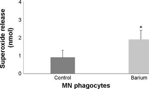
MN phagocytes treated with barium chloride exhibited higher superoxide release than spontaneous release () and lower intracellular calcium. Treating colostrum phagocytes with TMB-8 also reduced intracellular Ca2+ release by colostrum phagocytes, which was similar to that of cells treated with barium chloride ().
Table 2 Release of intracellular Ca2+ by colostral MN phagocytes exposed to barium
Figure 1 Superoxide release by blood mononuclear phagocytes (mean ± SD, N=6 in each treatment).
Notes: Colostral mononuclear cells were treated or not with barium. F=14.8023, P<0.0001. *Differences between phagocytes treated with or without barium.
Abbreviations: MN, mononuclear; SD, standard deviation.
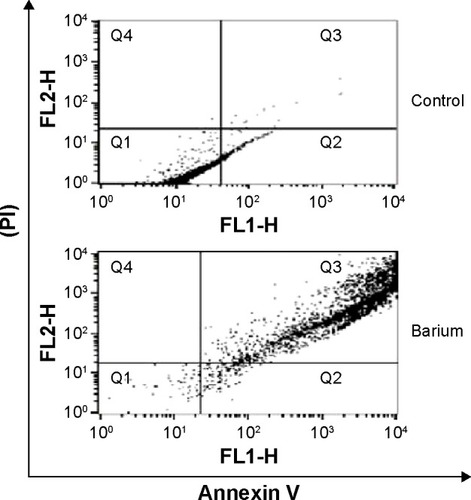
The annexin V assay detected barium-induced apoptosis of colostral MN phagocytes (). In the treatment without barium chloride, MN phagocytes showed a lower apoptosis rate. The highest apoptosis index was observed in colostral MN phagocytes treated with barium chloride ( and ).
Table 3 Apoptosis (%) and necrosis (%) of colostral MN phagocytes exposed to barium chloride (1 ng/mL)
Figure 2 Apoptosis of colostral MN phagocytes exposed to barium (1 ng/nL) and staurosporine (Sigma-Aldrich Co., St Louis, MO, USA) indicated by fluorescence intensity.
Notes: Cells were stained with Annexin V-FITC (Sigma-Aldrich Co.). Immunofluorescence analyses were carried out by flow cytometry (FACScalibur; BD Biosciences, San Jose, CA, USA).
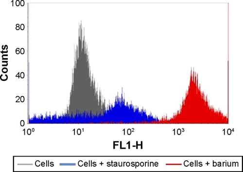
Figure 3 Barium chloride induces apoptosis in colostral MN phagocytes.
Notes: Different types of cell death were assessed using flow cytometry with annexin V PI staining. The sum of the upper-right (Q3) and lower-right (Q2) quadrants represents total apoptosis percentage. The upper-left (Q4) quadrant is the percentage of necrosis and lower-left (Q1) quadrant corresponds to viable cells. Data represent one experiment.
Abbreviations: MN, mononuclear; PI, propidium iodide.
Discussion
The present study showed that nanoparticles of barium chloride are toxic for colostral MN phagocytes, reducing cell viability and intracellular calcium release and increasing apoptosis rate.
Barium toxicity is likely caused by K+ channel blockade, changing cell membrane permeability to potassium.Citation19,Citation25,Citation34 It may also interact with calcium.Citation35 The increase in superoxide levels modifies intracellular calcium release and phosphorylation during oxidative metabolism.Citation6 In the present study, barium chloride exposure increased superoxide release and reduced calcium release by MN phagocytes. The formation of superoxide anion (O2−) is a physiological mechanism for microorganism elimination.Citation11,Citation36 However, during oxidative stress, cells can produce a large amount of superoxide radicals.Citation9,Citation10
Intracellular calcium release promotes cellular activationCitation31,Citation32,Citation37 through self-regulation system.Citation38 Even minor changes in plasma membrane alter Ca2+ permeability, triggering physiological responses and significant changes in the cytosolic concentration of this element.Citation39
Monitoring intracellular calcium has become a useful tool in assessing cell activity.Citation40 Interestingly, barium chloride also inhibited intracellular Ca2+ release in colostral MN phagocytes. In addition, we detected a synergistic effect of barium chloride plus TMB-8, since either combined or separately, they similarly inhibit intracellular Ca2+ release.
The precise mechanisms regarding the action of barium on intracellular calcium homeostasis have yet to be elucidated, and studies of barium action on human cells are scarce. Other toxic metalsCitation41–Citation44 have been found to trigger different responses in order to maintain intracellular calcium homeostasis, enhancing or suppressing intracellular calcium mobilization, eventually causing apoptosis.Citation44
Trace elements play an important role in regulating cell growth and metabolism, including the control of apoptosis.Citation45 In the present study, barium chloride exposure induced apoptosis in colostral MN phagocytes. Other studies describe the mechanisms involved in the death of other cell types due to barium chloride exposure.Citation46 Other toxic metals were found to induce apoptosis and/or necrosis due to changes in intracellular calcium homeostasis and promotion of microvascular endothelial dysfunction.Citation41,Citation44,Citation47 Our findings indicate that the barium chloride induced higher apoptosis rate but lower number of necrotic cells.
Here, the results suggest that trace amounts of barium chloride can change the function of colostral phagocytes and induce apoptosis. However, other investigations that test different barium chloride concentrations and incubation periods are needed to produce dose-dependent curves.
Conclusion
Nanoparticles of barium chloride have immunotoxic effects on colostral MN phagocytes. Barium toxicity likely extends to other cell types, showing the importance of avoiding exposure to this chemical agent, especially for nursing mothers. Accordingly, health education programs should be developed to avoid exposure to barium and other chemical agents.
Author contributions
All authors contributed toward data analysis, drafting and critically revising the paper, and agree to be accountable for all aspects of the work. L Mores carried out the assay, participated in the sequence alignment, and drafted the manuscript. EL França and EA Suchara participated in the design of the study, coordination, and helped to draft the manuscript. NA Silva participated in the collection of samples, carried out the assays, and helped to draft the manuscript. AC Honorio-França carried out the assay, conceived the study, carried out the assays, participated in its design and coordination, and helped to draft the manuscript.
Acknowledgments
This work was supported by the Conselho Nacional de Desenvolvimento Científico e Tecnológico (CNPq), Brazil, Fundação de Apoio a Pesquisa de Mato Grosso (FAPEMAT), Brazil, and Coordenação de Aperfeiçoamento de Pessoal de Nível Superior, CAPES, Brazil.
Disclosure
The authors declare no conflicts of interest in this work.
References
- FieldCJThe immunological components of human milk and their effect on immune development in infantsAm Soc Nutr Sci200513514
- HansonLASession 1: feeding and infant development breast-feeding and immune functionProc Nutr Soc20076638439617637091
- ChiricoGMarzolloRCortinovisSFonteCGasparoniAAntiinfective properties of human milkJ Nutr20081381801S1806S18716190
- HolmlundUAmoudruzPJohanssonMAMaternal country of origin, breast milk characteristics and potential influences on immunity in offspringClin Exp Immunol201016250050920942805
- FrançaELNicomedesTRCalderonIMPHonorio-FrançaACTime-dependent alterations of soluble and cellular components in human milkBiol Rhythm Res201041543547
- CarrichonLPicciocchiADebeurmeFCharacterization of superoxide overproduction by the D-Loop(Nox4)-Nox2 cytochrome b(558) in phagocytes – differential sensitivity to calcium and phosphorylation eventsBiochim Biophys Acta20111808789020708598
- Honorio-FrançaACCarvalhoMPIsaacLTrabulsiLRCarneiro-SampaioMMColostral mononuclear phagocytes are able to kill enteropathogenic Escherichia coli opsonized with colostral IgAScand J Immunol19974659669246209
- França-BotelhoACHonorio-FrançaACFrancaELGomesMACosta-CruzJMPhagocytosis of Giardia lamblia trophozoites by human colostral leukocytesActa Paediatr20069543844316720491
- FrançaELBittercourtRFijimoriMMoraesTCCalderonIMPHonorio-FrançaACHuman colostral phagocytes eliminate enterotoxigenic Escherichia coli opsonized by colostrum supernatantJ Microbiol Immunol Infect2011441721531345
- FrançaELMorceliGFagundesDLGRudgeMVCCalderonIMPHonorio-FrançaACSecretory IgA-FCαR receptor interaction modulating phagocytosis and microbicidal activity by phagocytes in human colostrum of diabeticsAPMIS201111971071921917008
- Honorio-FrançaACHaraCCPOrmondeJVSNunesGTFrançaELHuman colostrum melatonin exhibits a day-night variation and modulates the activity of colostral phagocytesJ Appl Biomed201311153162
- NickersonKEnvironmental contaminants in breast milkJ Midwifery Womens Health200651263416399607
- AlmeidaAALopesCMSilvaAMBarradoETrace elements in human milk: correlation with blood levels, inter-element correlations and changes in concentration during the first month of lactationJ Trace Elem Med Biol20082219620518755395
- ChristensenJMHuman exposure to toxic metals: factors influencing interpretation of biomonitoring resultsSci Total Environ1995166891357754357
- PronczukJAkreJMoyGVallenasCGlobal perspectives in breast milk contamination: infectious and toxic hazardsEnviron Health Persp2002110A349A351
- KhlifiRHamza-ChaffaiAHead and neck cancer due to heavy metal exposure via tobacco smoking and professional exposure: a reviewToxicol Appl Pharmacol2010248718820708025
- FalomirPAlegríaABarberáRFarréRLagardaMJDirect determination of lead in human milk by electrothermal atomic absorption spectrometryFood Chem199964111113
- ConiEBoccaBGaloppiBAlimontiACaroliSIdentification of chemical species of some trace and minor elements in mature breast milkMicrochem J200067187194
- PurdeyMChronic barium intoxication disrupts sulphated proteoglycan synthesis: a hypothesis for the origins of multiple sclerosisMed Hypotheses20046274675415082100
- DóreaJGDonangeloCMEarly (in uterus and infant) exposure to mercury and leadClin Nutr20062536937616307830
- KaziTGJalbaniNBaigJÁAssessment of toxic metals in raw and processed milk samples using electrothermal atomic absorption spectrophotometerFood Chem Toxicol2009472163216919500636
- ÖrünEYalçinSSAykutOBreast milk lead and cadmium levels from suburban areas of AnkaraSci Total Environ20114092467–24722011
- BleackleyMRMacgillivrayRTATransition metal homeostasis: from yeast to human diseaseBiometals20112478580921479832
- ThangNDYajimaIKumasakaMYBarium promotes anchorage-independent growth and invasion of human HaCaT keratinocytes via activation of c-SRC kinasePLoS One2011618
- OscarssonAReevesALBariumNordbergGFFowlerBANordbergMFribergLFHandbook on the Toxicology of MetalsAmsterdamElsevier2007407414
- MillourSNoelLChekriRStrontium, silver, tin, iron, tellurium, gallium, germanium, barium and vanadium levels in foodstuffs from the Second French Total Diet StudyJ Food Compos Anal201225108129
- SyedIBHosainFDetermination of LD50 of barium chloride and allied agentsToxicol Appl Pharmacol1972221501525034984
- RozaOBermanLBThe pathophysiology of barium: hypokalemic and cardiovascular effectsJ Pharm Exp Ther1971177433439
- SchroederHATiptonIHNasonAPTrace metals in man: strontium and bariumJ Chronic Dis1972254915174647214
- Agency for Toxic Substances and Disease Registry (ATSDR)Toxicological Profile for Barium and Barium CompoundsAtlantaU.S. Department of Health and Human Services20071179
- MorceliGHonorio-FrançaACFagundesDLGCalderonIMPFrançaELAntioxidant effect of melatonin on the functional activity of colostral phagocytes in diabetic womenPLoS One20138e5691523437270
- FagundesDLGFrançaELHaraCCPHonorio-FrançaACImmunomodulatory effects of poly (ethylene glycol) microspheres adsorbed with cortisol on activity of colostrum phagocytesInt J Pharmacol20128510518
- PundtNPetersMAWunrauCSusceptibility of rheumatoid arthritis synovial fibroblasts to FasL- and TRAIL-induced apoptosis is cell cycle-dependentArth Res Ther200911110
- DiazPVallejosCGuerreroIRiquelmeGBarium, TEA and sodium sensitive potassium channels are present in the human placental syncytiotrophoblast apical membranePlacenta20082988389118708253
- WonesRGStadlerBLFrohmanLALack of effect of drinking water barium on cardiovascular risk factorsEnviron Health Persp199085355359
- PessoaRSFrançaELRibeiroEBAbudNGHonorio-FrançaACMicroemulsion of babassu oil as a natural product to improve human immune system functionDrug Des Dev Ther201492131
- FagundesDLGFrançaELMorceliGRudgeMVCCalderonIMPHonorio-FrançaACThe role of cytokines in the functional activity of phagocytes in blood and colostrum of diabetic mothersClin Dev Immunol2013201359019024489577
- RasmussenHBarrettPSmallwoodJBollagWIsalesCCalcium ion as intracellular messenger and cellular toxinEnviron Health Persp1990841725
- CarafoliEIntracellular calcium homeostasisAnn Rev Biochem1987563954333304139
- NovakEJRabinovitchPSImproved sensitivity in flow cytometric intracellular ionized calcium measurement using Fluo-3/Fura Red fluorescence ratiosCytometry1994171351417835163
- SuriyoTWatcharasitPThiantanawatASatayavivadJArsenite promotes apoptosis and dysfunction in microvascular endothelial cells via an alteration of intracellular calcium homeostasisToxicol In Vitro20122638639522244921
- SchanneFAXMoskalJRGuptaRKEffect of lead on intracellular free calcium ion concentration in a presynaptic neuronal model: 19F-NMR study of NG108-15 cellsBrain Res19895033083112605523
- OkamotoYKagayaAMotohashiNYamawakiSInhibitory effects of lithium ion on intracellular Ca2+ mobilization in the rat hippocampal slicesNeurochem Int1995262332387787770
- BiagioliMPifferiSRagghiantiMBucciSRizzutoRPintonPEndoplasmic reticulum stress and alteration in calcium homeostasis are involved in cadmium-induced apoptosisCell Calcium200843184–1952008
- KoudrineAVTrace elements and apoptosisJ Trace Elem Med Biol19981265769760414
- Cano-AbadMFGarcíaAGSánchez-GarcíaPLópezMGBa2+ induced chromaffin cell death: cytoprotection by Ca2+ channel antagonistsEur J Pharmacol2000402192910940353
- CatelasIPetitAValiHQuantitative analysis of macrophage apoptosis vs. necrosis induced by cobalt and chromium ions in vitroBiomaterials2005262441245315585247