
Abstract
This study centered on an innovative application of Porphyra yezoensis polysaccharide (PPS) with cationic modification as a safe and efficient nonviral gene vector to deliver a plasmid encoding human Wnt3a (pWnt3a) into human umbilical cord mesenchymal stem cells (HUMSCs). After modification with branched low-molecular-weight (1,200 Da) polyethylenimine, the cationized PPS (CPPS) was combined with pWnt3a to form spherical nanoscale particles (CPPS-pWnt3a nanoparticles). Particle size and distribution indicated that the CPPS-pWnt3a nanoparticles at a CPPS:pWnt3a weight ratio of 40:1 might be a potential candidate for DNA plasmid transfection. A cytotoxicity assay demonstrated that the nanoparticles prepared at a CPPS:pWnt3a weight ratio of 40:1 were nontoxic to HUMSCs compared to those of Lipofectamine 2000 and polyethylenimine (25 kDa). These nanoparticles were further transfected to HUMSCs. Western blotting demonstrated that the nanoparticles (CPPS:pWnt3a weight ratio 40:1) had the greatest transfection efficiency in HUMSCs, which was significantly higher than that of Lipofectamine 2000; however, when the CPPS:pWnt3a weight ratio was increased to 80:1, the nanoparticle-treated group showed no obvious improvement in translation efficiency over Lipofectamine 2000. Therefore, CPPS, a novel cationic polysaccharide derived from P. yezoensis, could be developed into a safe, efficient, nonviral gene vector in a gene-delivery system.
Introduction
Porphyra yezoensis, widely distributed in eastern Asia and on the west coast of the USA, has been praised as an “affinal drug and diet” for its various biological activities. Edible P. yezoensis, commonly referred to as nori,Citation1 has become a promising source of nutrients, owing to its richness in polysaccharides, which have antioxidative,Citation2–Citation5 anti-inflammatory,Citation6,Citation7 hyperglycemic,Citation8 and immunostimulatory effects.Citation9 More recently, several studies have revealed that sulfated polysaccharides from P. yezoensis can inhibit tumor growth both in vitro and in vivo.Citation10,Citation11 However, despite its biological and pharmacological functions, little attention has been paid to P. yezoensis polysaccharide (PPS) as a functional material for gene transfection.
With the tremendous progress in biotechnology, gene therapy has been proposed as a strategy for the treatment of some diseases, such as incurable cancers. Successful delivery of a specific functional gene in a simple manner is vital in gene therapy. Given the chance of degradation of naked therapeutic genes by nucleases and the low efficiency of the cellular uptake, the development of efficient and safe gene vectors is an urgent need.Citation12,Citation13 Recombinant viruses, such as lentivirus,Citation14–Citation18 retrovirus,Citation19–Citation22 and adenovirus,Citation23–Citation25 have been considered ideal gene carriers because of their considerable cellular uptake. Even though their high transfection efficiency might make clinical applications of recombinant viruses possible in gene therapy, obstacles, such as abnormal gene expressionCitation26–Citation28 and immune responses,Citation29–Citation31 are still unresolved for the use of viral vectors.
Nonviral vectors, which have been regarded as alternative gene carriers for functional gene delivery, have received a great deal of attention, due to their advanced properties, such as low immunogenicity, safety, and specificity, as well as long-term gene expression. Cationic lipids, such as LipofectamineCitation32,Citation33 and 1,2-dioleoyl-3-trimethylammonium,Citation34,Citation35 are popular nonviral vectors that are widely used for target-gene delivery because of their high in vitro transfection efficiency. In addition, cationic polymers that contain amino groups in their side chains or backbones have become another promising nonviral gene carrier, not only because of their ease of production, low risk of side effects, and non-immunogenicityCitation36 but also for their compact combination with DNA,Citation37 which is an issue of crucial importance during gene transfection. However, these cationic polyplexes also have inevitable drawbacks that restrict their clinical application. For example, cytotoxicity and macrophage activation caused by cationic liposomes may induce cell shrinkage and vacuolization of the cytoplasm.Citation38 To overcome these difficulties, polysaccharides, a natural product and an ideal material, have been modified and reformed to produce gene vectors. Ethylenediamine-modified polysaccharide from mulberry leavesCitation39 and Lycium babarumCitation40 has high efficiency for the transfer of a DNA plasmid encoding TGFβ1 into rat bone marrow mesenchymal stem cells. Moreover, Angelica sinensis polysaccharide modified with polyethylenimineCitation41 and Pleurotus eryngii polysaccharide cationized by spermineCitation42 could become potential candidate nonviral vectors for gene transfection, because of their low cytotoxicity and excellent transfection efficiency compared to Lipofectamine 2000.
The objective of this study was to establish a novel gene vector, cationized PPS (CPPS) nanoparticles, for the nonviral gene delivery of a plasmid DNA encoding Wnt3a into human umbilical cord mesenchymal stem cells (HUMSCs) in vitro. Specifically, we optimized the extraction process for PPS and successfully developed a CPPS via chemical modification with low-molecular-weight polyethylenimine (PEI; 600 Da). Moreover, HUMSCs, which normally would otherwise be discarded after birth, have emerged as a desirable source of multipotent progenitor cells that can differentiate into several lineage-specific cell types to form bone, fat, tendon, and muscle tissue.Citation39 Meanwhile, HUMSCs have been ultimately converted into induced pluripotent stem cells via the code-livery of Yamanaka factors (c-Myc, Klf4, Sox2, and Oct4) using calcium phosphate nanoparticles,Citation43 which illustrated the remarkable inducibility of HUMSCs based on a nonviral gene-delivery system. In addition, the overexpression of Wnt3aCitation44 can direct the differentiation of neural stem cells into neurons both in vitro and in vivo by activating Wnt signaling, which is also a requirement for the maintenance, proliferation, and survival of neural progenitors when cultured under neurosphere conditions.Citation45 Previous studies have demonstrated that the overexpression of Wnt3a in highly self-renewable cellsCitation46,Citation47 might contribute to the formation and proliferation of neural cells, which could be a new and safe method for the recovery of spinal cord injury.Citation48,Citation49 The novelty of this study is that we established a CPPS for the first time as a nonviral vector for the delivery of plasmid Wnt3a (pWnt3a) into HUMSCs, which might facilitate the effective application of P. yezoensis to the area of gene therapy.
Materials and methods
Materials
PPS was obtained in our laboratory.Citation8 Branched PEI (molecular weight 600 Da) was purchased from Sigma-Aldrich (St Louis, MO, USA). Dulbecco’s modified Eagle’s medium (DMEM)/F12 medium was purchased from Thermo Fisher Scientific (Waltham, MA, USA), as were penicillin–streptomycin, trypsin, fetal bovine serum (FBS), Lipofectamine 2000, and MTT. Antibodies, including mouse polyclonal anti-GAPDH, were purchased from Santa Cruz Biotechnology Inc (Dallas, TX, USA). Rabbit polyclonal anti-Wnt3a primary antibody was obtained from Abcam (Cambridge, UK). Horseradish peroxidase-conjugated secondary antibodies were obtained from Bioworld Technology, Inc., (Wuhan, People’s Republic of China [PRC]). All other chemicals and reagents were of an analytical grade or better, and were used without further purification. All of the solutions were prepared with highly purified water. The experimental protocol was approved by the University Ethics Committee for the Use of Experimental Animals, and it conformed to the Guide for Care and Use of Laboratory Animals. The experiments involving human cell lines were approved by the Institutional Review Board of Jiangsu University (Zhenjiang, China).
Preparation of CPPS
Extraction and purification of PPS
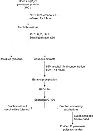
The purification of PPS in our laboratoryCitation8 was conducted according to a procedure illustrated in . In brief, dried P. yezoensis powder (100 g) was homogenized and dispersed in 1 L of 95% ethanol and refluxed in a hot water bath at 70°C for 1 hour, followed by filtration to obtain the residue. Then, the residue was decentralized in double-distilled water (DDW; solid:liquid ratio 1:30, pH 11) at 90°C for 3 hours, followed by filtration. This process was repeated once. The filtrates from each run were combined and concentrated under reduced pressure at 80°C and precipitated with 95% (v/v) ethanol until the final ethanol concentration reached 80% (v/v). The resulting precipitate was dissolved into 150 mL DDW and reprecipitated with absolute ethanol. Finally, the ultimate precipitate was lyophilized to generate a crude extract, which was dissolved in DDW and applied to a diethylaminoethanol cellulose (DE-52; Shanghai YuanYe Biotechnology Co Ltd, Shanghai, PRC) column (diameter 2.6 cm × length 40 cm) and a Sephadex G-100 gel resin (Shanghai Richu Bioscience Co Ltd, Shanghai, PRC) column (diameter 2.6 cm × length 40 cm) in sequence. The fractions that contained polysaccharides were detected with a phenol–sulfuric acid assay. A single elution peak was chosen, collected, concentrated, dialyzed, and lyophilized to obtain purified PPS.
Figure 1 Flowchart for Porphyra yezoensis polysaccharide extraction and purification.
Abbreviation: DEAE, diethylaminoethanol.
Preparation of CPPS
As shown in , 112 mg of PPS was dissolved in 10 mL of phosphate-buffered saline (PBS; pH 7.0) in a round-bottom flask that was degassed with N2. N,N′-carbonyldiimidazole (80 mg) was dissolved in 4 mL of methylene chloride. After the addition of 0.15 mL of tri-ethylamine (Et2N) to the reaction, the methylene chloride– carbonyldiimidazole solution was added to the reaction slowly with gentle magnetic stirring. After 60 minutes of reaction time, a PEI (600 Da) solution (1,200 mg PEI dissolved in 15 mL PBS, pH 7.0) was slowly added to the reaction mixture and magnetically stirred in a dark room for 24 hours. Finally, the mixture was dialyzed (molecular weight cutoff 3,000 Da; Genia Biotech, Beijing, PRC) against DDW for 2 days and then freeze-dried to yield CPPS.
Figure 2 Flowchart for cationized Porphyra yezoensis polysaccharide preparation.
Abbreviations: PBS, phosphate-buffered saline; CDI, carbonyldiimidazole; PEI, polyethylenimine.

Cell culture
HUMSCs, generously provided by the Beike Jiangsu Stem Cell Bank (Taizhou, Jiangsu, PRC), were cultured in DMEM/F12 supplemented with 10% FBS, 0.1 mM nonessential amino acids (NEAAs; Sigma-Aldrich), FGFβ (PeproTech, Rocky Hill, NJ, USA), and 0.1 mM β-mercaptoethanol (β-ME; Toyoda Bio, Beijing, PRC). The cells were passaged with 0.25% trypsin and 0.1% ethylenediaminetetraacetic acid (EDTA) and incubated at 37°C in 5% humidified CO2.
Preparation of CPPS-pWnt3a complexes
CPPS-pWnt3a complexes were prepared by coacervation,Citation50 a technique based on the electrostatic interaction of two oppositely charged compounds. Briefly, 20 mg of PPS-PEI was fully dissolved in 1 mL of sterile water to obtain a PPS-PEI stock solution (20 mg/mL). The working pWnt3a solution (20 μg/mL) was prepared with sterile water. Aliquots (100 μL) of CPPS solution and pWnt3a were heated separately at 55°C for 30 minutes. Equal volumes of these two solutions were immediately mixed together and vortexed for 30 seconds to obtain CPPS-Wnt3a nanoparticles and then incubated at room temperature for 30 minutes.
Electrophoresis of PPS-Wnt3a nanoparticles
The plasmid DNA retardation effect of the CPPS-Wnt3a nanoparticles was analyzed using 1% agarose gel electrophoresis. Different PPS:Wnt3a weight ratios (5:1, 10:1, 20:1, 40:1, 80:1, 100:1, and 150:1) of CPPS-Wnt3a nanoparticles were prepared. The PPS-Wnt3a nanoparticle solution (5 μL) was added to 1 μL of loading buffer (0.1% sodium dodecyl sulfate, 5% glycerol, and 0.005% bromophenol blue) and applied to a 1% agarose gel in a Tris/borate/EDTA (TBE) buffer solution (pH 8.0) containing 0.1 mg·mL−1 ethidium bromide. Meanwhile, 5 μL of a naked pWnt3a solution that was diluted with sterile water was used as a negative control. Electrophoresis of the nanoparticles was performed in TBE solution at 80 V for 90 minutes. The gel was imaged with an ultraviolet transilluminator (Gel Doc 2000; Bio-Rad Laboratories Inc, Hercules, CA, USA).
Cytotoxicity assay
The in vitro cytotoxicity of the CPPS-Wnt3a complex was measured by the MTT assay. Briefly, the HUMSCs were seeded in 96-well plates at a density of 30,000 cells per well and incubated at 37°C. The medium was subsequently removed and replaced with serum-free medium until cell confluence reached 80%. After 12 hours of serum-free incubation, CPPS-Wnt3a nanoparticles (weight ratios of 40:1, 80:1, 100:1, 150:1, 200:1, 250:1, and 300:1) were transferred, and the Lipofectamine 2000-Wnt3a and PEI-Wnt3a groups (multiple dilutions of PEI: 50-fold, 100-fold, 200-fold, 400-fold, and 800-fold) were used as positive controls according to the manufacturer’s protocol under the same conditions. The viability percentage was relative to untreated cells. The cell viability (%) was calculated by the formula cell viability (%) = Abssample/Abscontrol ×100.
Zeta potential
The zeta potential of the nanoparticle suspensions was measured with a Nano ZS90 Zetasizer (Malvern Instruments, Malvern, UK). The zeta potential of the free plasmid and the original CPPS were determined under the same conditions.
Determination of nanoparticle-size distribution
The nanoparticle-size distribution was determined by a dynamic light-scattering technique, performed at 25°C with the Nano ZS90 Zetasizer. The measured scattering intensity was then analyzed by the software provided by Malvern.
Transmission electron microscopy
Transmission electron microscopy (TEM; Tecnai 12; Philips, Amsterdam, the Netherlands) was used to investigate the shape and size of the PPS-PEI/Wnt3a. Briefly, 1 μL of nanoparticle suspension was applied to a copper screen and air-dried. The air-dried samples were then observed directly under TEM.
Gene transfection
GFP transfection
HUMSCs were seeded in 24-well plates at a density of 100,000 cells/well in 0.5 mL of DMEM/F12 containing 10% FBS without antibiotics and cultured for 24 hours to reach a confluence of 80% before gene transfection. The medium was subsequently replaced with 0.5 mL of serum-free DMEM/F12 at 12 hours before transfection. After removal of the medium, 0.5 mL of the nanoparticle suspension (pGFP 800 ng/well) prepared in three different CPPS:pGFP weight ratios (40:1, 80:1, and 100:1) was added to each well. The Lipofectamine 2000–pGFP complex was used as a positive control according to the manufacturer’s instructions. The medium was replaced with fresh growth medium (DMEM/F12 containing 10% FBS, 0.1 mM NEAA, 0.1 mM β-ME, and FGF-β) 5 hours after transfection. All cells were cultured at 37°C for 72 hours after transfection.
pWnt3a transfection
The HUMSCs were seeded in six-well plates at a density of 400,000 cells per well in 2 mL DMEM/F12 containing 10% FBS without antibiotics and cultured for 24–48 hours to reach a confluence of 80% before gene transfection. The medium was subsequently replaced with 2 mL of serum-free DMEM/F12 at 12 hours before transfection. After removal of the medium, 2 mL of the nanoparticle suspension (pWnt3a 4 μg/well) diluted with serum-free medium was added to each well. The Lipofectamine 2000–Wnt3a complex was used as a positive control according to the manufacturer’s instructions. Wnt3a free plasmid in a serum-free nutrient solution was used as a negative control. Four hours after transfection, the medium was replaced with fresh growth medium (DMEM/F12 containing 10% FBS, 0.1 mM NEAA, 0.1 mM β-ME, and FGF-β). All cells were cultured at 37°C for 72 hours after transfection.
Western blotting
The transfected HUMSCs were lysed in radioimmunoprecipitation assay lysis buffer with a protease-inhibitor cocktail (Santa Cruz Biotechnology) in an ice bath for 30 minutes, then washed with PBS and sonicated for 1 minute. The cell lysates were harvested from the supernatant after centrifugation at 12,000 ×g for 5 minutes. The protein extracts were mixed with an equal volume of loading buffer and then heat-denatured at 100°C for 5 minutes. An equal amount of protein from each sample was loaded onto 12% sodium dodecyl sulfate polyacrylamide gels, separated electrophoretically, and transferred to a polyvinylidene difluoride membrane (EMD Millipore, Billerica, MA, USA) by electrophoresis. The polyvinylidene difluoride membrane was incubated with antibodies against human Wnt3a and GADPH (standard control), followed by horseradish peroxidase-conjugated secondary antibodies. Protein bands were visualized using a Pierce Eclipse Plus substrate (Thermo Fisher Scientific) and scanned with a ChemiScope 3200 mini (Shanghai Clinx Science Instruments Co Ltd, Shanghai, PRC).
Statistical analyses
All data are reported as means ± standard deviation. Two-way analysis of variance was used to analyze all data in this study with GraphPad Prism version 5.01 for Windows (GraphPad Software Inc, La Jolla, CA, USA). P<0.05 was considered statistically significant.
Results and discussion
CPPS characteristics
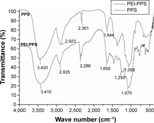
Purified PPS was made in our laboratory. Fourier-transform infrared (FTIR) analysis was carried out to characterize the CPPS through at least three repeated operations. A qualitative functional group analysis by FTIR (KBr) of PEI-modified PPS yielded peaks at 3,410, 1,650, 1,250, and 1,070 cm−1. In , compared with the spectrum of PPS, the strong absorption peak at 3,410 cm−1 was assigned to –NH stretching. The absorption peak at 2,935 cm−1 of PEI-PPS, which was stronger than that in PPS, was the absorption peak of methylene. The absorption peak at 1,650 cm−1 indicated the in-plane and out-of-plane bending vibration of β-amides. In addition, the absorption peaks at 1,250 and 1,070 cm−1 were significantly stronger than those in PPS, which we attributed to C–N stretching. All of the absorption peaks demonstrated that PPS was successfully cationized by PEI through the connection between –CO–NH– and R2NH of PEI.
Figure 3 Fourier-transform infrared spectra of Porphyra yezoensis polysaccharide (PPS) and polyethylenimine (PEI)-PPS.
Agarose gel electrophoresis assay
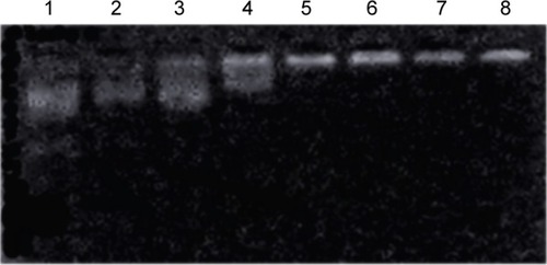
The combination of CPPS and plasmid DNA was evaluated at various CPPS:pWnt3a weight ratios (5:1, 10:1, 20:1, 40:1, 80:1, 100:1, and 150:1) with an agarose gel retardation assay, which was conducted in triplicate (). An increase in the CPPS:pWnt3a weight ratio retarded the DNA migration. No retardation was detected for the complexes at CPPS:pWnt3a weight ratios of 5:1 or 10:1 (lanes 2 and 3 in ). Partial migration of the plasmids was observed with the complex that was prepared at a CPPS:pWnt3a weight ratio of 20:1 (lane 4 in ). In contrast, no plasmid migration occurred when the CPPS:pWnt3a weight ratio reached 40:1, which illustrated that the plasmids were retarded completely. This result indicated that the firmness of the combination of CPPS and DNA plasmid was highly associated with the content of the CPPS in the CPPS-pWnt3a nanoparticles. A higher CPPS:pWnt3a weight ratio resulted in a superior combination of CPPS and pWnt3a.
Figure 4 Electrophoretic mobility of plasmid in cationized Porphyra yezoensis polysaccharide (CPPS)–plasmid Wnt3a (pWnt3a) nanoparticles at various ratios.
Notes: Lane 1, Wnt3a naked plasmid; lanes 2–8, CPPS:pWnt3a weight ratios of 5:1, 10:1, 20:1, 40:1, 80:1, 100:1, and 150:1, respectively.
Cytotoxicity assay of CPPS-pWnt3a nanoparticles
Low cytotoxicity is crucial for evaluating the safety of a gene carrier during in vitro transfection. Based on the results of the gel retardation assay, the CPPS:pWnt3a weight ratio for testing was set from 40:1 to 300:1. In , the MTT assay revealed that a higher CPPS:pWnt3a ratio led to increased cytotoxicity. No obvious differences in cytotoxicity occurred at CPPS:pWnt3a weight ratios of 40:1 and 80:1 compared to the free pWnt3a group, which was used as a negative control. However, in the Wnt3a group, cell viability was slightly higher than 100%, which may have been due to experimental errors. Meanwhile, as for the MCPPS/MpWnt3a (40:1) group, the increased cell viability might have been due to the excellent cell compatibility of the cationic polysaccharide, which may favor cell proliferation to a certain extent. In addition, the percentage of viable cells at a CPPS:pWnt3a ratio of 100:1 and 150:1, higher ratios than for the positive control Lipofectamine 2000, was distinctly lower than that in CPPS:pWnt3a weight ratios of 40:1 and 80:1. In addition, the PEI group displayed a continuous increase in cell viability with a reduction in PEI concentration, which indicated that PEI cannot be used alone as a gene vector for gene delivery because of its high cytotoxicity.
Figure 5 Cytotoxicity assay.
Notes: Bar 1, Lipofectamine 2000 (Lip2000)–plasmid Wnt3a (pWnt3a); bars 2–8, cationized Porphyra yezoensis polysaccharide (CPPS)-pWnt3a nanoparticles at ratios of 40:1, 80:1, 100:1, 150:1, 200:1, 250:1, and 300:1 from left to right, respectively; bar 9, free pWnt3a (control group); bars 10–14, multiple dilutions of PEI – 50-fold, 100-fold, 200-fold, 400-fold, and 800-fold from left to right, respectively. Data presented as average values (means) ± standard deviation of measurements from three replicates.
Abbreviation: PEI, polyethylenimine.
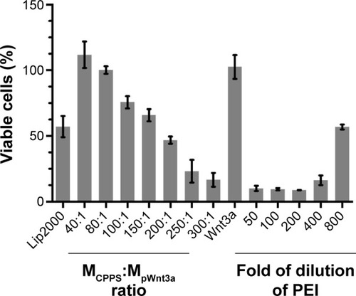
A possible reason for this result is that PEI, which is frequently utilized for cationic modification, might induce the overexpression of caveolin 1, which could partially determine the transfection efficiency and the cytotoxicity of PEI.Citation51 Similarly, with an increase in the CPPS:pWnt3a weight ratio, the concentration of PEI also increased during the gene transfection, which led to a gradual increase in cytotoxicity.
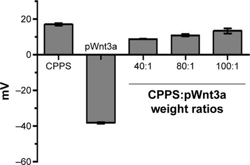
Zeta-potential measurement
Zeta-potential measurement is a common method to test the ability of a gene vector to combine with the negatively charged plasmid. In addition, positively charged nanoparticles may easily come into contact with negatively charged cytomembrane. According to the results of the gel retardation assay and the cytotoxicity test, the zeta potential of the nanoparticles, which were prepared at three different CPPS:pWnt3a weight ratios (40:1, 80:1, and 100:1), was measured with the Nano ZS90 Zetasizer. The zeta potential of the original CPPS and the free pWnt3a was also investigated under the same conditions. shows that the zeta potential of the original CPPS and Wnt3a plasmids was +16.9±0.6 and −38.7±1.5 mV, respectively. After cationic modification, the zeta potential of the nanoparticles, which were prepared at three different CPPS:pWnt3a weight ratios (40:1, 80:1, and 100:1), changed from the negative value of −38.7±1.5 mV to a positive value, and increased to 8.7±0.1, 10.8±0.3, and 13.3±1.0 mV, respectively. An increase in the amount of CPPS leading to an increased positive charge density provided solid evidence for the successful cationization of PPS.
Figure 6 Zeta-potential results.
Notes: Bar 1, cationic Porphyra yezoensis polysaccharide (CPPS); bar 2, naked plasmid Wnt3a (pWnt3a); bars 3–5, CPPS-pWnt3a nanoparticles prepared at various CPPS:pWnt3a weight ratios – 40:1, 80:1, and 100:1 from left to right, respectively (means ± standard deviation of measurements from three replicates).
Morphology and particle size of CPPS-pWnt3a nanoparticles
The size of the nanoparticles with CPPS:pWnt3a weight ratios of 40:1, 80:1, and 100:1 was investigated with the ZS90. The particle diameters, as shown in , increased from 167.5±6.2 to 341.7±7.2 nm when the CPPS:pWnt3a weight ratio was increased from 40:1 to 100:1. The complex that was prepared at a CPPS:pWnt3a ratio of 40:1 obviously had the smallest particles. This could be attributed to the excessive amount of CPPS that resulted in a loose combination with the plasmid, which enlarged the nanoparticles. When the nanoparticles were prepared at a CPPS:pWnt3a weight ratio of 40:1, the particle size was significantly smaller than for the other ratio groups, which indicated that a CPPS:pWnt3a weight ratio of 40:1 might be optimal for gene transfection.
Figure 7 Characterization of cationized Porphyra yezoensis polysaccharide (CPPS)–plasmid Wnt3a (pWnt3a) nanoparticles.
Notes: (A) Transmission electron microscopy image of the nanoparticles at a CPPS:pWnt3a weight ratio of 40:1. (B) Size distribution of the nanoparticles at CPPS:pWnt3a weight ratios of 40:1, 80:1, and 100:1, respectively. (C) Particle-size distribution of the nanoparticles at a CPPS:pWnt3a weight ratio of 40:1.
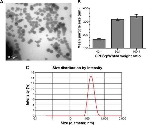
TEM was used to analyze the morphology and particle size of nanoparticles at a CPPS:pWnt3a weight ratio of 40:1. As shown in , most of the nanoparticles fell within a narrow range of 100–200 nm, which agreed with particle size range 167.5±6.2 nm by DLS, as show. The CPPS-Wnt3a nanoparticles prepared at a CPPS:pWnt3a weight ratio of 40:1 exhibited a uniform spherical shape, which may be conducive to cell endocytosis during transfection.Citation52,Citation53
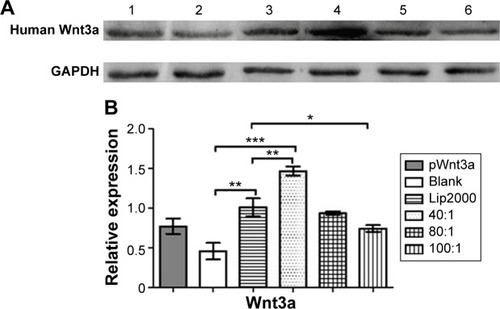
In vitro transfection experiment
To assess transfection efficiency, CPPS-pWnt3a and CPPS-pGFP nanoparticles were prepared at three different ratios (40:1, 80:1, and 100:1). The result of pGFP transfection was analyzed by fluorescence microscopy (TI-U; Nikon, Tokyo, Japan). Lipofectamine 2000 was set as a positive control based on the manufacturer’s instructions, while HUMSCs with and without free pGFP transfection acted as negative controls. Meanwhile, the Wnt3a expression was tested by Western blotting. Lipofectamine 2000 was used as a positive control according to the manufacturer’s instructions, and the Wnt3a naked plasmid (4 μg/well) was employed as the negative control.
From , it is clear that the fluorescence intensity was at the top in the 40:1 group. Meanwhile, as shown in , ample cell debris indicated that the cytotoxicity of the Lipofectamine 2000 group was also higher than the other groups involved, which agreed with the results of cytotoxicity assay. As demonstrate, the gene-transfection efficiency and Wnt3a expression were different for the various CPPS:pWnt3a weight ratios. The optimal Wnt3a-expression level was observed at a CPPS:pWnt3a weight ratio of 40:1. Furthermore, the transfection efficiency of the CPPS/pWnt3a nanoparticles prepared at a 40:1 weight ratio was significantly higher than that for both the 80:1 and 100:1 weight ratios (both P<0.0001, Student’s t-test). One possible reason for this result is that excess CPPS reduced the dissociation of the plasmid due to the strong bonds between the cationic carrier and the plasmid. In addition, the high cytotoxicity of a high dose of PEI could also have caused low transfection efficiency. Damage to the cell membrane caused by cationic carriers can also impact endocytosis, which is an important mechanism for gene delivery. Moreover, the transfection efficiency of Lipofectamine 2000, which was not significantly different from CPPS/pWnt3a at a weight ratio of 80:1, was obviously lower than that of CPPS/pWnt3a at a weight ratio of 40:1. This result indicated that CPPS/pWnt3a, rather than Lipofectamine 2000, could be a potential gene carrier for gene delivery.
Figure 8 Observation of human umbilical cord mesenchymal stem cells transfected with plasmid GFP (pGFP).
Notes: (A) Free pGFP; (B) untreated cells (blank control); (C) Lipofectamine 2000–plasmid Wnt3a (pWnt3a); (D–F) cationized Porphyra yezoensis polysaccharide-pWnt3a nanoparticles prepared at 40:1, 80:1, and 100:1, respectively.
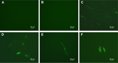
Figure 9 Protein-expression measurement.
Notes: (A) Western blotting indicated the expression of the Wnt3a protein, using GAPDH protein as the control. Lane 1, free plasmid Wnt3a (pWnt3a); lane 2, human umbilical cord mesenchymal stem cells (HUMSCs) without transfection; lane 3, Lipofectamine 2000 (Lip2000)-pWnt3a; lanes 4–6, HUMSCs transfected with cationized Porphyra yezoensis polysaccharide-pWnt3a nanoparticles prepared at 40:1, 80:1, and 100:1, respectively. (B) A quantitative analysis of the relative expression levels of Wnt3a (means ± standard deviation of measurements from three replicates). *P<0.05; **P<0.01; ***P<0.001; Student’s t-test.
Abbreviation: GAPDH, glyceraldehyde-3-phosphate dehydrogenase.
Conclusion
In this study, we successfully created CPPS-pWnt3a nanoparticles, a novel nonviral gene vector, based on a PEI-modified polysaccharide from P. yezoensis. FTIR analyses and gel retardation assay confirmed the successful cationic modification of the original PPS. In addition, a cytotoxicity assay also showed that the 40:1 CPPS-pWnt3a nanoparticles were the least cytotoxic compared to Lipofectamine 2000 and PEI (25 kDa). The result of the cell-transfection experiment demonstrated that the optimal CPPS:pWnt3a weight ratio for gene delivery was 40:1, which was also consistent with the particle size and shape as revealed by TEM. Together, these findings indicated that CPPS could become a promising nonviral gene vector for gene delivery.
Acknowledgments
This work was supported by the National Natural Science Foundation of China (81072586, 81273470, and 81473172), the Doctoral Fund of the Ministry of Education of China (20123227110009), Special Funds for 333 Projects (BRA2013198), and industry–university–research institution cooperation (JHB2012-37, GY2012049, and GY2013055) in Jiangsu Province and Zhenjiang. This was a project funded by the Priority Academic Program Development of Jiangsu Higher Education Institutions.
Disclosure
The authors report no conflicts of interest in this work.
References
- TakahashiKHiranoYArakiSHattoriMEmulsifying ability of porphyran prepared from dried nori, Porphyra yezoensis, a red algaJ Agric Food Chem20004872721272510898612
- IsakaSChoKNakazonoSAntioxidant and anti-inflammatory activities of porphyran isolated from discolored nori (Porphyra yezoensis)Int J Biol Macromol201574687525499893
- WangJZhangQZhangZSongHLiPPotential antioxidant and anticoagulant capacity of low molecular weight fucoidan fractions extracted from Laminaria japonicaInt J Biol Macromol201046161219883681
- RupérezPAhrazemOLealJAPotential antioxidant capacity of sulfated polysaccharides from the edible marine brown seaweed Fucus vesiculosusJ Agric Food Chem200250484084511829654
- MayerAMSHamannMTMarine pharmacology in 1999: compounds with antibacterial, anticoagulant, antifungal, anthelmintic, anti-inflammatory, antiplatelet, antiprotozoal and antiviral activities affecting the cardiovascular, endocrine, immune and nervous systems, and other miscellaneous mechanisms of actionComp Biochem Physiol C Toxicol Pharmacol2002132331533912161166
- SakaiSKomuraYNishimuraYSugawaraTHirataTInhibition of mast cell degranulation by phycoerythrin and its pigment moiety phycoerythrobilin, prepared from Porphyra yezoensisFood Sci Technol Res2011172171177
- ShinESHwangHJKimIHNamTJA glycoprotein from Porphyra yezoensis produces anti-inflammatory effects in liposaccharide-stimulated macrophages via the TLR4 signaling pathwayInt J Mol Med201128580981521701768
- CaoJWangSYaoCXuZXuXHypolipidemic effect of porphyran extracted from Pyropia yezoensis in ICR mice with high fatty dietJ Appl Phycol. Epub201565
- KitajimaHKomizuYIchiharaHImmunostimulatory effects of extract from nori (Porphyra yezoensis) in vitro and in vivoKagaku Kogaku Ronbunshu2013394359362
- KwonMJNamTJChromatographically purified porphyran from Porphyra yezoensis effectively inhibits proliferation of human cancer cellsFood Sci Biotechnol2007166873878
- ParkSJRyuJKimIHChoiYHNamTJActivation of the mTOR signaling pathway in breast cancer MCF627 cells by a peptide derived from Porphyra yezoensisOncol Rep2015331192425333576
- KlinkDSchindelhauerDLanerAGene delivery systems – gene therapy vectors for cystic fibrosisJ Cyst Fibros20043Suppl 220321215463959
- HanSMahatoRISungYKKimSWDevelopment of biomaterials for gene therapyMol Ther20002430231711020345
- de Carvalho RodriguesDAsensiKDVairoLHuman menstrual blood-derived mesenchymal cells as a cell source of rapid and efficient nuclear reprogrammingCell Transplant201221102215222422776164
- FujikuraJNakaoKSoneMInduced pluripotent stem cells generated from diabetic patients with mitochondrial DNA A3243G mutationDiabetologia20125561689169822396012
- KaneNMNowrouziAMukherjeeSLentivirus-mediated reprogramming of somatic cells in the absence of transgenic transcription factorsMol Ther201018122139214520978477
- ShaoLFengWSunYGeneration of iPS cells using defined factors linked via the self-cleaving 2A sequences in a single open reading frameCell Res200919329630619238173
- WangWWWeiWYanJReprogramming of mouse renal tubular epithelial cells to induced pluripotent stem cellsCytotherapy201315557858523415920
- MoriguchiHChungRTMiharaMSatoCGeneration of human induced pluripotent stem cells from liver progenitor cells by only small moleculesHepatology2010523116920812362
- TakahashiKTanabeKOhnukiMInduction of pluripotent stem cells from adult human fibroblasts by defined factorsCell2007131386187218035408
- TakahashiKYamanakaSInduction of pluripotent stem cells from mouse embryonic and adult fibroblast cultures by defined factorsCell2006126466367616904174
- YuJVodyanikMASmuga-OttoKInduced pluripotent stem cell lines derived from human somatic cellsScience200731858581917192018029452
- ChoiJWJungSJKasalaDpH-sensitive oncolytic adenovirus hybrid targeting acidic tumor microenvironment and angiogenesisJ Control Release201520513414325575865
- LeeCHKimHWKimTLeeSWRecombinant adenovirus infection suppresses hTERT expression through virus-associated RNA-mediated induction of type 1 interferonBiochem Biophys Res Commun2015458483083525689720
- Rodríguez-GarcíaAGiménez-AlejandreMRojasJJSafety and efficacy of VCN-01, an oncolytic adenovirus combining fiber HSG-binding domain replacement with RGD and hyaluronidase expressionClin Cancer Res20152161406141825391696
- PendariesVGascGTiteuxMImmune reactivity to type VII collagen: implications for gene therapy of recessive dystrophic epidermolysis bullosaGene Ther201017793093720376098
- McmenaminMMWoodMJProgress and prospects: immunobiology of gene therapy for neurodegenerative disease: prospects and risksGene Ther201017444845820147982
- SvahnMGLundinKEGeRAdding functional entities to plasmidsJ Gene Med20046Suppl 1S36S4414978749
- BacharachEGonskyJLimDGoffSPDeletion of a short, untranslated region adjacent to the polypurine tract in Moloney murine leukemia virus leads to formation of aberrant 5′ plus-strand DNA ends in vivoJ Virol200074104755476410775614
- SomanathanSAAV vectors avoid inflammatory signals necessary to render transduced hepatocyte targets for destructive T cellsMol Ther201018597798220234342
- AtkinsonHChalmersRDelivering the goods: viral and non-viral gene therapy systems and the inherent limits on cargo DNA and internal sequencesGenetica2010138548549820084428
- YuGBorlonganCVOuYIn vitro non-viral Lipofectamine delivery of the gene for glial cell line-derived neurotrophic factor to human umbilical cord blood CD34+ cellsBrain Res20101325114715420171195
- GadiJRuthalaKKongKAParkHWKimMHThe third helix of the Hoxc8 homeodomain peptide enhances the efficiency of gene transfer in combination with LipofectamineMol Biotechnol2009421414818991027
- KimHKDavaaEMyungCSParkJSEnhanced siRNA delivery using cationic liposomes with new polyarginine-conjugated PEG-lipidInt J Pharm20103921–214114720347025
- DavaaEKangBSHanJHCombined delivery of the adiponectin gene and rosiglitazone using cationic lipid emulsionsInt J Pharm20154831–212413025681724
- TanasienkoIVYemetsAIFiniukNSStoikaRRBlumeYBDMAEM-based cationic polymers as novel carriers for DNA delivery into cellsCell Biol Int201539324324525234366
- SlitaAVKasyanenkoNANazarovaOVDNA-polycation complexes: effect of polycation structure on physico-chemical and biological propertiesJ Biotechnol2007127467969316934901
- FilionMCPhillipsNCMajor limitations in the use of cationic liposomes for DNA deliveryInt J Pharm19981621–2159170
- DengWWCaoXWangMEfficient gene delivery to mesenchymal stem cells by an ethylenediamine-modified polysaccharide from mulberry leavesSmall20128344145122213679
- WangMDengWFuMEfficient gene transfer into rat mesenchymal stem cells with cationized Lycium barbarum polysaccharides nanoparticlesCarbohydr Polym201186415091518
- DengWFuMCaoYAngelica sinensis polysaccharide nanoparticles as novel non-viral carriers for gene delivery to mesenchymal stem cellsNanomedicine2013981181119123727125
- DengWWCaoXWangMDelivery of a transforming growth factor β-1 plasmid to mesenchymal stem cells via cationized Pleurotus eryngii polysaccharide nanoparticlesInt J Nanomedicine201271297131122457592
- CaoXDengWQuRNon-viral co-delivery of the four Yamanaka factors for generation of human induced pluripotent stem cells via calcium phosphate nanocomposite particlesAdv Funct Mater2013234354035411
- YangXTBiYYChenETFengDFOverexpression of Wnt3a facilitates the proliferation and neural differentiation of neural stem cells in vitro and after transplantation into an injured rat retinaJ Neurosci Res201492214816124254835
- DavidsonKCJamshidiPDalyRHearnMTPeraMFDottoriMWnt3a regulates survival, expansion, and maintenance of neural progenitors derived from human embryonic stem cellsMol Cell Neurosci200736340841517822920
- Lee-KubliCALuPInduced pluripotent stem cell-derived neural stem cell therapies for spinal cord injuryNeural Regen Res2015101101625788906
- LiXSaikiCIdeRStem cell therapy for central nerve system injuries: glial cells hold the keyNeural Regen Res20149131253126025221575
- NicolasGFranklinRJJefferyNDCell therapy for spinal cord injuries: what is really going on?Neuroscientist201420662363824415275
- MatsuiTAkamatsuWNakamuraMOkanoHRegeneration of the damaged central nervous system through reprogramming technology: basic concepts and potential application for cell replacement therapyExp Neurol20142605121823036600
- XuXCapitoRMSpectorMDelivery of plasmid IGF-1 to chondrocytes via cationized gelatin nanoparticlesJ Biomed Mater Res A2008841738317600330
- YangHJPeiFLeiWCaveolin-1 mediates gene transfer and cytotoxicity of polyethyleneimine in mammalian cell linesMol Cell Biochem20154021–220321125626893
- XuXCapitoRMSpectorMPlasmid size influences chitosan nanoparticle mediated gene transfer to chondrocytesJ Biomed Mater Res A200884A41038104817685397
- MacLaughlinFCMumperRJWangJChitosan and depolymerized chitosan oligomers as condensing carriers for in vivo plasmid deliveryJ Control Release1998561–32592729801449