
Abstract
Obstructive jaundice as an initial manifestation of uterine cancer is extremely rare. We present a case of a 72-year-old female who presented with obstructive jaundice, supposedly for pancreatic cancer. After detailed diagnostic investigation, the cause of the jaundice was attributed to a metastatic compression of the common bile duct, from the primary neoplasm of the uterus. This case highlights the importance of including uterine cancer in the differential diagnosis of woman presenting with obstructive jaundice, even though it is very rare.
Introduction
The majority of the tumors that can cause obstructive jaundice originate from pancreatic, biliary, or periampullary sites.Citation1–Citation2 There are other tumors that cause external compression of the biliary channels resulting in obstructive jaundice, the most frequent are primary carcinomas of the stomach, colon, rectum, esophagus, kidney, and lung.Citation1–Citation3 Pancreatic metastasis from a primary cancer of the uterus cervix is extremely rare with few cases reported.Citation4–Citation9
In this report, we present a case of obstructive jaundice initially attributed to pancreatic cancer. Detailed radiological, pathological, and laboratory investigation clarify that the cause of the obstructive jaundice was metastases from a primary malignant cancer of the uterus.
Case report
A 72-year-old female with no history of cancer or gallstones presented with complaints of pain in the upper right abdomen and yellowish discoloration of the eyes and skin, for the past 6 months. She reported weight loss of 10 kg. On clinical examination, vitals signs were stable, icterus was present, and no peripheral lymphadenopathy was observed. The abdomen was slightly distended but smooth, and the gall bladder was palpable on the right upper quadrant of the abdomen.
Laboratory data were total bilirubin 4.8 mg/dL; direct bilirubin 4.3 mg/dL; indirect bilirubin 0.5 mg/dL; alkaline phosphatase 434 U/L; gamma-glutamyltransferase 744 U/L; alanine aminotransferase 303 U/L; aspartate aminotransferase 604 U/L; lipase 78 U/L; and amylase 101 U/L. Tumor markers were CA125 122 U/mL; CA15.3 60 U/mL; CA19.9 394 U/mL; and CA 72.49 U/mL.
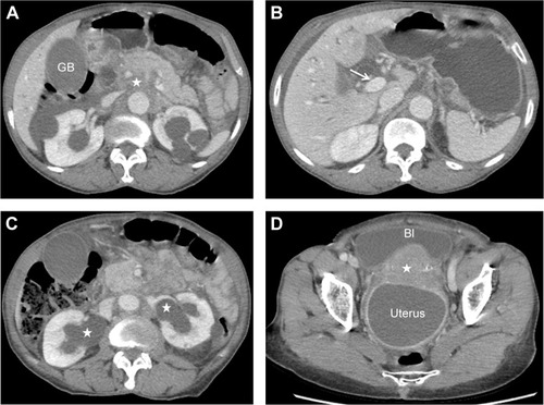
Abdominal and pelvis computed tomography (CT) scan revealed retropancreatic and periaortic images suggestive of lymphadenomegaly involving the distal choledocus (). A solid mass in the uterus cervix without cleavage plane with the posterior wall of the bladder was also observed. This tumor was involving the distal ureters resulting in bilateral hydronefrosis (). A cystic formation with hypodense content matching with distended uterine cavity containing mucus/old hematic material, causing displacement of the bladder, and compression on the upper rectum and distal sigmoid (), was also noted. There was no evidence of peritoneal carcinomatosis or involvement of other organs.
Figure 1 Abdominal and pelvis computed tomography scans.
Vaginal examination confirmed a large mass in the uterine cervix. Transvaginal ultrasound revealed a heterogeneous cervix without endocervical canal evidence and a uterine cyst with a thick content inside. The biopsy from the cervix showed an invasive squamous-cell carcinoma, moderately differentiated. A diagnosis of primary neoplasm of the cervix stage IIIB/IV was established.
An endoscopic retrograde cholangiopancreatography (ERCP) and drainage of the choledocus with endoprothesis was performed. The patient’s cholestasis improved and she was referred for further oncological treatment.
Discussion
Obstructive jaundice can be caused by compression of the bile ducts due to intra- or extra-hepatic lesions. Extra-hepatic causes are divided into intra-ductal and extra-ductal etiologies. Neoplasms, choledocholithiasis, biliary strictures, parasites, and primary sclerosing cholangitis lead the intra-ductal obstruction causes. Tumors involving the pancreas, biliary, or periampullary region and cystic duct stone lead the extra-ductal obstruction causes. The majority of the tumors involving the pancreas are primary, or have biliary or periampullary origins.Citation1–Citation8 Metastatic pancreatic cancer is rare, with a reported frequency ranging from 2% to 5% of all pancreatic malignant tumors.Citation1–Citation3,Citation8–Citation10
Metastasis to the pancreas from uterine cancer is an extremely rare cause of obstructive jaundice,Citation4–Citation9 obstructive jaundice as initial manifestation of uterine cancer is the rarest.Citation4–Citation9 In this case, the patient presented due to the jaundice, this sign can confound the diagnosis, mimicking primary pancreatic lesion. Distinguishing primary pancreatic cancer from pancreatic metastasis of cancers arising elsewhere in the body is not easy.Citation2,Citation4 Further investigation including ultrasound imaging, CT scan, magnetic cholangioresonance, ERCP, percutaneous cholangiography, and endoscopic ultrasound biopsy may be required.Citation3,Citation4,Citation8–Citation10 In this case, the abdominal ultrasound performed at admission was inconclusive. The abdominal CT performed was essential for the diagnosis of pancreatic metastasis and to identify the primary tumor. Although surgical resection of pancreatic metastasis have been reported, there are no guidelines for the management of these patients.Citation2–Citation10 Surgical resection is often advocated for single lesion and for patients with clinical condition to perform a pancreatectomy. The usefulness of pancreatic resection is mainly linked to the biology of the primary tumor metastasizing to the pancreas.Citation2,Citation8 Endoscopic biliary drainage is a palliative approach when surgery is not possible.Citation4 Our patient was submitted to endoscopic biliary drainage and improved of the cholestasis, thus creating a better clinical condition in order to start adjuvant oncological therapy.
Conclusion
The current case clearly shows the importance of high suspicion of uterine cancer in woman presenting with obstructive jaundice, eventhough it is uncommon. Abdominal CT plays a key role in the diagnosis of the primary lesion.
Acknowledgments
The ethics committee of Hospital das Clinicas, School of Medicine, University of São Paulo, approved the study. The participant provided informed written consent.
Disclosure
The authors report no conflicts of interest in this work.
References
- AdsayNVAndeaABasturkOSecondary tumors of the pancreas: an analysis of a surgical and autopsy database and review of the literatureVirchows Archiv20044445273515057558
- SpertiCMolettaLPatanèGMetastatic tumors to the pancreas: the role of surgeryW J Gastro Oncology20146381
- MoonSGHanJKKimTKBiliary obstruction in metastatic disease: thin-section helical CT findingsAbdom Imaging200328455212483383
- LeveyJMEndoscopic biliary drainage for metastatic squamous cell carcinoma of the cervixGastro Endoscopy200051979
- WastellCA solitary secondary deposit in the pancreas from a carcinoma of the cervixPostgraduate Med J1966425961
- MackayBOsborneBMWhartonJTSmall cell tumor of cervix with neuroepithelial features: ultrastructural observations in two casesCancer197943113845427717
- KuwataniMKawakamiHAsakaMPancreatic metastasis from small cell carcinoma of the uterine cervix demonstrated by endoscopic ultrasonography-guided fine needle aspirationDiagn Cytopathol200836840218831029
- OgawaHTsujieMMiyamotoAIsolated pancreatic metastasis from uterine cervical cancer: a case reportPancreas201140797821673546
- NishimuraCNaoeHHashigoSPancreatic metastasis from mixed adenoneuroendocrine carcinoma of the uterine cervix: a case reportCase Rep Oncol201362566223741220
- SmithALOdronicSISpringerBSReynoldsJPSolid tumor metastasis to the pancreas diagnosed by FNA: a single-institution experience and review of the literatureCancer Cytopathology20151233475525828394