Abstract
Objective
Our objective was to determine the accuracy of ultrasound at the time of the fetal anatomy survey in the diagnosis of velamentous cord insertion (VCI).
Study design
This retrospective case–control study identified placentas with VCI (cases) and randomly selected placentas with normal placental cord insertion (PCI) (controls) as documented by placental pathology for mothers delivered from 2002 through 2015. Archived ultrasound images for PCI at the time of the fetal anatomy survey were reviewed. Data analysis was by calculation of sensitivity, specificity, and accuracy and their 95% CI for the ultrasound diagnosis of VCI.
Results
The prevalence of VCI was 1.6% of placentas submitted for pathologic examination. There were 122 cases of VCI and 347 controls with normal PCI. The performance criteria calculated for the diagnosis of VCI at the time of fetal anatomy survey were as follows: sensitivity 33.6%; 95% CI: 25.3, 42.7; specificity 99.7%; 95% CI: 98.4, 99.9 and accuracy 82.5; 95% CI: 80.5, 82.9.
Conclusion
The identification of a VCI at the time of fetal anatomy survey is highly specific for the presence of a VCI as documented by placental pathology. The sensitivity in this study was less than expected. Sensitivity could be improved by reducing the number of nonvisualized PCIs, creating an awareness of risk factors for VCI, and obtaining more detailed images in the case of an apparent marginal PCI.
Introduction
A velamentous cord insertion (VCI) is one in which the umbilical cord inserts into the fetal membranes, rather than directly on the placental disk, causing the umbilical arteries and vein to travel unprotected for varying distances before reaching the placenta. It has been identified in ~1.5% of singleton deliveries when the gross examination is performed by an obstetrician or midwife at the time of delivery.Citation1 The frequency of VCI is much higher in multiple gestations, with a prevalence of 7.6%, 34.7%, and 28.2% in dichorionic twins, monochorionic twins, and triplet pregnancies, respectively.Citation2,Citation3
VCI is associated with both perinatal and maternal morbidity. A VCI coursing over the internal cervical os, vasa previa, estimated to occur in 1 in 50 VCIs, can lead to fetal exsanguination.Citation4 There are increased risks for preterm delivery, small for gestational age (SGA) infants, manual removal of the placenta, postpartum hemorrhage, and intrauterine fetal demise.Citation1,Citation5,Citation6 There are also associations with birth defects, placenta previa, and single umbilical artery (SUA).Citation6–Citation8 These risks highlight the importance of identifying the VCI in the antepartum period.
Ultrasound detects VCI with variable success. In one prospective study of 832 women referred to ultrasound ≥16 weeks, the placental cord insertion (PCI) was able to be identified in 825; 8 VCIs were identified and of these 7 proved to have a VCI at delivery.Citation9 In another prospective study, Hasegawa et alCitation10 identified 25 out of 40 VCIs (sensitivity 62.5%) among 3,446 routine ultrasounds performed at 18–20-week gestation with no false positives. In our maternal-fetal medicine unit PCI identification is a routine part of every fetal anatomy survey; thus, a large number of archived images are available for ultrasound and placental pathology correlation. We chose to study the accuracy of ultrasound for the diagnosis of VCI in a retrospective case–control study of placentas that had been previously submitted for examination to pathology where the site of the PCI was well documented. This design differs from prospective studies but was necessary as clinician-performed gross placental examination is not done with sufficient detail nor reliably and consistently documented at our institution. VCI recorded at the time of pathologic examination of the placenta was considered the gold standard and this would also allow us to obtain a large sample of VCIs to study. We hypothesized, based on the studies referenced above and our own clinical experience, that fetal anatomy survey would have high specificity but lower sensitivity for the diagnosis of VCI.
Methods
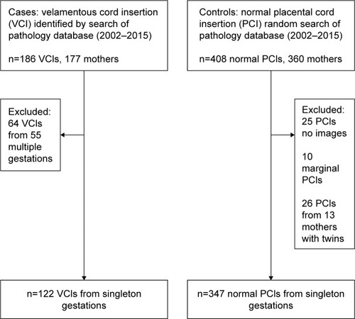
This was a retrospective case–control study of placentas submitted for pathologic examination. The study was approved by the Research Subjects Review Board at the Penn State Milton S Hershey Medical Center (IRB protocol no. 43017EP). A waiver of informed consent was granted, in accordance with U.S. Federal regulation 45 CFR Part 46.116(d): the research involved minimal risk to subjects; the waiver would not adversely affect the rights and welfare of the subjects; the research could not practically be carried out without the waiver. A waiver was granted for authorization to access protected health information, in accordance with U.S. Federal regulation 45 CFR Part 164. These waivers were in accordance with the Declaration of Helsinki document on human research ethics. The gold standard for VCI diagnosis was placental pathology. The study began with the identification of the cases, VCI group, recorded on the pathology report from placentas examined from deliveries between 2002 and 2015 at Milton S. Hershey Medical Center. The indication for pathologic placental examination was at the obstetrician’s discretion, which in general followed established guidelines.Citation11 Briefly, these are College of American Pathologists’ guidelines that list indications for placental examination in three broad categories: maternal, fetal/neonatal, and placental. Preeclampsia, multiple gestation, and VCI, as examples, would fall into the categories of maternal, fetal, and placental indications, respectively, for examination of the placenta. The obstetrician-recorded indication on the pathology report was categorized into maternal, fetal/neonatal, placenta, or other, if it did not meet the three criteria for examination, for each patient. Pregnancies that have pathologic examination at our institution are predominantly those that are classified as high risk; every placenta is not submitted for evaluation. Historically, at Penn State Milton S Hershey Medical Center 40%–50% of all placentas have a pathologic examination. Placental examination search was performed with the use of Cerner CoPath Plus Anatomic Pathology (Cerner, Kansas City, MO, USA). The controls, the normal PCI group, were selected randomly during the same time period. We extracted all placental report requisition numbers in the pathology database from 2002 to 2015 into a numbered spread sheet followed by the use of a random number generator to select the normal PCI group. We evaluated the placental pathology report’s gross description section for a thorough documentation of PCI. A cord insertion directly onto the placental disk and >1 cm from the nearest placental edge was considered a normal PCI and classified into the normal PCI group. A clear description of the umbilical cord insertion site off of the placental disk and into the fetal membranes qualified as a VCI, and the distance the cord inserted off of the placental disk in centimeters was also sought, when available, but an exact measurement was not required for a diagnosis. Cord insertions described as marginal on gross pathology of the placenta were excluded from the study. Also excluded were cases and controls with no archived ultrasound studies available for review. We ultimately chose to exclude multiple gestations from the final analysis.
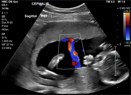
Ultrasound images and reports were accessed through the Philips IntelliSpace PACS Enterprise (Koninklijke Philips NV, Amsterdam, the Netherlands). Clinical data were extracted through the use of the electronic medical record, Cerner Millennium (Cerner Health Systems, Malvern, PA, USA). Ultrasound identification of the PCI at the fetal anatomic survey was performed with 2–5 MHz curvilinear transducers using the Philips iU22 or EPIQ machines (Koninklijke Philips NV). All ultrasounds were performed by experienced sonographers in maternal-fetal medicine. Identification and imaging of the PCI were part of all fetal anatomy surveys regardless of whether they were for routine screening or a detailed, also referred to as targeted, examination. Images were acquired in sagittal and transverse planes with the aid of color flow Doppler. All images were reviewed by two of the researchers (WMC, JMH). Images showing the insertion of the umbilical cord beyond the placental edge and into the extraplacental membranes verified the diagnosis of VCI. We measured the distance off the placental disk where possible. Images showing the PCI >1 cm from the placental edge were designated normal PCI and images showing the PCI ≤1 cm from the placental edge but still on the placental disk were designated as marginal PCI.Citation12 Representative examples are given in and 2. If there was a follow-up ultrasound study where the PCI was subsequently visualized that had not been visualized at the prior fetal anatomy survey this was included in the data. It was not the usual practice to reimage the PCI on subsequent studies if it had already been identified. Studies where the umbilical cord was not imaged were considered indeterminate.
Figure 1 (A) Color flow Doppler ultrasound showing velamentous umbilical cord inserting into fetal membranes, 2.19 cm off of placental disk for twin B in a monochorionic, diamniotic twin pregnancy identified at 20-week gestation. (B) Delivered monochorionic, diamniotic twin placenta at 30 weeks from same pregnancy as (A). There is a velamentous insertion of the umbilical cord for twin B (two clamps). Note the division of cord vessels within the fetal membranes and before they join the placental disk at multiple sites along the margin; insufficient imaging can confuse these vessels with a marginal cord insertion. The umbilical cord insertion site of twin A (one clamp) is inserted at the margin of the disk. This pregnancy was complicated by late-onset twin-to-twin transfusion syndrome with twin B, the donor, and twin A, the recipient.

Figure 2 Color flow Doppler ultrasound from singleton pregnancy during a routine fetal anatomy survey showing the PCI to be centrally located on the placental disk.

Maternal variables collected included age, parity, race/ethnicity, assisted reproduction, smoking, diabetes, hypertension, and antepartum bleeding after the first trimester. Additional ultrasound variables collected included gestational age at the time of anatomy survey or follow-up study that subsequently successfully imaged the cord, and placental location. Intrapartum variables collected included the occurrence of abnormal fetal heart rate patterns, retained placenta, gestational age, and mode of delivery. Birth information collected included livebirth/stillbirth, birth weight, SGA (<10th percentile for gestational age), Apgar scores, and the presence of major birth defects. We recorded the presence of a SUA noted on the placental examination.
Data were analyzed between cases and controls by Student’s t-tests for independent comparisons, chi-square tests, OR with 95% CI as appropriate. Statistical analysis was performed using SPSS (Chicago, IL, USA). Significance was set at P<0.05. Multivariate analyses were performed where applicable. The sensitivity, specificity, and accuracy with their 95% CIs were calculated for ultrasound as a diagnostic test for the detection of VCI using placental pathology as the gold standard. We used the Standards for Reporting of Diagnostic Accuracy (STARD) checklist guidelines for the study.Citation13 Using the data from Hasegawa et alCitation10 we hypothesized sensitivity around 63%. We hypothesized a specificity of 90%. We used the tables reported by Flahault et alCitation14 for sample size calculation in diagnostic studies. According to these tables a hypothesized sensitivity of 65% with a lower 95% CI of 50% required 119 VCIs; a hypothesized specificity of 91% with a lower 95% CI of 85% required 319 normal PCIs. We planned to have two normal PCIs for each VCI.
Results
The prevalence of VCI among pathologically examined placentas, including multiple gestations, at our institution was ~186/11,618 (1.6%). There were seven cases of vasa previa (3.8%) among the VCIs. The selection of cases and controls is given in . Overall there were 469 study subjects consisting of 122 singleton pregnancies with VCI (cases) and 347 singleton pregnancies with normal PCI (controls). Indications recorded for placental examination in the 122 case mothers were as follows: 48 (39.3%) maternal, 23 (18.9%) fetal/neonatal, 46 (62.2%) placental, and other 5 (4.1%). Indications recorded for placental examination in the 347 control mothers fell into the following categories: 231 (66.2%) maternal, 68 (19.6%) fetal/neonatal, 28 (8.1%) placenta, and other 20 (5.8%), P<0.001 for all. Maternal comparisons of the VCI and the control groups are given in . The VCI group delivered at a lower mean gestational age and was more likely to be preterm. Pregnancies in the VCI group were more likely to have been conceived by assisted reproduction, to have had vaginal bleeding beyond the first trimester and a retained placenta. VCI was not significantly associated with preterm delivery after adjustment for bleeding beyond the second trimester, parity, smoking, hypertension, diabetes, and race/ethnicity (adjusted OR 1.34; 95% CI: 0.80, 2.22). The cesarean section rate was high among both groups, which is much higher than our institution’s overall rate. This finding reflects that placentas submitted to pathology tend to be from high-risk pregnancies, which have already high cesarean section rates, and that physicians may be more likely to submit placentas for examination from cesarean deliveries than vaginal deliveries.Citation15
Table 1 VCI vs normal PCI, pregnancy comparisons
Newborn and placental data for the two groups are given in . Birth weights were lower in the VCI group but the infants were not more likely to be SGA. There was a higher incidence of birth defects in the VCI group. Information regarding the distance of the cord insertion off the placental disk, as recorded at the time of placental examination, was recorded for 80 VCIs, 5.11±3.69 cm.
Table 2 VCI vs normal PCI, neonatal and placental comparisons
gives the ultrasound data and PCI designation at the time of the fetal anatomic survey. The average gestational age at the time of the fetal anatomy survey for the VCI group was marginally more than for the normal PCI group. The proportion of placentas noted to be low-lying or placenta previa at the time of the fetal anatomy survey were greater in the VCI group. The VCI group was less likely than the control group to have had successful imaging of the PCI location. The classification of the archived images for the VCI group was as follows: 41 VCI, 24 marginal, 44 normal, and 13 indeterminate. In the control group the classification was as follows: 1 VCI, 6 marginal, 328 normal, and 12 indeterminate. The number of PCIs able to be visualized on the initial fetal anatomy survey and those on a subsequent follow-up scan were as follows: VCI group 98 and 11 and controls 325 and 10 (OR 0.27; 95% CI: 0.10, 0.72). PCIs were not reimaged if they had been successfully identified on the initial fetal anatomy survey. A misclassification of marginal PCI on the fetal anatomy survey was more likely in the VCI group than the normal PCI group (OR 13.92; 95%: CI: 5.23, 39.22; P<0.001). Overall, ultrasound detected 41 (33.6%) of 122 VCIs. There was one false-positive VCI among the normal PCIs. We measured the distance of the cord insert off the placental disk for the 43 VCIs that were observed on ultrasound, 3.13±1.58 cm. Of the 28 cases that had both an ultrasound measurement and a placental measurement of this distance the correlation was rs =0.455, P=0.015.
Table 3 Characteristics of umbilical cord identification at the time of fetal anatomy survey
gives the performance characteristics of ultrasound at the time of the fetal anatomy survey for the diagnosis of VCI on placental examination. The sensitivity of the identification of a VCI at the time of fetal anatomy survey is limited but the specificity is high. Overall, the location of the PCI is correctly classified 82.5% of the time. The adjusted positive predictive and negative predictive values were calculated based on a 1.6% population prevalence of VCI.
Table 4 Performance characteristics of fetal anatomy survey in the diagnosis of VCI
Discussion
VCI is an abnormal PCI that can be identified at the time of the fetal anatomy survey. In this retrospective case–control study, where PCI was a routine part of the fetal anatomy survey, the detection rate of VCI was 33.6%. This is significantly lower than expected based on previous prospective studies specifically targeting the PCI that report 60%–90% sensitivity.Citation9,Citation10 The reason for the discrepancy of the results between ours and that of the others is likely related to the latter’s prospective design specifically focusing on the PCI, the limited numbers of VCIs, and the relatively short time periods (8–24 months) of the studies. One studyCitation10 utilized physicians in the technical performance of the ultrasound. We do not feel that our results represent a lack of technical ability on the part of our sonographers. Our reported sensitivity may represent a more realistic value when the PCI is attempted to be visualized in the context of a fetal anatomy survey, where the PCI is just one item in a number of structures to be visualized, with limited time for the overall examination. Guidelines for identification of the PCI on fetal anatomy survey have changed over time. The American Institute of Ultrasound in Medicine (AIUM) document, Guidelines for the Performance of Obstetric Ultrasounds, recommended identification of PCI when technically feasible for the standard fetal examination in the second and third trimesters.Citation16 Eddleman et alCitation17 found that routine nontargeted ultrasound detected none of their 82 cases of VCI at delivery in a four-year period. More recent guidelines from the AIUM recommend identification of the PCI, when possible, on the basic fetal anatomy survey, and PCI identification is now considered to be an integral component of the detailed (targeted) fetal anatomy survey.Citation18 The number of VCI cases identified subsequent to publication of the 2014 AIUM guidelines was small. Although the detection of VCI after the 2014 AIUM guidelines was higher, the results were not significant, 7/16 (44%) vs 36/105 (34%) (P=0.461).
VCI is associated with adverse fetal outcomes; therefore, its antenatal identification could be useful for identification of risk, management of the pregnancy, and timing and conduct of delivery. We found an association of VCI with birth defects in our study. One population-based study showed an increased risk for birth defects with VCI.Citation6 We noted an increased frequency of retained placentas in the VCI group. This happens most likely because of cord avulsion and may be able to be prevented by limited use of cord traction to deliver the placenta. Others have noted the increased rate of manual removal of the placenta, as well as an increased frequency of postpartum hemorrhage.Citation5,Citation19
Antenatal detection of VCI is a worthy endeavor as it has the potential to reduce adverse fetal outcomes. Guidelines recommending the identification of the PCI at the time of all fetal anatomy surveys, whether basic or detailed, may increase the detection of VCI overall. It does appear that there is zero to coincidental detection of the VCI when the PCI is not part of routine screening.Citation17,Citation20 In our study, even with the intent of identifying the PCI, the sensitivity of ultrasound was low; however, the specificity was high. Efforts to improve the detection rate of VCI with routine screening could consist of, first, identifying known risk factors for VCI and second, getting more detailed images when a marginal cord insertion is identified. Multiple gestations and in particular monochorionic placentation have high rates of VCI.Citation2,Citation6 Other risk factors for VCI that we identified in the antenatal period that, if known at the time of the ultrasound, could direct more attention to the PCI are assisted reproduction, bleeding after the first trimester, and failure to identify the PCI. Associations between VCI and assisted reproduction and bleeding during pregnancy have been previously reported.Citation6 The sensitivity of ultrasound for diagnosis of VCI might be improved if further attention is given to imaging in the case of a marginal PCI. The VCI has a fixed insertion into the fetal membrane, and the arteries and veins ramify within the membranes, eventually entering or leaving the placental disk at the margin, which is also fixed. These velamentous branching vessels at the margin of the disk may be mistaken for a marginal cord insertion. Ideally, when a marginal cord insertion is identified the vessels should be followed off the disk until they clearly join the portion of the umbilical cord that floats freely in the amniotic fluid to exclude the presence of a VCI. Kuwata et alCitation21 made an analogy to a mangrove tree, where the trunk of the tree (umbilical cord) is connected to the roots located above ground (velamentous vessels) before they finally enter the soil (placental disk). They called this the “mangrove sign” and provided color Doppler and gross images of the VCI aside an image of a mangrove tree to illustrate their point. Regardless of whether a PCI has a velamentous or marginal insertion, they have a similar risk of adverse perinatal outcomes, albeit to a lesser degree in the case of a marginal insertion, so the ultrasound identification of either prior to delivery would be clinically relevant.Citation6
There are two theories on how a VCI develops: 1) primary abnormal implantation or polarity theory – with implantation of the blastocyst, the embryonic pole is oriented away from the endometrium and the cord insertion occurs in what ultimately becomes the chorion laeve; or 2) trophotropism – the implantation site is unfavorable, for example, a placenta previa, the placenta atrophies at one pole and if the umbilical cord had inserted there it then becomes velamentous.Citation22 The findings we observed of more frequent low-lying placental locations at the time of the fetal anatomy survey and the occurrence of bleeding beyond the first trimester lend support to the theory of trophotropism in the genesis of VCI, whereas the higher frequency of assisted reproduction may support the polarity theory.
Strengths and limitations
The strength of the study was the large numbers of VCIs present that were documented by placental pathology over a number of years. We had a large archive of placental images for correlation with placental pathology. This study may more closely simulate what results would be obtained in a busy ultrasound practice where the PCI is one component, among many, of a fetal anatomy survey. The weaknesses of the study were the usual biases present in retrospective studies, lack of blinding of the ultrasound study reviewers, and only having placentas that underwent pathologic examination, as this group would tend to be a high-risk group. Although ideally it may have been preferable to have had a complete gross description of the placenta and the PCI at the time of deliveries for all deliveries, this data is not routinely collected nor reported upon in a consistent manner at our institution. We also chose to calculate the sensitivity and specificity of ultrasound for VCI without dropping out the cases where the location of the PCI was indeterminate from the images that were archived. This could be perceived as inappropriately lowering the diagnostic ability of ultrasound since in strictest terms this is not a missed diagnosis; however, nonvisualization of the PCI is almost fourfold more likely with a VCI in comparison to a normal PCI and may be an important clue in the diagnosis.
Conclusion
VCI occurred iñ1.6% of all placentas submitted for pathologic examination at our institution. VCI was associated with perinatal and maternal morbidity; thus, its antenatal detection is important. The identification of a VCI at the time of fetal anatomy has high specificity (99.7%) for the presence of VCI at delivery. The sensitivity of fetal anatomy survey for VCI in our study was less than expected (33.6%) and represents an opportunity for improvement. Awareness of risk factors for VCI, such as multiple gestation and assisted reproduction, decreasing the incidence of nonvisualized PCIs, and allowing more technical time in distinguishing a marginal PCI from a VCI, may increase the frequency of antenatal diagnosis and mitigate the associated morbidity.
Disclosure
The authors report no conflicts of interest in this work.
References
- EbbingCJohnsenSLAlbrechtsenSSundeIDVeksethCRasmussenSVelamentous or marginal cord insertion and the risk of spontaneous preterm birth, prelabor rupture of the membranes, and anomalous cord length, a population-based studyActa Obstet Gynecol Scand2017961788527696344
- Costa-CastroTZhaoDPLipaMVelamentous cord insertion in dichorionic and monochorionic twin pregnancies – does it make a difference?Placenta201642879227238718
- FeldmanDMBorgidaAFTrymbulakWPBarsoomMJSandersMMRodisJFClinical implications of velamentous cord insertion in triplet gestationsAm J Obstet Gynecol2002186480981111967512
- QuekSPTanKLVasa praeviaAust N Z J Obstet Gynaecol19721232062094511749
- EsakoffTFChengYWSnowdenJMTranSHShafferBLCaugheyABVelamentous cord insertion: is it associated with adverse perinatal outcomes?J Matern Fetal Neonatal Med201528440941224758363
- EbbingCKiserudTJohnsenSLAlbrechtsenSRasmussenSPrevalence, risk factors and outcomes of velamentous and marginal cord insertions: a population-based study of 634,741 pregnanciesPLoS One201387e7038023936197
- AlbalawiABrancusiFAskinFPlacental characteristics of fetuses with congenital heart diseaseJ Ultrasound Med201736596597228258617
- SuzukiSKatoMClinical significance of pregnancies complicated by velamentous umbilical cord insertion associated with other umbilical cord/placental abnormalitiesJ Clin Med Res201571185385626491497
- SepulvedaWRojasIRobertJASchnappCAlcaldeJLPrenatal detection of velamentous insertion of the umbilical cord: a prospective color Doppler ultrasound studyUltrasound Obstet Gynecol200321656456912808673
- HasegawaJMatsuokaRIchizukaKSekizawaAFarinaAOkaiTVelamentous cord insertion into the lower third of the uterus is associated with intrapartum fetal heart rate abnormalitiesUltrasound Obstet Gynecol200627442542916479618
- LangstonCKaplanCMacphersonTPractice guideline for examination of the placenta: developed by the Placental Pathology Practice Guideline Development Task Force of the College of American PathologistsArch Pathol Lab Med199712154494769167599
- di SalvoDNBensonCBLaingFCBrownDLFratesMCDoubiletPMSonographic evaluation of the placental cord insertion siteAJR Am J Roentgenol19981705129512989574605
- BossuytPMReitsmaJBBrunsDETowards complete and accurate reporting of studies of diagnostic accuracy: the STARD initiativeBMJ20033267379414412511463
- FlahaultACadilhacMThomasGSample size calculation should be performed for design accuracy in diagnostic test studiesJ Clin Epidemiol200558885986216018921
- CurtinWMKraussSMetlayLAKatzmanPJPathologic examination of the placenta and observed practiceObstet Gynecol20071091354117197585
- American Institute of Ultrasound in MedicineAIUM practice guideline for the performance of obstetric ultrasound examinationsJ Ultrasound Med20133261083110123716532
- EddlemanKLockwoodCBerkowitzGLapinskiRBerkowitzRClinical significance and sonographic diagnosis of velamentous umbilical cord insertionAm J Perinatol1992921231261590867
- WaxJMinkoffHJohnsonAConsensus report on the detailed fetal anatomic ultrasound examination: indications, components, and qualificationsJ Ultrasound Med201433218919524449720
- EbbingCKiserudTJohnsenSLAlbrechtsenSRasmussenSThird stage of labor risks in velamentous and marginal cord insertion: a population-based studyActa Obstet Gynecol Scand201594887888325943426
- YerlikayaGPilsSSpringerSChalubinskiKOttJVelamentous cord insertion as a risk factor for obstetric outcome: a retrospective case-control studyArch Gynecol Obstet2016293597598126498602
- KuwataTSuzukiHMatsubaraSThe “mangrove sign” for velamentous umbilical cord insertionUltrasound Obstet Gynecol201240224124222241676
- BenirschkeKAnatomy and pathology of the umbilical cordBenirschkeKBurtonGJBaergenRNPathology of the Human Placenta6th edBerlinSpringer-Verlag2012332336

