
Abstract
Premature ovarian failure (POF) is a common cause of infertility in women, characterized by amenorrhea, hypoestrogenism, and elevated gonadotropin levels in women under the age of 40. Many genes have been identified over the past few years that contribute to the development of POF. However, few genes have been identified that can explain a substantial proportion of cases of POF. The unbiased approaches of genome-wide association studies and next-generation sequencing technologies have identified several novel genes implicated in POF. As only a small proportion of genes influencing idiopathic POF have been identified thus far, it remains to be determined how many genes and molecular pathways may influence idiopathic POF development. However, owing to POF’s diverse etiology and genetic heterogeneity, we expect to see the contribution of several new and novel molecular pathways that will greatly enhance our understanding of the regulation of ovarian function. Future genetic studies in large cohorts of well-defined, unrelated, idiopathic POF patients will provide a great opportunity to identify the missing heritability of idiopathic POF. The identification of several causative genes may allow for early detection and would provide better opportunity for early intervention, and furthermore, the identification of specific gene defects will help direct potential targets for future treatment.
Introduction
The end of a woman’s reproductive lifespan is marked by the occurrence of menopause, defined as being the last menstruation that occurs for a woman, but is caused by the exhaustion of the ovarian reserve.Citation1 In the general female population, across many ethnicities and over recent human history, the average age of natural menopause has remained at 50–52 years. However, aberrations in ovulatory processes, as outlined in , may cause a pathogenic early depletion of ovarian follicles, which may result in premature menopause. Menopause before the age of 40 years is defined as premature ovarian failure (POF), also known as primary ovarian insufficiency.Citation2 POF is described as the premature cessation of ovarian function and is characterized by 4–6 months of amenorrhea, a rise in serum follicle-stimulating hormone (FSH) levels to greater than 40 mIU/L, and hypoestrogenism.Citation2,Citation3 POF is a common disease, occurring in 1% of all women and 0.1% of women below the age of 30 years.Citation2 As a consequence of hypoestrogenism, POF is associated with a greater risk of osteoporosis, osteoarthritis, and cardiovascular disease.Citation4,Citation5 Women suffering from POF experience similar symptoms to natural menopause; however, these symptoms are also accompanied by an earlier loss of fertility. Therefore, women at risk of POF who delay childbearing until after their 30s may experience problems conceiving and carrying a pregnancy to full term.Citation1 This loss of fertility can be due to an accelerated loss of follicles, an inability of the remaining follicles to respond to ovulatory signals, an initially reduced ovarian reserve at the time of birth, or a combination of all.Citation2 As the development of POF occurs without any early discernible signs or symptoms, the diagnosis of POF currently occurs at the end stage of the disease, with a large proportion of these patients having not previously conceived before the time of diagnosis.Citation2
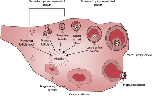
Figure 1 A schematic representation of the process of folliculogenesis in the mammalian ovary from primordial follicles through to follicle ovulation and subsequent corpus luteum formation.
POF is a heterogeneous disease that can develop secondary to many factors such as infection, metabolic disease, autoimmune disorders, or iatrogenic causes such as radiation, chemotherapy, or physical damage to the ovary.Citation6 Additionally, some POF cases are syndromic, where the POF phenotype accompanies a specific disease, such as Turner’s syndrome or blepharophimosis–ptosis–epicanthus inversus syndrome (BPES) type 1. However, 50%–90% of cases are idiopathic and likely involve a substantial genetic contribution.Citation7 A genetic basis of menopause is supported by heritability estimates of menopausal age between mother–daughter pairs ranging from 44% to 65% and recent reports of several genetic loci with significant associations with age at natural menopause identified through genome-wide association studies (GWAS).Citation1,Citation8–Citation10 The importance of genetics in the etiology of POF is supported by the observation that in approximately 10%–30% of idiopathic cases, a first-degree relative is also affected, as well as the observation that a woman with an affected mother is six times more likely to develop POF.Citation4
Genetic analyses of POF patients have identified several chromosomal abnormalities, single gene mutations, and genetic polymorphisms, from multiple different biological pathways, associated with POF development.Citation11 However, the genetic defects investigated thus far have been shown to contribute only a small percentage toward the development of POF.Citation4 The diverse etiology of POF is in line with these findings and suggests that the pathogenesis of nonsyndromic POF is unlikely to be caused by a single gene or genetic defect, but rather supports the view of POF being a heterogeneous genetic disease involving the interaction of multiple genetic defects and environmental factors.Citation1,Citation12 Therefore, current research is focused on increasing the understanding of the genetic basis of idiopathic POF. This information will provide a greater insight into POF pathogenesis and allow for the identification of biomarkers, which may have the ability to predict early onset, providing women at risk with the opportunity to plan their families earlier.
In this review, we aim to provide an overview of the current knowledge of the genetic basis of POF development. To review current literature, we did a descriptive review of the literature based on our existing knowledge in the field and by searching publication databases with the terms “premature ovarian failure”, “primary ovarian insufficiency”, “genetics”, and “mutation”. In addition, we will provide an update on the recent advances in identifying novel genetic factors associated with POF through means of GWAS and next-generation sequencing (NGS).
X-chromosome defects
X-chromosome abnormalities have long been recognized as the most common genetic cause of POF, estimated to account for as much as 12% of cases.Citation13–Citation15 These abnormalities usually involve either whole or partial deletions, duplications, or translocations of the X-chromosome. X monosomy (45X), known as Turner’s syndrome, has an incidence of 1:2,500.Citation16 In phenotypic women, Turner’s syndrome is associated with short stature, gonadal dysgenesis, and primary amenorrhea.Citation16 In women with Tuner’s syndrome, oocyte loss usually begins early in childhood as a result of accelerated follicle atresia.Citation16 Trisomy X (47XXX) is the most common form of aneuploidy, occurring in 1:1,000 females and may manifest in many mosaic forms, ie, 45X/47XXX, 46XX/47XXX, or 47XXX/48XXXX.Citation17 Although most cases do not present with major medical problems, the 47XXX genotype has been associated with hypergonadotropic POF in some cases.Citation17 Cytogenetic analysis in POF patients have identified a “critical region” on the long arm of the X-chromosome, necessary for ovarian development and function.Citation7 Deletions are commonly localized at Xq21.3-q27 (POF1) in POF patients, whereas balanced X: autosomal translocations occur frequently at Xq13.3-q21.1 (POF2).Citation2,Citation18 Although no single gene has been shown to be consistently associated with POF within this critical region, the strong association of POF with X-chromosome abnormalities suggests that X-chromosome genes are critical for normal ovarian function, and any structural abnormality in this region may contribute toward POF development.Citation2,Citation6
Aside from Turner’s syndrome, a premutation in the fragile-X mental retardation (FMR1) gene, located at Xq27.3, is the most commonly known congenital cause of POF.Citation15 The 5′UTR region of the gene contains an expandable CGG repeat region, whereby <45 repeats is considered normal, while >200 repeats is considered a full mutation and is associated with fragile-X syndrome in male carriers.Citation12 Repeats within the range of 55–199 are known as a premutation and are associated with an increased risk of POF in women.Citation19,Citation20 FMR1 premutations have been identified in 6% of sporadic POF cases and up to 13% in familial forms.Citation20 The FMR1 protein is highly expressed in fetal ovary germ cells, as well as in the granulosa cells of maturing follicles.Citation21 There are two prevailing explanations for the association of the FMR1 premutation and POF. The FMR1 protein is a RNA-binding protein, which has a suppressive effect on the translation of a subset of mRNAs, the overexpression of which may impair the expression of oocyte development genes, resulting in a decrease in the size of the initial follicular pool.Citation7,Citation12 Alternatively, it has been proposed that an accumulation of mutated FMR1 mRNA may have a long-term toxic effect on the ovary and lead to excessive follicle atresia.Citation22 Some recent studies have suggested that there is no association between CGG repeats of intermediate size and POF compared with controls; therefore, some of this data may need to be re-evaluated in light of recent findings.Citation23
Candidate genes
Given that the main phenotype of POF is infertility, identifying the causal gene in nonsyndromic POF families has proven difficult, as the majority of these cases have small or no family histories.Citation6 Therefore, researchers have relied on candidate gene-driven studies in order to identify potentially causative genes involved in POF development.Citation2 By screening genes known to play roles in folliculogenesis and ovarian function, as well as genes identified in the use of genetically modified mouse models, several POF candidate genes have been recognized. These genes are summarized in . However, the field has been limited by many underpowered studies, especially when only a single gene or a few gene variants have been studied at once, without providing adequate statistical analysis for the likelihood of the increased chance of identifying a false positive by not correcting for multiple testing.Citation24 In addition, the identification of relevant candidate genes is restricted by our current understanding of disease, which in many cases is limited.
Table 1 POF candidate genes and their functions in relation to POF pathogenesis
Oocyte-specific transcription factors
The initiation of primordial follicle formation and their subsequent differentiation into primary follicles is dependent on the expression of oocyte-specific genes, which are regulated by specific transcription factors.Citation14 The screening of transcription factors regulating folliculogenesis in multiple POF populations has led to the identification of several causative mutations in the NR5A1, NOBOX, FIGLA, and FOXL2 genes.Citation25–Citation27 The nuclear receptor subfamily five group A member 1 (NR5A1), also known as steroidogenic factor 1, is a nuclear receptor located at 9q33.3 and is implicated in early gonadal differentiation.Citation27,Citation28 NR5A1 modulates steroidogenesis via the regulation of genes involved in the hypothalamic–pituitary–steroidogenic axis, such as STAR, CYP11A1, CYP17A1, CYP19A1, LH, and INHA.Citation28 The conditional knockout of NR5A1 in mouse granulosa cells causes infertility, as a result of hypoplastic ovaries and a reduced number of oocytes.Citation29 To date, several NR5A1 mutations have been identified in four families with a history of 46XX POF, as well as in 2/25 idiopathic POF patients, which were absent in the 700 controls.Citation27 All of these mutations resulted in impaired transactivational activity of gonadal promoters regulating follicle growth, thus leading to altered folliculogenesis and subsequent POF.Citation30
Both newborn ovary homeobox (NOBOX) and factor in germline alpha (FIGLA) encode oocyte-specific transcription factors, which regulate oocyte-specific genes necessary for the early stages of folliculogenesis.Citation31 Through the use of knockout mouse models, NOBOX is thought to regulate the expression of a variety of oocyte-specific genes including BMP-15 and GDF-9.Citation31 The deletion of NOBOX expression in these mice results in an accelerated loss of oocytes post-natally, representing a phenotype similar to nonsyndromic POF in humans.Citation28 Two separate studies in Caucasian POF patients have identified several NOBOX mutations, which were absent in the control populations.Citation26 Functional analysis of these mutations revealed a significant decrease in NOBOX DNA binding activity, resulting in the impaired regulation of genes critical for folliculogenesis.Citation26 However, no NOBOX mutations were identified in studies of Chinese and Japanese POF patients, suggesting that NOBOX mutations may be rare in these populations.Citation32,Citation33 FIGLA regulates the expression of zona pellucida genes, the knockout of which in mice also mimics the phenotype of nonsyndromic POF, with an accelerated postnatal loss of primordial follicles.Citation34 A study by Zhao et alCitation35 in 2008 identified two novel FIGLA coding variants in a cohort of 100 Chinese POF women. These variants were absent in the 304 Chinese controls.Citation35 The suggested mechanisms through which these variants led to POF are through haploinsufficiency (p.G6sfsX66), and a dominant negative effect, through altered heterodimerization (p.140delN).Citation35
The forkhead box L2 (FOXL2) gene, located at 3q23, is constitutively expressed primarily in granulosa cells in the ovaries from embryogenesis through to adult life.Citation25 Mutations in the FOXL2 gene are responsible for BPES type 1, of which female carriers also present with POF.Citation6,Citation36 FOXL2 is believed to play a role in granulosa cell differentiation, which is supported by in vitro studies localizing FOXL2 expression to undifferentiated granulosa cells, and through knockout mouse models that show the failure of granulosa cell differentiation, leading to the premature activation and depletion of primordial follicles.Citation37 In addition, FOXL2 mutations have been identified in ~5% of nonsyndromic POF patients (without BPES), suggesting that FOXL2 mutations may also be associated with idiopathic POF.Citation25
More recent studies have highlighted the involvement of the spermatogenesis and oogenesis-specific basic helix–loop–helix (SOHLH) 1 and 2 transcription factors in POF development. SOHLH 1 and 2 are expressed exclusively in primordial follicles up until the primary follicle stage, and are master regulators of oocyte-specific genes critical for early follicle growth and differentiation, including NOBOX, FIGLA, BMP-15, and GDF-9.Citation14 Both SOHLH 1 and 2 knockout female mouse models present with infertility and accelerated follicle loss, due to defects in primordial cell differentiation, with both models exhibiting atrophied ovaries devoid of oocytes by 6 weeks of age.Citation38 A recent study by Qin et alCitation14 identified four novel potentially deleterious heterogeneous SOHLH2 variants in a large cohort of Chinese and Serbian patients with POF. The study suggested that as two of these variants, −210G > T and 530+6T > G, resulted in reduced mature protein production, the primordial to primary follicle transition will be altered, leading to accelerated follicle loss and contributing to the POF phenotype in these patients.Citation14 Additionally, a study conducted by Zhao et alCitation39 identified three novel SOHLH1 variants potentially causative to POF in 364 Chinese that were absent in 400 controls. However, replication studies in larger populations, especially those in different ethnicities, are required to validate a causative association of SOHLH1 and 2 variants with POF.
Folliculogenesis growth factors
Bone morphogenetic protein 15 (BMP-15), located at Xq11.2, is a member of the transforming growth factor β (TGF-β) superfamily and encodes an oocyte-specific growth and differentiation factor.Citation40 BMP-15 is involved in stimulating folliculogenesis and promoting follicle maturation by regulating granulosa cell differentiation and proliferation.Citation41 The essential role of BMP-15 in folliculogenesis is illustrated by knockout mice models that present with subfertility due to defective ovulation processes.Citation42 Many heterozygous variants in the BMP-15 gene proregion, essential for proprotein dimerization, have been identified in several populations of POF patients with variable frequencies.Citation43–Citation46 These variants result in either a reduced production of mature BMP-15 protein or the production of variants with impaired activity.Citation47 Although the mechanism through which BMP-15 variants influence POF development is presently unknown, it is proposed that BMP-15 haploinsufficiency may lead to defective granulosa cell signaling and impaired antiapoptotic mechanisms resulting in increased follicle atresia.Citation47 However, an association of BMP-15 variants and POF has not been found in all studies.Citation48 Additionally, the variants have also been identified in a small number of control participants, which may diminish their significance. Conversely, it may suggest that these are relatively common variants that contribute to an individual’s predisposition toward POF in combination with other predisposing factors.Citation42
The growth differentiation factor 9 (GDF-9) gene, a homologue of BMP-15, is also a member of the TGF-β superfamily and is located at 5q23.2. Like BMP-15, GDF-9 is oocyte specific and regulates primordial follicle development and stimulates granulosa cell proliferation and follicle maturation.Citation49 GDF-9 is also thought to play a role in steroido-genesis and the modulation of FSH sensitivity in granulosa cells.Citation43 GDF-9 null mice are completely infertile, suggesting that GDF-9 stimulation is essential for primordial follicles to progress.Citation50 To date, several GDF-9 variants have been identified in POF cohorts of varying ethnicities, all of which are heterozygous and located in the proregion.Citation43,Citation46 However, several other studies have found no association between GDF-9 variants and POF, suggesting GDF-9 variants may be a rare contributor to POF.Citation43,Citation48,Citation51
Inhibin alpha (INHA) encodes the alpha subunit of inhibin A and inhibin B, which together with their corresponding beta units (INHBA and INHBB), a class of dimeric glycoproteins, also belonging to the TGF-β superfamily.Citation52 Inhibins are produced primarily by granulosa cells and act on the pituitary by inhibiting FSH production.Citation53 Decreased inhibin production is associated with increased FSH production, leading to increased follicle recruitment and an increased depletion of the follicular pool.Citation13 Indeed, women with idiopathic POF have lower serum levels of inhibins and higher serum FSH than that of age-matched fertile controls, suggesting that inhibins play an important role in normal ovarian function.Citation52 Given the important biological role of inhibins in regulating folliculogenesis, INHA has long been a candidate gene for POF.Citation13 Genetic studies on INHA variants have identified three polymorphisms associated with POF.Citation52 One such polymorphism, a missense 769G > A variation, has shown a significant association in multiple POF populations across several ethnicities.Citation13,Citation54,Citation55 Functional studies show this polymorphism results in a decrease in inhibin activity, suggesting that women carrying this variant have reduced FSH inhibition and an increased rate of follicular depletion, predisposing these women to POF.Citation56 However, the association of INHA variants with POF is controversial due to a lack of association found in a larger Italian and German cohort consisting of 611 POF patients and 1,084 controls, and in recent meta-analysis.Citation56,Citation57 As the 769G > A variant is rare in some ethnicities, it is possible this variant may increase the risk of POF in some ethnicities more than others, thus contributing toward the discrepancies in the literature.Citation49,Citation52
Ovarian steroidogenesis
The FSH and luteinizing hormone receptors (FSHR and LHR) and their corresponding ligands, FSH and LH, play a critical role during the gonadotropic-dependent stages of follicle growth and development. FSH and LH are secreted by the pituitary gland in response to the pulsatile stimulation of gonadotropin-releasing hormone from the hypothalamus.Citation58 The binding of FSH to its receptor, located on the granulosa cells of small antral follicles, causes the upregulation of granulosa cell estrogen production, increased follicle growth, and the recruitment of follicles into a pool of growing preovulatory follicles.Citation59 LHR activation is involved in oocyte maturation, the promotion of ovulation, and ovarian steroidogenesis.Citation60 The hypothalamic–pituitary–gonadal loop is closed by the negative feedback of estradiol and inhibins on the pituitary.Citation61 Because of its central role in follicle recruitment, FSHR has long been considered a candidate gene for POF.Citation6 The first FSHR mutation, a homozygous C566T variant, was reported by Aittomäki et alCitation62 which was found to be causative for primary amenorrhea in six Finnish POF families.Citation62 However, the FSHR C566T variant is rare outside of the Finnish population.Citation63,Citation64 Mutations in the FSHR gene are associated with FSH resistance resulting in a raised basal serum FSH and estrogen deficiency, indicative of a reduced ovarian reserve. Women with complete FSH resistance have hypoplastic ovaries and primary amenorrhea, similar to FSHR knockout mice, which are sterile due to a lack of puberty and incomplete follicular development.Citation65 Several other FSHR variants have been identified in POF women with secondary amenorrhea, including a novel heterozygous substitution found recently in a New Zealand woman with mild ovarian dysfunction and a family history of POF.Citation66 However, FSHR variants are rare and have not been shown to have a direct role in POF development.Citation6,Citation11 LH resistance is characterized by oligomenorrhea or secondary amenorrhea due to the failure of ovulation.Citation27,Citation67 However, the occurrence of inactivating LHR variants in POF women is rare, probably because the degree of LH resistance must be severe in order to produce the POF phenotype.Citation28
Estrogen receptor 1 (ESR1), located at 6q25.1, encodes ER-α and is one of the two estrogen receptor subtypes; the other of which is ER-β, encoded by ESR2.Citation68 The estrogen receptors are ligand-dependent transcription factors located on thecal and granulosa cells, as well as a variety of other tissues, which regulate the expression of a variety of genes involved in cellular growth and proliferation.Citation69 Estrogenic signaling plays a critical role in follicular growth and maturation by maintaining FSH-induced follicular growth, regulating FSH activity, and inhibiting follicle apoptosis.Citation70 Indeed, ESR1 knockout mice models are born fertile, but gradually lose their fertility due to the inability of follicles to progress to the antral stage, resulting in the presence of multiple large atretic follicles around 20–22 days of age.Citation71 Several studies have investigated the relationship between the two most common ESR1 variants, PvuII (397T > C) and XbaI (351A > G), and POF development; however, the results are conflicting. Some studies reported an association between these variants and an increased risk of POF,Citation70,Citation72,Citation73 while others have found either no associationCitation74 or that the XbaI variant in particular is associated with a reduced risk of POF.Citation69,Citation75 Additionally, the data on PvuII/XbaI haplotypes are also inconsistent, for instance, Cordts et alCitation73 found the C/A haplotype to be associated with an increased risk of POF, whereas a study by Yang et alCitation5 identified an association with a decrease in risk. In an unpublished New Zealand study conducted in a predominantly Caucasian cohort of 245 women, our team identified an association between the C/A haplotype and a reduced risk of low ovarian reserve, supporting a protective effect against POF development.
Genome-wide association studies
The genes mentioned previously were chosen as candidate genes for POF due to their known roles in folliculogenesis or ovarian function. Although the candidate gene approach has yielded some significant findings, it fails to identify novel genes from other pathways, which may be associated with POF development.Citation76 A more recent strategy for identifying novel causative genes is to perform GWAS. In contrast to the candidate gene approach, which assesses one or a few genetic regions, GWAS studies employ an unbiased approach by investigating the entire genome, allowing for the identification of genetic variants without any assumptions as to their underlying mechanisms or biological pathways.Citation77 GWAS identify potential causal genes by analyzing the association of genetic markers or single nucleotide polymorphisms (SNPs), between large case-control groups of unrelated individuals.Citation78 GWAS have identified novel gene–disease associations, opening up new areas of research, often where previously there had been limited understanding of the disease etiology.Citation24 A significant amount of interest has developed around identifying the genes governing menopausal age, with several loci significantly associated with age at natural menopause identified through multiple large-scale GWAS studies.Citation8–Citation10 These studies identified 17 novel susceptibility loci, over several chromosomes, involved in hormonal regulation, immune function, and DNA repair, which account for 2.5%–4.1% of the variation in the age at natural menopause.Citation8–Citation10,Citation76 It has always been assumed that genes that regulate the age at natural menopause would also be involved in the more pronounced POF phenotype. To date, only a few GWAS studies have been conducted in POF patients, and these have often been in only small populations. The results of these studies will need to be replicated in larger studies, and across different ethnicities, to confirm their importance and validity.
Kang et alCitation79 reported the first two-stage POF GWAS study in 101 cases and 87 controls of Korean descent. This study identified a strong association between the parathyroid hormone-responsive B1 (PTHB1) gene and POF. The physiological function of PTHB1 and its role in ovarian function remain unknown.Citation80 However, PTHB1 variants have been identified in patients with Bardet–Biedl syndrome, a rare multisystemic genetic condition that can sometimes be associated with POF, suggesting that PTHB1 may be a novel candidate gene for POF.Citation80 Another GWAS study by Knauff et alCitation81 in 99 unrelated idiopathic POF patients and 235 unrelated controls of Caucasian descent identified a significant association between the ADAMTS19 gene and POF.Citation81 ADAMTS19 encodes a zinc-dependent metalloprotease, which has been shown to be upregulated in female mice gonads during sexual differentiation, suggesting that ADAMTS19 is a biologically plausible POF candidate gene.Citation81 However, this finding was not confirmed in an independent replication study of 60 POF patients and 90 controls,Citation77 and so its role is currently unclear.
Following these initial POF GWAS studies, researchers have investigated the risk of early menopause (<45 years of age) and POF, for the 17 genetic variants known to be associated with the natural age of menopause.Citation8–Citation10 Genetic variants in four loci, 20q12.3 (MCM8), 19q13.42 (BRSK1/TMEM150B), 5q35.2 (UIMC1/HK3), and 6q24.2 (SYCP2L), previously shown to be associated with variations in the age at natural menopause, were shown to increase the risk of both early menopause and POF.Citation1,Citation82,Citation83 These findings suggest that normal menopause and POF are interrelated traits, which are influenced by common underlying genetic variants, with POF representing the extreme end of the normal distribution of the age at natural menopause.Citation1 In support of this hypothesis, our team identified an association between a TMEM150B variant and a BRSK1/TMEM150B haplotype, with an increased risk of low ovarian reserve in an unpublished study of 245 New Zealand women, hence predisposing carriers toward POF.
Little is known about the physiological functions of these newly identified variants, which is a common problem associated with GWAS, as genes are identified based on their statistical significance, and not their biological relevance. However, it is believed MCM8 is involved in the initiation of DNA replication and cellular proliferation, BRSK1 in oocyte polarity and growth, and TMEM150B in damage-induced apoptosis.Citation8,Citation9 The overlap of the association of natural menopause variants with POF risk highlights a significant influence of DNA damage and repair pathways in the natural process of folliculogenesis. As an accumulation of DNA damage is a major contributor to the aging process, the association of these ubiquitously expressed DNA repair genes with earlier onset menopause aligns with the hypothesis that early menopause and POF are symptoms of an individual who suffers from an overall premature aging phenotype.Citation10,Citation22 However, the lack of functional characterization of these novel variants limits the ability to interpret GWAS findings, as they provide no biological insight into disease pathogenesis. Therefore, further studies, including replication in larger independent populations and the development of animal models, are required to provide functional support to the GWAS candidate gene associations and further improve our knowledge of the mechanisms regulating ovarian aging and possibly aging in general.Citation82 Currently, the ReproGen Consortium is undertaking the largest GWAS study on early menopause and POF to date. The results of this project will help illuminate whether common genetic variants regulating normal menopausal age variation also influence the phenotypic extremes of the menopausal age distribution, as well as provide novel insights into the mechanisms involved in regulating ovarian aging.Citation76
While GWAS studies have identified some important and relevant genes, future studies will require larger datasets and meta-analysis of combined GWAS to provide even greater statistical power to identify smaller genetic associations. To date, for other diseases, GWAS studies have only been able to explain a small amount of the genetic variation related to that disease. This suggests that there are either more genes to be identified, other genetic mechanisms involved, or the GWAS technology is unable to identify all the relevant variants.Citation24 It is unlikely that any really significant common SNPs will be present in POF; therefore, it is more likely that there will be more importance placed on individual rare SNPs that will require new sequencing technology to identify.
Copy number variants
In addition to candidate gene and SNP-based GWAS studies, genome-wide structural variation technologies, such as array comparative genomic hybridization, have been applied to complex diseases such as POF for the detection of genetic imbalances, such as deletions and duplications, also known as copy number variations (CNVs).Citation84 CNVs are DNA regions >1 kb that vary in number between individuals and contribute toward phenotypic variation and disease susceptibility by altering transcriptional and translational levels, disrupting regulatory elements, or influencing gene dosage levels of adjacent or nearby genes.Citation85 Aboura et alCitation86 conducted the first study to assess the presence and prevalence of CNV in both sporadic and familial POF cases. The researchers identified five statistically significant potential candidate CNVs in genes involved in reproductive disease (DNAH5 and NAIP), reproductive endocrinology (DUSP22 and NUPR1), and folliculogenesis (AKT1).Citation86 Another CNV study, conducted with higher resolution, identified a further 44 potentially causative microdeletions and microduplications in 74 POF patients.Citation87 Interestingly, the rearrangements affected genes involved in meiosis (PLCB1, RB1CC1, and MAP4K4), DNA repair (RBBP8), and folliculogenesis (IMMP2L, FER1L6, and MEIG1),Citation87 which are biological processes already implicated in POF pathogenesis, as identified in other POF genetic studies. McGuire et alCitation88 conducted a SNP array-based study in 88 POF women and discovered 25 novel microdeletions and microduplications. The large majority of these CNVs were located on autosomes, suggesting that autosomal gene defects may have a larger contribution toward POF development than X-chromosome abnormalities, which coincides with the genetic heterogeneity and complex etiology of POF.Citation77,Citation88 Two of the genes associated with the autosomal microdeletions are involved in DNA double-strand break repair during meiosis (SYCE1) and mitotic cell progression (CPEB1). SYCE1 disruption leads to germ cell apoptosis in knockout mice, whereas CPEB1-deficient mice lack follicles as a result of oocyte loss.Citation88 Therefore, these two genes are possible candidate POF genes, whereby reduced copy number/haploinsufficiency may lead to accelerated follicle loss and POF in humans.Citation88 The consistent association of genetic defects in DNA repair and replication genes with POF suggests that this pathway significantly influences POF development and highlights a potential area to focus on for possible targeted treatments. Further studies in much larger POF cohorts are warranted to validate these findings and to identify novel CNVs. Animal studies should also be utilized to elucidate the role of these novel variants in ovarian development and aging; however, creating animal models of gene duplications is more technically challenging to perform than simple gene deletions.
MicroRNAs
The analysis of microRNAs (miRNAs) is another promising strategy recently applied to POF gene research. miRNAs are small noncoding single-stranded RNA molecules, approximately 22–24 nucleotides in size, that are involved in posttranslational gene regulation through repression or cleavage of specific RNA targets.Citation89 miRNAs regulate cell differentiation, cell cycle progression, and apoptosis and, through the use of mouse models, have been shown to play a role in oocyte maturation and folliculogenesis.Citation90 The first POF miRNA microarray analysis, by Zhaou et al demonstrated a differential miRNA expression profile in a very small cohort of three POF patients, compared to three normal women. One of the upregulated miRNAs, mir-23a, was demonstrated to promote granulosa cell apoptosis via the repression of X-linked inhibitor of apoptosis (XIAP) expression, suggesting the differential expression of mir-23a may be a potential contributor to POF development.Citation91 However, because of the extremely small sample size in this study, the role of miRNAs in the etiology of POF is unclear. More recently, Dang et alCitation92 investigated the differential expression of miRNAs in a large cohort of Chinese women, and identified 22 significantly upregulated and 29 significantly downregulated miRNAs in 140 POF patients, compared to 140 controls. Among them, mir-22-3p was significantly downregulated in POF patients, and a negative association between serum mir-22-3p and FSH levels was identified.Citation92 The researchers suggest mir-22-3p may regulate pituitary FSH secretion, as its expression has been identified in the pig pituitary, whereby the decreased expression may contribute toward POF pathogenesis.Citation92 These findings highlight how defects in the miRNA regulatory networks can impact on folliculogenesis. Further studies with larger sample sizes and different ethnicities are warranted to investigate the role of miRNAs in POF pathogenesis.
NGS and future directions
Over the last two decades, considerable efforts have been made to identify genetic causes and risk factors of idiopathic POF; however, only a limited number of genes have been validated as causative. The failure thus far to identify the genetic basis of idiopathic POF is due, in part, to the lack of familial cases large enough to allow causal gene detection through traditional genetic linkage analysis approaches. Additionally, ovarian development and function is influenced by several different molecular pathways, each regulated by many genes, making candidate gene selection difficult. The selection of relevant candidate genes is limited by our current understanding of POF, and it is likely many biologically relevant molecular pathways are underrepresented in our current genetic studies. Although the application of GWAS in POF research has accelerated the identification of novel candidate genes and genetic risk factors through unbiased genome analysis, it has not identified any genes that have a significant role in the development of POF. Sequencing-based approaches to gene identification have previously been limited due to its expense, limited throughput, and also due to technical limitations, as traditional Sanger sequencing can only analyze ~700 base pairs per reaction.Citation93 Recently, significant advances in NGS technologies have provided a more efficient means of large-scale analysis of the entire human genome.Citation61 NGS, such as whole-exome sequencing (WES), targeted NGS and whole-genome sequencing are high throughput DNA sequencing technologies that allow for the analysis of millions of base pairs in parallel across the entire exome or genome in a single reaction.Citation61 Since the initial introduction of NGS in 2005, the cost of sequencing entire genomes has greatly reduced, resulting in NGS being used for both the discovery of novel disease-related genes and improving diagnostic genetic screening for both monogenic and polygenic diseases.Citation94
To date, four studies using WES have mainly been conducted in consanguineous families with inherited POF, of which pathogenic variants in the stromal antigen 3 (STAG3), HFM1, MCM8, and MCM9 genes were identified.Citation95–Citation98 Interestingly, these genes are implicated in DNA replication and repair, meiosis, and chromosome stability, further supporting the importance of these pathways in idiopathic POF pathogenesis. Although the exact mechanism of how defects in DNA repair pathways and genomic instability contribute toward POF development is not yet known, it is likely that an accumulation of DNA damage and chromosomal instability in the ovary would lead to accelerated follicle atresia, therefore predisposing women to POF.Citation96 Indeed, these findings coincide with several knockout mouse models of meiotic and DNA repair genes, such as DMC1, MSH5, STAG3, and SYCE1, which present with accelerated follicle loss, due in part to increased double-stranded DNA breaks.Citation96 Additionally, a more recent targeted NGS study of 70 candidate POF genes by Fonseca et alCitation93 reported an association between mutations in the ADAMTS19 and BMP receptor 2 (BMPR2) genes with POF pathogenesis, in 12 unrelated idiopathic POF women. Both of these genes are plausible candidate genes for POF. ADAMTS19 has previously been reported as associated with POF,Citation81 and defects in BMPR2 may disrupt the downstream signaling of its ligands, BMP-15 and GDF-9, therefore disrupting folliculogenesis and contributing to POF pathogenesis.Citation93
Most NGS studies conducted thus far have only been performed in small cohorts and are usually consanguineous POF families. This focus on consanguineous families has been partially to ensure a high likelihood for success, as the identified genes would be recessive, and therefore would be homozygously mutant in the affected individuals. As NGS technology gets cheaper, it is expected that there will be more studies in larger cohorts of unrelated idiopathic POF patients, which will identify novel rare gene variants and will identify further genetic regions contributing to POF development. Any new identified gene variants will produce a larger suite of candidate genes for more thorough analysis. Additionally, as the role of these newly identified variants play in POF pathogenesis is not known, future functional studies, both in in vitro human cell lines and in vivo mouse models, are necessary to elucidate the mechanisms through which these variants influence POF risk. Nevertheless, these preliminary findings suggest NGS technologies are powerful new tools, useful for identifying novel genetic regions and pathways involved in POF pathogenesis. The continued discovery and validation of POF-related genetic variants will allow for the development of new diagnostic and predictive tests, as well as the potential for novel targeted treatments.
Conclusion
As a result of recent technological advances, knowledge of the molecular basis and pathophysiology of idiopathic POF is rapidly growing. The unbiased approaches of GWAS and NGS technologies have highlighted defects in the folliculogenesis and DNA damage and repair pathways as significant contributors toward POF pathogenesis, providing a tangible focus for potential targeted treatments. However, it is unclear at present how we might design targeted therapies to defects affecting molecular mechanisms such as DNA damage and repair. Because of POF’s diverse etiology and genetic heterogeneity, we expect to see the contribution of many new and novel molecular pathways that will greatly enhance our understanding of the regulation of ovarian function. As only a small proportion of genes influencing idiopathic POF have been identified thus far, it remains to be determined how many genes and molecular pathways may influence idiopathic POF development. Future NGS studies in large cohorts of well-defined, unrelated, idiopathic POF patients will provide a great opportunity to identify the missing heritability of idiopathic POF.
Disclosure
The authors report no conflicts of interest in this work.
References
- PerryJRBCorreTEskoTA genome-wide association study of early menopause and the combined impact of identified variantsHum Mol Genet20132271465147223307926
- ShellingANPremature ovarian failureReproduction2010140563364120716613
- BachelotARouxelAMassinNPhenotyping and genetic studies of 357 consecutive patients presenting with premature ovarian failureEur J Endocrinol200916117918719411303
- QinYSunMYouLESR1, HK3 and BRSK1 gene variants are associated with both age at natural menopause and premature ovarian failureOrphanet J Rare Dis20127522248077
- YangJChoLLimYEstrogen receptor-1 genetic polymorphisms for the risk of premature ovarian failure and early menopauseJ Women’s Health2010192297304
- WoadKJWatkinsWJPrendergastDShellingANThe genetic basis of premature ovarian failureAust N Z J Obstet Gynecol2006463242244
- CordtsEChristofoliniDAmaro dos SantosABiancoBBarbosaCGenetic aspects of premature ovarian failure: a literature reviewArch Gynecol Obstet201128363564321188402
- HeCKraftPChenCGenome-wide association studies identify loci associated with age at menarche and age at menopauseNat Genet200941672472819448621
- StolkLGuangjuZvan MeursJLoci at chromosomes 13, 19 and 20 influence age at natural menopauseNature20094164564719194415
- StolkLPerryJChasmanDMeta-analyses identify 13 loci associated with age at menopause and highlight DNA repair and immune pathwaysNat Genet201244326026822267201
- PuDXingYGaoYGuLWuJGene variation and premature ovarian failure: a meta-analysisEur J Obstet Gynecol Reprod Biol201418222623725445105
- DixitHRaoLPadmalathaVGenes governing premature ovarian failureReprod Biomed Online20102072474020382564
- ShellingANBurtonKAChandALInhibin: a candidate gene for premature ovarian failureHum Reprod200015122644264911098038
- QinYJiaoXDalgleishRNovel variants in the SOHLH2 gene are implicated in human premature ovarian failureFertil Steril2014101411041109.e624524832
- GoswamiDConwayGSPremature ovarian failureHum Reprod200511391410
- BiancoBNunes LipayMVGuedesADVerreschiITClinical implications of the detection of the Y-chromosome mosaicism in Turner’s syndrome: report of 3 casesFertil Steril20089041197.e171197.e2018295215
- VillanuevaALRebarRWTriple-X syndrome and premature ovarian failureObstet Gynecol1983623 Suppl70s73s6410314
- ThermanELaxovaRSusmanBThe critical region on the human XqHum Genet1990854554612227929
- Allingham-HawkinsDJBabul-HirjiRChitayatDFragile X premutation is a significant risk factor for premature ovarian failure: the International Collaborative POF in Fragile X study–preliminary dataAm J Med Genet19998332232510208170
- AllenEGSullivanAKMarcusMExamination of reproductive aging milestones among women who carry the FMR1 premuationHum Reprod2007222142215217588953
- HergersbergMMatsuoKGassmannMTissue-specific expression of a FMR1/beta-galactosidase fusion gene in transgenic miceHum Mol Genet199543593667795588
- WoodMARajkovicAGenomic markers of ovarian reserveSemin Reprod Med20133139941524101221
- VoorhuisMOnland-MoretNCJanseFThe significance of fragile X mental retardation gene 1 CGG repeat sizes in the normal and intermediate range in women with primary ovarian insufficiencyHum Reprod20142971585159324812319
- ShellingANProgress in the study of genetic disease: bringing new light to complex problemsPostgrad Med J200985100850550719789186
- HarrisSChandAWinshipIGersakKAittomakiKShellingAIdentification of novel mutations in FOXL2 associated with premature ovarian failureMol Hum Reprod2002872973312149404
- QinYChoiYZhaoHSimpsonJLChenZJRajkovicANOBOX homeobox mutation causes premature ovarian failureAm J Hum Genet20078157658117701902
- LourencoDBraunerRLinLMutations in NR5A1 associated with ovarian insufficiencyN Engl J Med20093601200121019246354
- PersaniLRossettiRCacciatoreCGenes involved in human premature ovarian failureJ Mol Endocrinol20104525727920668067
- JeyasuriaPIkedaYJaminSPCell-specific knockout of steroidogenic factor 1 reveals its essential roles in gonadal functionMol Endocrinol2004181610161915118069
- BashambooAMcElreaveyKNR5A1/SF-1 and development and function of the ovaryAnn Endocrinol (Paris)20107117718220394914
- RajkovicAPangasSABallowDSuzumoriNMatzukMMNOBOX deficiency disrupts early folliculogenesis and oocyte-specific gene expressionScience20043051157115915326356
- ZhaoXXSuzumoriNYamaguchiMSuzumoriKMutational analysis of the homeobox region of the human NOBOX gene in Japanese women who exhibit premature ovarian failureFertil Steril2005831843184415950662
- QinYShiYZhaoYCarsonSASimpsonJLChenZJMutation analysis of NOBOX homeodomain in Chinese women with premature ovarian failureFertil Steril2009911507150918930203
- LiangLSoyalSMDeanJFIGalpha, a germ cell specific transcription factor involved in the coordinate expression of the zona pellucida genesDevelopment1997124493949479362457
- ZhaoHChenZJQinYTranscription factor FIGLA is mutated in patients with premature ovarian failureAm J Hum Genet2008821342134818499083
- CrisponiLDeianaMLoiAThe putative forkhead transcription factor FOXL2 is mutated in blepharophimosis/ptosis/epicanthus inversus syndromeNat Genet20042715916611175783
- SchmidtDOvittCEAnlagKThe murine winged-helix transcription factor FOXL2 is required for granulosa cell differentiation and ovary maintenanceDevelopment200413193394214736745
- ChoiYYuanDRajkovicAGerm cell-specific transcriptional regulator Sohlh2 is essential for early mouse folliculogenesis and oocyte-specific gene expressionBiol Reprod20087961176118218753606
- ZhaoHLiGDalgleishRTranscription factor SOHLH1 potentially associated with primary ovarian insufficiencyFertil Steril20151032548553.e525527234
- HashimotoOMooreRKShimasakiSPosttranslational processing of mouse and human BMP-15: potential implication in the determination of ovulation quotaPNAS20051025426543115809424
- DubeJLWangPElvinJLyonsKMCelesteAJMatzukMMThe bone morphogenetic protein 15 gene is X-linked and expressed in oocytesMol Endocrinol199812180918179849956
- TiotiuDAlvaro MercadalBImbertRVariants of the BMP15 gene in a cohort of patients with premature ovarian failureHum Reprod20102561581158720364024
- ChandALPonnampolamAHarrisSEWinshipIMShellingANMutational analysis of GDF9 and BMP15 as candidate genes in premature ovarian failureFertil Steril20068641009101217027369
- Di PasqualeERossettiRMarozziAIdentification of new variants of human BMP15 gene in a large cohort of women with premature ovarian failureJ Clin Endocrinol Metab20069151976197916464940
- DixitHRaoLPadmalathaVMissence mutations in the BMP15 gene are associated with ovarian failureHum Genet2006119440841516508750
- LaissuePChristin-MaitreSTourainePMutations and sequence variants in GDF9 and BMP15 in patients with premature ovarian failureEur J Endocrinol2006154573974416645022
- RossettiRDi PasqualeEMarozziABMP15 mutations associated with primary ovarian insufficiency cause a defective production of bioactive proteinHum Mutat200930580481019263482
- TakebayashiKTakakuraKWangHKimuraFKasaharaKNodaYMutation analysis of the growth differentiation factor-9 and -9B genes in patients with premature ovarian failure and polycystic ovary syndromeFertil Steril20007497697911056243
- PersaniLRossettiRCacciatoreCFabreSGenetic defects of ovarian TGF-B-like factors and premature ovarian failureJ Endocrinol Invest20113424425121297384
- DongJAlbertiniDFNishimoriKRajendra KumarTLuNMatzukMMGrowth differentiation factor is required during early ovarian folliculogenesisNature19963835315358849725
- WangBWenQNiFZhouSWangJCaoYMaXAnalyses of growth differentiation factor 9 (GDF9) and bone morphogenetic protein 15 (BMP15) mutation in Chinese women with premature ovarian failureClin Endocrinol201072135136
- RahHJeonYJKoJJAssociation of inhibin α gene promoter polymorphisms with risk of idiopathic primary ovarian insufficiency in Korean womenMaturitas201477216316724269065
- RobertsonDMCahirNFindlayJKBurgerHGGroomeNBiological and immunological characterization of inhibin forms in human follicular fluid and plasmaJ Clin Endocrinol199782889896
- DixitHDeendayalMSinghLMutational analysis of the mature peptide region of inhibin genes in Indian women with ovarian failureHum Reprod20041981760176415205401
- MarozziAPortaCVegettiWMutation analysis of the inhibin alpha gene in a cohort of Italian women affected by ovarian failureHum Reprod20021771741174512093833
- ChandALHarrisonCAShellingANInhibin and premature ovarian failureHum Reprod Update2010161395019752047
- CorreTSchuettlerJBioneSA large-scale association study to assess the impact of known variants of the human INHA gene on premature ovarian failureHum Reprod20092482023202819363042
- MohiyiddeenLNewmanWGMcBurneyHMulugetaBRobertsSANardoLGFollicle-stimulating hormone receptor gene polymorphisms are not associated with ovarian reserve markersFertil Steril201297367768122265040
- DiasJACohenBDLindau-ShepardBNechamenCAPetersonAJSchmidtAMolecular, structural, and cellular biology of follitropin and follitropin receptorVitam Horm20026424932211898394
- AscoliMFanelliFSegaloffDLThe lutropin/choriogonadotropin receptor, a 2002 perspectiveEndocr Rev20022314117411943741
- LaissuePEtiological coding sequence variants in non-syndromic premature ovarian failure: from genetic linkage analysis to next generation sequencingMol Cell Endocrinol201541124325725960166
- AittomäkiKDieguez LucenaJPakarinenPMutation in the follicle-stimulating hormone receptor gene causes hereditary hypergonadotropic ovarian failureCell19958269599687553856
- da Fonte KohekMBBatistaMCRussellAJNo evidence of the inactivating mutation (C566T) in the follicle-stimulating hormone receptor gene in Brazilian women with premature ovarian failureFertil Steri199870565567
- LaymanLCAmdeSCohenDPJinMXieJThe Finnish follicle-stimulating hormone receptor gene mutation is rare in North American women with 46,XX ovarian failureFertil Steril1998693003029496345
- AbelMHWoottonANWilkinsVHuhtaniemiIKnightPGCharltonHMThe effect of a null mutation in the follicle-stimulating hormone receptor gene on mouse reproductionEndocrinology20001411795180310803590
- WoadKJPrendergastDWinshipIMShellingANFSH receptor gene variants are rarely associated with premature ovarian failureReprod Biomed Online201326439639923419799
- LatronicoACChaiYArnholdIJLiuXMendoncaBBSegaloffDLA homozygous microdeletion in helix 7 of the luteinizing hormone receptor associated with familial testicular and ovarian resistance is due to both decreased cell surface expression and impaired effector activation by the cell surface receptorMol Endocrinol1998124424509514160
- LeeSKangDHudgins-SpiveySTheca-specific estrogen receptor-α knockout mice lose fertility prematurelyEndocrinology20091508
- de MattosCSTrevisanCMPelusoCESR1 and ESR2 gene polymorphisms are associated with human reproduction outcomes in brazilian womenJ Ovarian Res2014711425526766
- M’RabetNMoffatRHelbingSKaechAZhangHde GeyterCThe CC-allee of the pvull polymorphic variant in intron 1 of the a-estrogen receptor gene is significanlty more prevalent amoung infertile woman at risk of premature agingFertil Steri2012984965972
- DrummondAEFindlayJKThe role of estrogen in folliculogenesisMol Cell Endocrinol19991511–2576410411320
- BretherickKLHannaCWCurrieLMFlukerMRHammondGLRobinsonWPEstrogen receptor α gene polymorphisms are associated with idiopathic premature ovarian failureFertil Steril200889231832417706202
- CordtsESantosAPelusoCBiancoBBarbosaCChristofoliniDRisk of premature ovarian failure is associated with the PvuII polymorphism at estrogen receptor gene ESR1J Assist Reprod Genet2012291421142523150099
- SilvaIVDias RezendeLCLanesSPEvaluation of PvuII and XbaI polymorphisms in the estrogen receptor alpha gene (ESR1) in relation to menstrual cycle timing and reproductive parameters in postmenopausal womenMaturitas20106736336720884142
- CorboRMUlizziLPiomboLMartinez-LabargaCDe StefanoGFScacchiREstrogen receptor alpha polymorphisms and fertility in populations with different reproductive patternsMol Hum Reprod200713853754017556378
- HeCMurabitoJGenome-wide association studies of age at menarche and age at natural menopauseMol Cell Endocrinol201438276777922613007
- StiglianiSAnseriniPNicolettiAJInsight into the genomics of premature ovarian failureMol Genet Med201373
- Christin-MaitreSTachdijanGGenome-wide association study and premature ovarian failureAnn Endocrinol201071218221
- KangHLeeSKKimMHParathyroid hormone-responsive B1 gene is associated with premature ovarian failureHum Reprod2008231457146518349106
- MukerjeeGDorfmanRHigh genetic heterogeneity of premature ovarian insufficiencyJ IHP20145863
- KnauffEAHFrankeLvan EsMAGenome-wide association study in premature ovarian failure patients suggests ADAMTS19 as a possible candidate geneHum Reprod20092492372237819508998
- ChenCTLFernández-RhodesLBrzyskiRGReplication of loci influencing ages at menarche and menopause in hispanic women: the women’s health initiative SHARe studyHum Mol Genet20112161419143222131368
- MurrayABennettCEPerryJRBCommon genetic variants are significant risk factors for early menopause: results from the breakthrough generations studyHum Mol Genet201120118619220952801
- ShellingANFergusonLRGenetic variation in human disease and a new role for copy number variantsMutat Res20076221–2334117555771
- KnauffEAHBlauwHMPearsonPLCopy number variants on the X chromosome in women with primary ovarian insufficiencyFertil Steril201195515841588.e121316664
- AbouraADupasCTachdijanGArray comparative genomic hybridization profiling analysis reveals deoxyribonucleic acid copy number variations associated with premature ovarian failureJ Clin Endocrinl Metab2009941145404546
- LedigSRopkeAWieackerPCopy number variants in premature ovarian failure and ovarian dysgenesisSex Dev201044–522523220606390
- McGuireMMBowdenWEngelNJAhnHWKovanciERajkovicAGenomic analysis using high-resolution single-nucleotide polymorphism arrays reveals novel microdeletions associated with premature ovarian failureFertil Steril20119551595160021256485
- BernsteinECaudyAAHammondSMHannonGJRole for a bidentate ribonuclease in the initiation step of RNA interferenceNature200140936336611201747
- MurchisonEPSteinPXuanZCritical roles for Dicer in the female germlineGenes Dev20072168269317369401
- YangXZhouYPengSDifferentially expressed plasma microR-NAs in premature ovarian failure patients and the potential regulatory function of mir-23a in granulosa cell apoptosisSoc Reprod Fertil2012144235244
- DangYZhaoSQinYHanTLiWChenZMicroRNA-22-3p is down-regulated in the plasma of Han Chinese patients with premature ovarian failureFertil Steril20151033802807.e125585503
- FonsecaDJPatinoLCSuarezYCNext generation sequencing in women affected by nonsyndromic premature ovarian failure displays new potential causative genes and mutationsFertil Steril2015104115416225989972
- BieseckerLGGreenRCDiagnostic clinical genome and exome sequencingN Engl J Med20143702418242524941179
- CaburetSArboledaVALlanoEMutant cohesin in premature ovarian failureN Engl J Med201437094394924597867
- Wood-TrageserMAGurbuzFYatsenkoSAMCM9 mutations are associated with ovarian failure, short stature, and chromosomal instabilityAm J Hum Genet20149575476225480036
- AlAsiriSBasitSWood-TrageserMAExome sequencing reveals MCM8 mutation underlies ovarian failure and chromosomal instabilityJ Clin Invest2015125125826225437880
- WangJZhangWJiangHWuBLMutations in HFM1 in recessive primary ovarian insufficiencyN Engl J Med201437097297424597873
- DixitHRaoLKPadmalathaVMutational screening of the coding region of growth differentiation factor 9 gene in Indian women with ovarian failureMenopause20051274975416278619
- ZhaoHQinYKovanciEAnalyses of GDF9 mutation in 100 Chinese women with premature ovarian failureFertility and Sterility2007881474147617482612