
Abstract
Introduction
Most pituitary adenomas are clinically inactive. In patients with long-standing compression of the optic chiasm, ganglion cells may undergo axonal degeneration. Spectral domain optical coherence tomography (SD-OCT) is able to identify retinal nerve fiber layer (RNFL) and ganglion cell loss in the retina. We present a case in which SD-OCT was used to diagnose an asymptomatic pituitary macroadenoma.
Clinical case
A 48-year-old female presented with progressive vision loss in both eyes. SD-OCT identified atrophy of the ganglion cell and nerve layers, with preservation of outer layers bilaterally. Magnetic resonance imaging of the brain showed a pituitary macroadenoma. The pathological diagnosis was nonfunctioning adenoma.
Discussion
As macroadenomas enlarge, they can induce uncrossed axon loss, resulting in nasal field defects and reduced visual acuity. In these cases, there is atrophy of the nasal and temporal portions of the optic disc, thus occupying a horizontal band across the disc. SD-OCT is able to identify RNFL loss in eyes with band atrophy of the optic nerve, which correlates with visual field defects found in perimetry. SD-OCT is a useful tool to assess the structural and functional damage of ganglion cells. In our case the SD-OCT demonstrated a symmetrical loss of the RNFL and the ganglion cell layer in both eyes, indicating important optic nerve damage.
Introduction
Pituitary adenoma is the most common cause of the chiasmal syndrome.Citation1 The tumor is classified based on size as microadenoma, smaller than 10 mm, or as macroadenoma when it exceeds 10 mm in diameter.Citation2 Some tumors secrete one or more hormones in excess, so-called secretory pituitary adenomas, but most are clinically inactive.Citation3 Classically, the nonsecretory tumors present with vision loss,Citation4 whereas patients with secretory tumors are usually referred to ophthalmologists for evaluation due to hormonal imbalances that affect bodily functions.Citation5 In patients with long-standing compression of the optic chiasm, ganglion cells may undergo axonal degenerationCitation4. Such axonal loss can sometimes be observed in the optic disc and retina. Optical coherence tomography (OCT) is able to identify retinal nerve fiber layer (RNFL) and ganglion cell loss in retina.Citation6,Citation7 We present a case in which the OCT (3D OCT-2000 Spectral Domain OCT, Topcon Corporation, Tokyo, Japan) was used to diagnose an asymptomatic pituitary macroadenoma.
The authors adhere to the International Standards developed in the 2nd Conference on Research Integrity in Singapore (2010), while performing this study. The patient agreed to have their case published, and all information presented is non-identifiable.Citation8
Clinical case
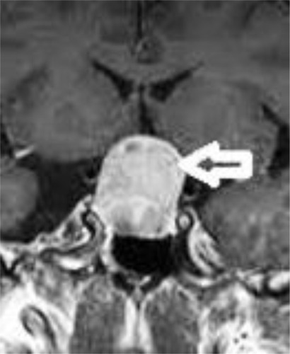
A 48-year-old, Caucasian female presented with progressive vision loss in both eyes for several months. Her past ocular, medical, and family history was non-contributory with no fatigue, loss of libido, no mood change, and no weight change. On examination, best-corrected visual acuity was hand motion in the right eye and 2/3 in the left eye. The anterior segment and intraocular pressure examination were normal in each eye. The macula and peripheral retinal fluorescein angiography were normal in both eyes. Spectral domain optical coherence tomography (SD-OCT) demonstrated diffuse atrophy of the ganglion cell and nerve layers, with preservation of outer layers bilaterally ( and ). Magnetic resonance imaging of the brain was performed and a pituitary macroadenoma with suprasellar extension was observed (). Resection was performed and the pathological diagnosis was nonfunctioning adenoma. Despite the surgery, the vision did not improve.
Figure 1 Spectral domain coherence tomography identifies symmetric retinal nerve fiber layer and ganglion layer loss in the left eye (arrow).
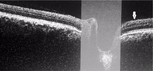
Figure 2 Spectral domain coherence tomography identifies symmetric retinal nerve fiber layer and ganglion layer loss in the right eye (arrow).
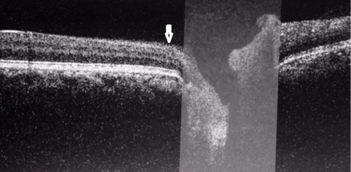
Figure 3 Coronal view, post-contrast T1 brain magnetic resonance imaging, showing a pituitary macroadenoma with compression of the optic chiasm (arrow).
Discussion
Pituitary adenomas are common benign tumors of the pituitary gland that account for 12% of all intracranial tumors.Citation9 People can develop pituitary adenomas at any age. Most pituitary adenomas are in the anterior lobe of the pituitary gland. Some adenomas secrete hormones in excess. Of the secretory tumors, the most common are prolactinomas.Citation5 They may be relatively small when detected. The most common symptoms include headaches, menstrual changes in females, sexual dysfunction in males, vision problems, and behavioral changes.Citation5 Diagnostic imaging may include a high-resolution, T1 weighted, gadolinium enhanced magnetic resonance imaging.Citation10 Other secretory tumors may secrete corticotropin, growth hormone, gonadotropins, or thyroid-stimulating hormone.Citation11 Adenomas remain confined to the pituitary gland, but when a pituitary adenoma is ≥10 mm it is called a macroadenoma. Macroadenomas can compress the rest of the pituitary gland and surrounding structures, such as the crossing retinal ganglion cell axons in the optic chiasm, resulting in bitemporal visual field loss.Citation4 As the tumor enlarges, it has been shown to induce uncrossed axons loss, resulting in nasal field defects and reduced visual acuity. In these cases, there is atrophy of the nasal and temporal portions of the optic disc with relative sparing of the superior and inferior parts where the majority of temporal fibers enter. The optic atrophy occupies a horizontal band across the disc, called bowtie or band atrophy.Citation12,Citation13 OCT is a noninvasive technique that allows cross-sectional imaging of the retina and quantifies the thickness of the RNFL around the optic nerve head. A number of studies have demonstrated that OCT is able to identify RNFL loss in eyes with band atrophy of the optic nerve.Citation6,Citation7 The degree of RNFL thickness reduction has been shown to correlate with that of visual field defects using Goldmann perimetryCitation4 and automated static perimetry.Citation14,Citation15 This fact gives OCT prognostic value regarding the visual outcome, as demonstrated in different studies.Citation16,Citation17 OCT is a useful tool to assess the structural and functional damage of ganglion cells, objectively and quantitatively.Citation4 In our case the SD-OCT demonstrates a symmetrical loss of the RNFL and the ganglion cell layer in both eyes, indicating important optic nerve damage.
Conclusion
SD-OCT can help in the diagnosis of adenomas in equivocal situations in which there is only subtle optic nerve pallor in the setting of nonspecific visual complaints or visual field defects, or when patients are referred to screen for subclinical compressive optic neuropathies.
Disclosure
The authors report no conflicts of interest in this work.
References
- ForoozanRChiasmal syndromesCurr Opin Ophthalmol200314632533114615635
- EzzatSAsaSLCouldwellWTThe prevalence of pituitary adenomasCancer2004101361361915274075
- Fernandez-BalsellsMMMuradMHBarwiseANatural history of nonfunctioning pituitary adenomas and incidentalomas: a systematic review and metaanalysisJ Clin Endocrinol Metab201196490591221474687
- AbouafLVighettoALebasMNeuro-ophthalmologic exploration in non-functioning pituitary adenomaAnn Endocrinol (Paris)201576321021926070465
- CooperOMelmedSSubclinical hyperfunctioning pituitary adenomas: the silent tumorsBest Pract Res Clin Endocrinol Metab201226444746022863387
- KanamoriANakamuraMMatsuiNOptical coherence tomography detects characteristic retinal nerve fiber layer thickness corresponding to band atrophy of the optic discOphthalmology20041112278228315582087
- MonteiroMLRLealBCRosaAAMBronsteinMDOptical coherence tomography analysis of axonal loss in band atrophy of the optic nerveBr J Ophthalmol20048889688915205233
- WagerEKleinertSResponsible research publication: international standards for authors. A position statement developed at the 2nd World Conference on Research Integrity, Singapore, July 22–24, 2010MayerTSteneckNPromoting Research Integrity in a Global EnvironmentImperial College Press / World Scientific PublishingSingapore2011
- MuslumanAMCanseverTYilmazASurgical results of large and giant pituitary adenomas with special consideration of ophthalmologic outcomesWorld Neurosurg20117614114821839965
- HallWALucianoMGDoppmanJLPatronasNJOldfieldEHPituitary magnetic resonance imaging in normal human volunteers: occult adenomas in the general populationAnn Intern Med1994120108178208154641
- DworakowskaDGrossmanABThe pathophysiology of pituitary adenomasBest Pract Res Clin Endocrinol Metab200923552554119945021
- JohanssonCLindblomBThe role of optical coherence tomography in the detection of pituitary adenomaActa Ophthalmol20098777677918771481
- UnsoldRHoytWFBand atrophy of the optic nerveArch Ophthalmol198098163716387425927
- JacobMRaverotGJouanneauEPredicting visual outcome after treatment of pituitary adenomas with optical coherence tomographyAm J Ophthalmol20091471647018774545
- Danesh-MeyerHVCarrollSCForoozanRRelationship between retinal nerve fiber layer and visual field sensitivity as measured by optical coherence tomography in chiasmal compressionInvest Ophthalmol Vis Sci2006484827483517065494
- SaxenaRGopalakrishnanKSinghDMahapatraAKMenonVRetinal nerve fiber layer changes: a predictor of visual function recovery in pituitary adenomasIndian J Med Specialities201564141145
- MoonCHHwangSCKimBTOhnYHParkTKVisual prognostic value of optical coherence tomography and photopic negative response in chiasmal compressionInvest Ophthalmol Vis Sci201152118527853321960556