
Abstract
Background
The presence of intraocular eyelashes following penetrating eye injury or ocular surgery is relatively uncommon. The response of the eye to intraocular eyelashes is variable. The eyelash may be symptomatic or may remain asymptomatic for long periods.
Objective
We report a case with two intraocular eyelashes and an iris cyst after 2 years of asymptomatic period following penetrating eye injury.
Case presentation
A 24-year-old male presented with decreased vision in the left eye which he had noticed for the previous 2 weeks. His visual acuity was 6/6 in the right eye and 6/18 in the left eye, improving to 6/9 with -2.5 DC × 140° correction. The intraocular pressure was 12 mmHg in both eyes. On slit-lamp examination, the left eye showed 8 mm linear peripheral corneal opacity nasally, two eyelashes in the superior anterior chamber, and an iris cyst measuring 4 mm × 4 mm in the superior iris. The right eye was normal. Dilated fundus examination of both eyes was normal. The eyelashes and cyst were removed surgically. There were no complications during the 3-month follow-up period.
Conclusion
Intraocular implantation of eyelashes following penetrating eye injury can remain asymptomatic for long periods; however, late development of iris cyst may occur.
Introduction
The presence of intraocular eyelashes following penetrating eye injury or ocular surgery is relatively uncommon. It has been reported to occur secondary to penetrating eye injury,Citation1–Citation3 ocular surgery,Citation4–Citation6 or even without apparent etiology.Citation7,Citation8 The response of the eye to intraocular eyelashes is variable. The eyelash may remain asymptomatic for long periodsCitation9,Citation10 or may be symptomatic.Citation4,Citation11 We report an uncommon case with coexistence of two intraocular eyelashes and a secondary iris cyst after 2 years of asymptomatic period following penetrating eye injury.
Case report
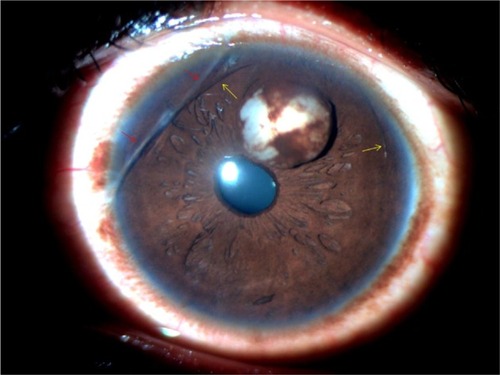
A 24-year-old Nepalese male presented with decreased vision in the left eye noticed for last 2 weeks. His visual acuity was 6/6 in the right eye and 6/18 in the left eye, improving to 6/9 with −2.5 DC × 140° correction. The intraocular pressure was 12 mmHg in both eyes. On slit-lamp examination, the left eye showed 8 mm linear peripheral corneal opacity nasally. In the anterior chamber, two curvilinear foreign bodies were seen superiorly resembling eyelashes, and an iris cyst measuring 4 mm × 4 mm was seen (). The anterior chamber was quiet, and there was no posterior synechia. Gonioscopy examination revealed normal angle structures and no foreign bodies. The pupil appeared slightly oval but was reacting normally to light. Lens was clear, and dilated fundus examination did not reveal any abnormality. The right eye was normal. Ocular history revealed past history of trauma in the left eye with a metallic wire while working in a construction site abroad 2 years back. No medical care was sought at that time. There was no history of past ocular surgeries.
Figure 1 Slit-lamp photograph of the anterior segment at presentation showing iris cyst, two eyelashes in the anterior chamber (yellow arrows) and linear corneal opacity (red arrows).
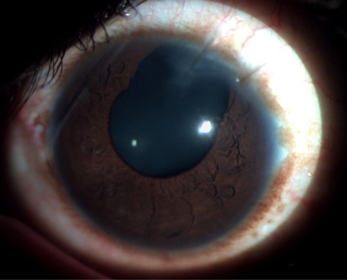
The eyelashes and the iris cyst were removed surgically. The anterior chamber was entered at the superotemporal limbus, and viscoelastic substance was introduced. The lashes were removed, and the cyst was excised in toto with a sectoral iridectomy. There was no collateral damage to other ocular structures. Postoperatively, there was transient anterior segment inflammation and microscopic hyphema, which subsided with topical antibiotic–steroid combinations. The visual acuity in the left eye eventually improved to 6/6 with −1.5 DC × 130° correction. The patient did not complain of glare despite the broad iridectomy (). There were no complications in the 3-month follow-up period.
Figure 2 Slit-lamp photograph of the anterior segment 1 week postoperatively showing the sectoral surgical iridectomy.
Discussion
The intraocular eyelashes are usually well tolerated and remain asymptomatic due to their relatively inert nature compared to other organic materials and the immune-privileged feature of the eye.Citation12 The posttraumatic intraocular cilia have been reported to remain silent for 50 years.Citation10 The intraocular eyelashes may be associated with corneal edema, granulomatous inflammation, cyst formation, intralenticular abscess, retinal detachment, endophthalmitis and even sympathetic ophthalmia.Citation3,Citation4,Citation12–Citation14 Various treatment modalities have been described for treatment of iris cyst with varying outcomes. Small and asymptomatic cysts may be closely observed, while the larger cysts require surgical management.Citation15 Conservative surgical approaches such as aspiration of the cyst have been described but are associated with a high rate of recurrence.Citation16 Intracystic injections of low-dose anti-mitotic agents have also been found useful in recurrent secondary iris cyst.Citation17 Needle aspiration and endodiathermy treatment of epithelial inclusion cyst of the iris have also been reported to have a good result.Citation18 Lasers play an important role in the management of iris cysts. Lasers can be used alone or in conjunction with other treatment modalities of the iris cyst. Laser iridotomy of the cyst offers a noninvasive method of therapy for posttraumatic iris inclusion cysts but is associated with a high rate of recurrence.Citation19 Viscoelastic dissection of the posttraumatic iris cyst has been described with an acceptable long-term outcome.Citation20 Viscoelastic-assisted endophotocoagulation has been documented to have an excellent response in the management of secondary iris cysts.Citation21 Various other forms of surgical excision include sector iridectomy, iridectomy plus cryotherapy, iridocyclectomy, iridectomy with corneal curettage and posterior corneal lamellar dissection.Citation22 In our case, sector iridectomy was done to excise the iris cyst in toto along with the intraocular eyelashes removal through the limbal approach. Viscoelastic substance was used so that the cyst could be excised with minimal trauma to the corneal endothelium and adjacent ocular structures. The postoperative visual outcome was good, and there were no significant complications associated with the procedure.
Conclusion
Intraocular implantation of eyelashes following penetrating eye injury can remain asymptomatic for a long period; however, late development of iris cyst may occur. Various surgical techniques, ranging from minimally invasive laser procedures to extensive surgical procedures, have been described with variable outcomes. We report a relatively uncommon case with coexistence of two intraocular eyelashes and a secondary iris cyst after 2 years of asymptomatic period following penetrating eye injury and its successful management with intraocular eyelashes removal and sector iridectomy.
Author contributions
SS examined and managed the patient, reviewed the literature and drafted the manuscript. LRP diagnosed and managed the case and supervised and drafted the manuscript. SKS critically analyzed the manuscript and offered valuable suggestions. All authors contributed toward data analysis, drafting and critically revising the paper, gave final approval of the version to be published, and agree to be accountable for all aspects of the work.
Acknowledgments
Written informed consent for the publication of the case report and images was obtained from the patient. The authors’ thanks go the patient for permitting them to report this case, and to Mr Dipesh Ram for helping with the patient interaction and clinical photography.
Disclosure
The authors report no conflicts of interest in this work.
References
- HohHMenageMIris cysts after traumatic implantation of an eyelash into the anterior chamberBr J Ophthalmol199377117417428280692
- KöseSKayikçioğluOAkkinCYağciABasdemirGCoexistence of intraocular eyelashes and anterior chamber cyst after penetrating eye injury: a case presentationInt Ophthalmol1994–1995185309311
- GopalLBankerASSharmaTParikhSBhendePSChopraSIntra-ocular cilia associated with perforating injuryIndian J Ophthalmol2000481333611271932
- GallowayGDAngGSShenoyRBeigiBRetained anterior chamber cilium causing endophthalmitis after phacoemulsificationJ Cataract Refract Surg200430252152215030854
- IslamNDabbaghAInert intraocular eyelash foreign body following phacoemulsification cataract surgeryActa Ophthalmol Scand20068434324
- WalkerNJHannJVTalbotAWPostoperative cilium entrapment by clear corneal incisionJ Cataract Refract Surg200733473373417397752
- OhKT-(Kean)OhKT-(Kong)SingermanLJAn eyelash in the vitreous cavity without apparent etiologyOphthalmic Surg Lasers19962732432458833131
- KertesPJAl-GhamdiAABrownsteinSCoupalDGilbergSBrittonWAJrAn intraocular cilium of uncertain originCan J Ophthalmol200439327928115180146
- KargiSHOzOErdincETekeMYFiratETolerated cilium in the anterior chamberOcul Immunol Inflamm2003111737812854030
- YalnizAkkayaZPosttraumatic cilia remaining inert in the anterior chamber for 50 years: a case reportJ Med Case Rep2011552722029734
- TanejaSAroraRYadavaUFingernail trauma causing corneal laceration and intraocular ciliaArch Ophthalmol199811645305319565056
- HumayunMde la CruzZMaguireADangelMEStarkWJGreenWRIntraocular cilia. Report of six cases of 6 weeks’ to 32 years’ durationArch Ophthalmol199311110139614018216021
- GottliebFFinestoneJAckermanJLIntravitreal cilia and retinal detachmentAnn Ophthalmol19821465415447114689
- DettorakiMAndreanosKDavouSNomikariosNMoschosMMBrouzasDIntravitreal cilium associated with retinal detachment 40 years following penetrating eye injury: a case reportBMC Ophthalmol2015152525884640
- HallerJAStarkWJAzabAThomsenRWGottschJDSurgical management of anterior chamber epithelial cystsAm J Ophthalmol2003135330931312614747
- ShieldsJAShieldsCLLoisNMercadoGIris cysts in children: classification, incidence, and managementBr J Ophthalmol199983333433810365043
- KawaguchiKYamamotoSNagaeYTreatment of recurrent giant iris cyst with intracyst administration of mitomycin CBr J Ophthalmol200084780080111032435
- TsaiJCArrindellELO’DayDMNeedle aspiration and endodiathermy treatment of epithelial inclusion cyst of the irisAm J Ophthalmol2001131226326511228309
- GuptaVRaoASinhaAKumarNSihotaRPost-traumatic inclusion cysts of the iris: a long term prospective case seriesActa Ophthalmol Scand200785889389617822446
- Al-GhadeerHAl-TowerkiAEAl-RajhiAAl-AwadALongterm follow-up and visual outcome after excision of a traumatic iris cyst by viscoelastic dissectionInt Ophthalmol201131652953122222716
- LockingtonDAltaieRMooreSMcGheeCNSuccessful management of secondary iris cysts with viscoelastic-assisted endophotocoagulationJAMA Ophthalmol2014132335435624626827
- RaoAGuptaVBhadangeYSharmaRShieldsJAIris cysts: a reviewSemin Ophthalmol2011261112221275599