
Abstract
Introduction
Nonprojectile penetrating skull base injuries as a result of falls have rarely been confronted in normal neurosurgery although a few nonmissile injuries have been reported. These kinds of injuries represent a life-threatening emergency.
Case presentation
We present an unusual case of a 25-year-old male construction worker who suffered an accidental penetrating skull base injury when he fell on a metal rod while he was walking on a 2-meter-high platform. He was clinically stable at presentation. Skull radiograph showed a solid metallic bar, 30 cm long, that penetrated through the right anguli oris eminence and was lodged low in the right occipital bone.
Conclusion
Penetrating injury to the head is considered a form of severe traumatic brain injury. Although case of penetrating head injuries as a result of fall from heights are very rare, we anticipate the construction works on high-rise buildings are at maximum risk. We advise that removal of this kind of foreign bodies be done in the theater and not outside because of risk of involvement of larger vessels leading to fatal hemorrhage. We further suggest that patients with nonprojectile injuries should undergo a preoperative computed tomography-angiography to rule out any vascular injury.
Introduction
Penetrating head injury arises when a projectile or nonprojectile object breaks through the cranium and its contents.Citation1–Citation5 They are relatively unusual wounds that constitute about only 0.4% of all head injuries.Citation2 These kinds of injuries usually occur as a result of gunshot, missiles or nonmissiles, or wooden or metal objects leading to closed or open wounds, although motor vehicle accidents or occupational accidents and suicidal attempts may also occur.Citation3,Citation5,Citation6
Whereas majority of penetrating injuries in western countries occurs as a result of gunshot wounds, surprisingly, this is completely different in developing and third-world countries. There are a wide range of nonmissile or nonprojectile penetrating injuries that have been reported, with weapons such as knifes, scissors, pencils, iron rods, nails, chopsticks, etc. Wounds resulting from these kinds of objects usually involve a smaller area with relatively low-velocity impact.Citation2,Citation3,Citation7 It must be recounted that most penetrating skull injuries occurring as a result of nonmissile or nonprojectile objects are rarely associated with major neurological symptoms regardless of the size of the penetrating bodies, except in high-velocity injuries where large amount of kinetic energy, contact with the skull, and the force acting for a shorter time probably cause local deformation resulting in penetration and skull fracture.Citation2,Citation8,Citation9 We present a case of a patient presenting with a penetrating iron rod from the maxillary to the posterior cranial fossa.
Clinical report
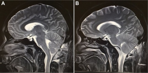
A 25-year-old construction worker suffered an accidental traumatic posterior cranial fossa injury after falling upon a metal rod while he was walking on a 2-meter-high platform.Citation10 On arrival at our department, he was alert and cooperative (Glasgow coma scale (GCS) score 15) (). There were no abnormal findings on neurologic exams. Skull radiographs showed that a solid metallic bar, 30 cm long, had penetrated through the right anguli oris eminence and was lodged low in the right occipital boneCitation10 (). The extent of structural damage could not be ascertained on the computed tomography (CT) scans as a consequence of severe artifacts associated with the metal barCitation10 (). CT angiography (CTA) illustrated that the metal rod steered clear of the internal carotid artery and narrowly missed the vertebral arteryCitation10 (). We used the rapid sequence intubation to pass the nasotracheal tube. A suboccipital craniotomy was unutilized to remove the rod. The patient was placed in a park-bench position (). A midline incision was made from two fingerbreadths above the inion to the mid-cervical region. After clearance of the musculature, the rod was visualized penetrating through the occipital squamaCitation10 (). Additional bone removal was achieved by using a high-speed drill and a Kerrison rongeur. The metal rod was further exposed to be going through the dura but left the brain stem aloneCitation10 (). A steady retraction force was used to pull the rod from the anterior side. Once the rod was clear, thorough hemostasis was achieved by bipolar coagulation. The ipsilateral vertebral artery was revealed to be intactCitation10 (). After repeated irrigation with hydrogen peroxide and saline water, the dura was closed in watertight fashion. We kept the patient on ceftriaxone (2 g q 8 h) and metronidazole (500 mg q 8 h) for 7 days postoperatively. Postoperative CT scan revealed no hematoma of edema (). The patient’s postoperative course was uneventful. MRI at the 3-month follow-up showed no abscess formation ().
Figure 1 Preoperative images (A–D) showing the iron rod both in the patient and on imaging studies. (E and F) are postoperative CT scan images.
Abbreviation: CT, computed tomography.
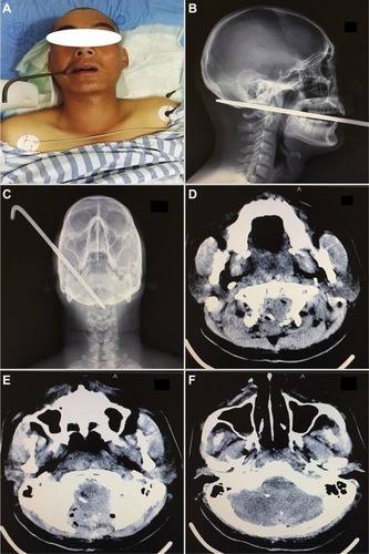
Figure 2 Digital subtraction angiography images showing the iron rod penetrating the bones but narrowly missing the right internal carotid artery (A–D).
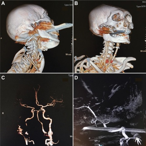
Figure 3 Intraoperative images showing the iron rod lodged in the posterior skull and in the brain tissues (A–D).
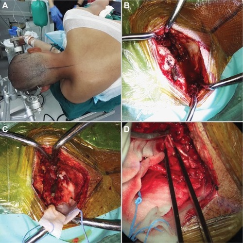
Figure 4 MRI taken during follow-up outpatient visit 3 months after the removal of the iron rod (A and B).
Discussion
Nonprojectile penetrating skull injuries as a result of fall from heights are very rare and seldom reported in literatures. A penetrating head injury occurs when the dura mater is breached as a result of either high-velocity objects like bullets and missiles or lower velocity objects such as knife (most common) and rarely nails, keys, pencils, and chopsticks and/orCitation1,Citation11–Citation14 wooden, stony, glassy, and other industrial or environmental objects.Citation3,Citation15–Citation18 In most cases, the bone fragments from a skull fracture are driven into the brain. Projectile objects (gunshots or missiles) are associated with high velocity, which makes them cause deep tissue cavitation. Furthermore, temporal cavitation is caused by missiles because tissue elasticity causes the cavity walls to pulsate in a violent waning fashion and eventually collapse, hence the name temporary cavity.Citation19 On the other hand, permanent cavitation is caused by penetration injuries, such as in our case, because there is always a formation of a permanent cavity. Their shock waves are the main causes of complex injuries and poor prognosis.Citation3,Citation20 Nonprojectiles objects on the other hand usually move with a velocity less than 100 m/s and are associated with direct disruption and laceration of tissues. These kinds of forces mainly cause localized primary injury and lead to better prognosis.Citation3,Citation20–Citation22 Our case is very unique because the solid metallic bar, 30 cm long, penetrated through the right anguli oris eminence, through the mylohyoid groove, sparing the right common carotid artery, and finally lodged low in the right occipital bone.
CT scan is the most accepted radiological modality of choice for the evaluation of patients with penetrating skull injuries. Many authors are of the view that patients with penetrating skull injuries should have unenhanced standard axial views with bone and soft tissue windows even with trivial scalp laceration,Citation3 and for accurate assessment of the injuries coronal and sagittal sections are very vital.Citation3,Citation23,Citation24 On the other hand, most neurosurgeons do not agree with the use of MRI in patients with penetrating skull injuries because it does not give any better visualization of bones and can be potentially dangerous in ferromagnetic objects.Citation3 However, patients with nonmetallic penetrating skull injuries can benefit immensely with MRI angiography, if the service is available at the facility they present. We suggest that patients with nonprojectile injuries should undergo a preoperative CTA to rule out any vascular injury.
The targets of surgical intervention in patients with these injuries are to remove the penetrating item from the brain parenchyma, remove necrotic tissue, debris, and other potential contaminants, evacuate any hematomas occurring from the injury, and secure hemostasis as well as ensure watertight closure of the dura to prevent cerebrospinal fluid leakage.Citation5 Notwithstanding the presence of well-written guidelines for managing these cases, surgical treatment requires an individualized approach tailored to the situation at hand.Citation3 Many authors including us suggest that the foreign body should never be removed outside the theater because of the possibility of injuring larger vessels leading to fatal hemorrhage when the object is forcefully pulled out.Citation3,Citation25 The bulging object should also be stabilized during transportation, evaluation, and treatment to prevent further injury.Citation3,Citation20 During the operation in the theater, the foreign object should be carefully removed in the direct reverse path of trajectory without swinging movements to prevent further damage.Citation3,Citation21,Citation26 The dura should be closed in a watertight manner, either by direct closure or by using autologous grafting materials, eg, pericranium. Synthetic grafts, being a foreign body, become a potential source of infection.Citation3,Citation27,Citation28
Some of the complications anticipated in the management of these kinds of injuries may be cerebrospinal fluid fistula and neuroendocrine dysfunction. Most deaths from penetrating skull injuries are as a result of damage to larger blood vessels leading to intracranial hematomas and ischemia, which can in turn lead to a biochemical cascade called the ischemic cascade.Citation3 Our patient was so fortunate the rod narrowly missed the external carotid artery, as seen in the angiogram (). Many authors with similar experiences have estimated vascular complications arising from penetrating skull injuries to be 5%–40% depending on the type of artery or sinus involve.Citation3,Citation29,Citation30 Patients who survive these complication most often develop vasospasm, pseudoor true traumatic aneurysm, arteriovenous malformation, subarachnoid hemorrhage.Citation3
Post-traumatic epilepsy is another complication experiences by many authors.Citation1,Citation3,Citation4,Citation29,Citation31,Citation32 They pinned direct cortical injury with subsequent scarring as the main cause of seizures after this traumatic event. This occurrence is further classified into immediate (<24 hours), early seizures (<1 week), and late seizures (>1 week).Citation3,Citation31,Citation33 Patients with penetrating skull injuries are also highly susceptible to infection, which may aggravate their condition as well as increase their mortality rates.Citation3,Citation34 Deep or multifocal brain injury, intracranial retained bone and metallic fragments, cerebrospinal fluid fistulas, air sinus wounds, transventricular injuries, or wounds crossing the midline are but the many possible way patients can develop post-traumatic infection.Citation3,Citation28 The most frequently associated organism is Staphylococcus aureus because this organism forms part of the normal flora of the skin and thus gets inoculated into the open wound,Citation3,Citation35 leading to scalp sepsis, osteomyelitis, epidural and subdural empyemas, meningitis, ventriculitis, cerebritis, and cerebral abscess.Citation2,Citation18
Antimicrobial agents have markedly decreased the rate of infections since their introduction. The choice of antimicrobial agents depends on the protocols of the institution or department and the type of organism anticipated. It is also imperative to give antitetanus shots to patients although the incidence of tetanus has reduced. Birch-Hirshfield has indicated in his report that there is about 67% mortality rate in the patients who developed tetanus infection, thus stressing the importance of tetanus immunization as required.Citation2
Conclusion
Penetrating injury to the head is considered a form of severe traumatic brain injury. Nonprojectile or nonmissile injuries are not uncommon in developing countries, especially in the rural setups and construction sites in the cities. Although cases of penetrating head injuries as a result of fall from heights are very rare, we anticipate the construction workers on high-rise building are at maximum risk. We advise that removal of this kind of foreign bodies be done in the operating room and not out outside because of risk of involvement of larger vessels leading to fatal hemorrhage. We further suggest that patients with nonprojectile injuries should undergo a preoperative CTA to rule out any vascular injury.
Ethics approval and consent to participate
The ethical committee of West China Hospital fully approved our case study. The patient and his relatives were informed about our intention to involve him in a case study and he/they agreed to partake in the study. He/they signed the consent form before the operation was carried out according to all surgical protocols and consent was also obtained for the case details and accompanying images to be published.
Author contributions
All authors contributed toward data analysis, drafting and critically revising the paper and agree to be accountable for all aspects of the work.
Disclosure
The authors report no conflicts of interest in this work.
References
- KothariKSinghAKDasSPenetrating skull injury with six inch fence rodNatl J Maxillofac Surg20123220723833500
- ArifinMZGillASFariedAPenetrating skull fracture by a wooden object: management dilemmas and literature reviewAsian J Neurosurg20127313123293668
- YounesWCraniocerebral injuries by impacted foreign objects: case series and literature reviewEgypt J Neurosurg2015305562
- XuFLiJSunSThe surgical management of a penetrating orbitocranial injury with a Bakelite foreign body reaching the brain stemBrain Injury2013277–895195623789869
- RegunathKAwangSSitiSPremanandaMTanWHaronRPenetrating injury to the head: case reviewsMed J Malaysia201267662262423770960
- BhaganagareANadkarniTGoelAPenetrating craniocerebral injury with nails: case reportIndian J Neurotrauma2007416364
- SchallerBSubcranial approach in the surgical treatment of anterior skull base traumaActa Neurochir2005147435536615726278
- GennarelliTAChampionHRSaccoWJCopesWSAlvesWMMortality of patients with head injury and extracranial injury treated in trauma centersJ Trauma Acute Care Surg198929911931202
- SchallerBJKleindienstAKruschatTSchliephakeHBuchfelderMMertenHAIndustrial nail gun injury to the anterior skull base: a case report and review of the literatureJ Trauma Acute Care Surg2008643E29E32
- HuangJLiDChenHSuccessful management of a penetrating iron-rod injury through the oral cavity involving the posterior cranial fossaNeurol India201765366666828488651
- PascualJNavasMCarrascoRPenetrating ballistic-like frontal brain injury caused by a metallic rodActa Neurochir2009151668919277462
- SalarGCostellaGBMottaranRMattanaMGazzolaLMunariMMultiple craniocerebral injuries from penetrating nails: case illustrationJ Neurosurg2004100596315137618
- KoestlerJKeshavarzRPenetrating head injury in children: a case report and review of the literatureJ Emerg Med200121214515011489404
- HerringCJLumsdenABTindallSCTranscranial stab wounds: a report of three cases and suggestions for managementNeurosurgery19882356586623200400
- BalakNAslanBSerefhanAElmaciIIntracranial retained stone after depressed skull fracture: problems in the initial diagnosisAm J Forensic Med Pathol200930219820019465819
- ChibbaroSTacconiLOrbito-cranial injuries caused by penetrating non-missile foreign bodies. Experience with eighteen patientsActa Neurochir2006148993794216763734
- GökçekCErdemYKöktekirEIntracranial foreign bodyTurk Neurosurg200717212112417935028
- KazimSFBhattiAGodilSSCraniocerebral injury by penetration of a T-shaped metallic spanner: a rare presentationSurg Neurol Int20134223493510
- StefanopoulosPFilippakisKSoupiouOPazarakiotisVWound ballistics of firearm-related injuries – Part 1: missile characteristics and mechanisms of soft tissue woundingInt J Oral Maxillofac Surg201443121445145825128259
- JandialRReichwageBLevyMDuenasVSturdivanLBallistics for the neurosurgeonNeurosurgery200862247248018382326
- KatariaRSinghDChopraSSinhaVLow velocity penetrating head injury with impacted foreign bodies in situAsian J Neurosurg2011613922059103
- ClarkWCMuhlbauerMSWatridgeCBRayMWAnalysis of 76 civilian craniocerebral gunshot woundsJ Neurosurg19866519143712033
- ColesJImaging after brain injuryBr J Anaesth2007991496017573394
- ZhangCWRichardSAWuCWangTLuiLXXieXDSuccessful treatment of iatrogenic vertebral artery pseudoaneurysm with coil embolization: case reportJ Vasc Med Surg20175342
- SchreckingerMOrringerDThompsonBGLa MarcaFSagherOTransorbital penetrating injury: case series, review of the literature, and proposed management algorithm: report of 4 casesJ Neurosurg20111141536120868210
- Prakash RaoGRaoNReddyPTechnique of removal of an impacted sharp object in a penetrating head injury using the lever principleBr J Neurosurg199812656957110070469
- EspositoDPWalkerJBContemporary management of penetrating brain injuryNeurosurg Q2009194249254
- SweeneyJMLebovitzJJEllerJLCoppensJRBucholzRDAbdulraufSIManagement of nonmissile penetrating brain injuries: a description of three cases and review of the literatureSkull Base Rep201111394623984201
- NathooNBoodhooHNadviSSNaidooSRGouwsETranscranial brainstem stab injuries: a retrospective analysis of 17 patientsNeurosurgery20004751117112311063104
- RichardSAWuMLinDTraumatic subdural effusion evolving into chronic subdural hematomaOpen J Modern Neurosurg20145112
- ChristensenJPedersenMGPedersenCBSideniusPOlsenJVestergaardMLong-term risk of epilepsy after traumatic brain injury in children and young adults: a population-based cohort studyLancet200937396691105111019233461
- RichardSAShresthaSSZhangCSuccessful treatment of a child with ruptured arteriovenous malformation using onyx embolization: a case reportOpen J Modern Neurosurg201774153
- KharatishviliIPitkänenAPosttraumatic epilepsyCurr Opin Neurol201023218318820125011
- Peek-AsaCMcArthurDHovdaDKrausJEarly predictors of mortality in penetrating compared with closed brain injuryBrain Injury200115980181011516348
- BaystonRDe LouvoisJBrownEJohnstonRLeesPPopleIUse of antibiotics in penetrating craniocerebral injuriesLancet200035592171813181710832851