
Abstract
Background
Peritoneal fistulization of a pyonephrosis is an extremely rare event which invariably leads to generalized peritonitis. This is a very rare case report on generalized peritonitis after spontaneous rupture of pyonephrosis.
Case presentation
A 28-year-old male patient from the rural part of Bale zone, Ethiopia, was admitted to Goba Referral Hospital with high-grade fever, diffused abdominal pain and abdominal distension. Initially, he experienced colicky and intermittent pain that made him stay at home for 2-3 days. He then started to develop constant left flank pain which gradually got worse and was associated with urinary frequency of approximately 5–6 times/day, high-grade intermittent fever, chills, rigors and loss of appetite. With the diagnosis of generalized peritonitis, we resuscitated him with two bags of normal saline and one bag of ringer lactate intravenously. During an abdominal ultrasound examination we identified that the left kidney was replaced by an abscess containing sac, and there was a huge intraperitoneal loculated abscess with internal septation and an associated free inter-loop and pelvic echo debris abscess. When we performed an exploratory laparotomy, 1 L-thick abscess from the general peritoneum was aspirated and early fibrinous inter-loop adhesion was identified. In addition, there was a large retroperitoneal cystic abscess containing sac extended from the spleen up to the pelvic brim crossing the midline to the right side and bulged intraperitoneally. Furthermore, a 1.5 cm wide perforation that pour abscess in to peritoneal cavity was found. A total of 4 L of puss was removed from the left kidney. As treatment, since the left kidney lost all function and became a pus-contacting sac, we performed a left-sided nephrectomy and abdominal lavage. Postoperatively, the patient had an uneventful recovery and was discharged from the hospital on the eighth day. We followed him for 6 months, and kidney function tests were normal and he did not develop any complications.
Conclusion
This case report highlighted the importance of recognizing the possibility of underlying kidney rupture in a patient with generalized peritonitis. Uretero-pelvic junction obstruction (UPJO) might be the possible cause of pyonephrosis in our case. As a treatment, nephrectomy is a preferable option when the affected kidney is not fully functional and the contralateral kidney is normal.
Introduction
The accumulation of purulent exudate in the hydronephrotic collecting system and abscess formation constitute the pathophysiology of pyonephrosis.Citation1 If it is not diagnosed early, it can worsen rapidly and cause the patient to die of septic shock.Citation2 Peritoneal fistulization of a pyonephrosis is an extremely rare event which invariably leads to generalized peritonitis.Citation3
The risk factors for pyonephrosis include an immunosuppressive status and an anatomic urinary tract obstruction, of which 75% is nephrolithiasis related.Citation4
Antibiotics have no effect in pyonephrosis unless the pus is surgically drained. Percutaneous nephrostomy and urethral catheter insertions are, therefore, necessary. In addition, studies showed that percutaneous drainage is a fast, trusted and effective diagnostic and therapeutic method.Citation5–Citation7
Radical nephrectomy can be the preferred treatment for a kidney that has lost most of its function if the contralateral kidney is normal. Nephrectomy has been found to have fewer complications compared to other treatments.Citation8,Citation9 The aim of this paper was to present an unusual case of generalized peritonitis after spontaneous rupture of pyonephrosis diagnosed in a 28-year-old man who was admitted to Goba Referral Hospital with high-grade fever, diffused abdominal pain and abdominal distension.
Case presentation
A 28-year-old male patient from the rural part of Bale zone, Ethiopia, was admitted to Goba Referral Hospital with high-grade fever, diffused abdominal pain and abdominal distension. He is a farmer and married with four children. The patient had experienced left flank pain for the past 6 years. Initially, he experienced colicky and intermittent pain that made him stay at home for 2-3 days. In addition, he had a history of reddish discoloration of urine during the flank pain episode. At the beginning of this condition, he did not visit any health care institution. He then started to develop constant left flank pain which gradually got worse and was associated with urinary frequency of around 5–6 times/day, high-grade intermittent fever, chills, rigors and loss of appetite. After 3 weeks of the abovementioned symptoms, he was admitted to Ginnir hospital (one of the rural hospitals in Ethiopia) for 7 days with the diagnosis of left pyelonephritis and was given ceftriaxone 1 g intravenous (IV) bid and diclofenac 75 mg intramuscular (im) and as needed (PRN). Despite this, after 6 days of stay in the aforementioned hospital, he developed generalized abdominal pain, abdominal distension, constipation and high-grade intermittent fever. Upon arrival to our hospital, Goba Referral Hospital, the patient was acutely sick looking and had a blood pressure (BP) of 80/50 mmHg, a pulse rate (PR) of 138 beats/minute, a respiratory rate (RR) of 34 breaths/minute and a temperature of 38.9°C. Besides, the patient had pink conjunctiva and dry tongue. He had clear chest on auscultation, and first heart sound or lub (S1) and second heart sound or dub (S2) were well heard when the heart was assessed. Regarding the abdominal assessment, his abdomen was distended, had no visible peristalsis, and it did not move with respiration. Also there was hypoactive bowel sound, guarding, rigidity, direct and rebound tenderness all over abdomen. The digital rectal examination showed empty rectum. He had a cold extremity. He had no history of diabetic mellitus, hypertension and he was neither a smoker nor an alcohol drinker. With the diagnosis of generalized peritonitis, we resuscitated him with two bags of normal saline and one bag of ringer lactate intravenously, causing his BP to became 100/60 mmHg. The laboratory investigation revealed the following: red blood cells, 201 g/dL; hemoglobin, 13.4 g/dL; white blood cells, 20,000/µL; granulocyte, 88%; platelet counts, 236,000/L; blood group, A+ and HIV antibody test, negative. In abdominal ultrasound examination, we identified that the left kidney was replaced by an abscess-containing sac, and there was huge intraperitoneal loculated abscess with internal septation and associated free inter-loop and pelvic echo debris abscess. After 2 hours of IV fluid administration, he produced 80 mL of urine. When we performed an exploratory laparotomy, 1 L-thick abscess from general peritoneum was aspirated, and early fibrinous inter-loop adhesion was identified. In addition, there was a huge retroperitoneal cystic abscess-containing sac extended from the spleen up to the pelvic brim crossing the midline to the right side and bulged intraperitoneally. Furthermore, about a 1.5 cm wide perforation that pour abscess in to peritoneal cavity was found. A total of 4 L of puss were removed out from the left kidney. As treatment, since the left kidney had lost full function () and became a pus-contacting sac, while the contralateral kidney was still normal, we performed a left-sided nephrectomy and abdominal lavage. When we mobilized the left colon during the intraoperative phase, adhesion and renal pedicle were identified. Therefore, double ligation of renal artery and vein together was done with silk number 1 as a result of inability to separate the renal artery and vein due to dense adhesion. Then, the ureter was ligated and divided. The abdominal wall was closed after the lavage drain was left inside. We did not find any stones in the affected kidney and nearby structure. Moreover, there was no evidence of tumor and tuberculosis from histopathologic examination of resected sample. Therefore, uretero-pelvic junction obstruction (UPJO) might be the possible cause of pyonephrosis of this case. Postoperatively, we put the patient on ceftriaxone 1 g IV bid, metronidazole 500 mg IV tid, maintenance fluid, tramadol 50 mg IV qid and diclofenac 50 mg im tid. He started sips on the third postoperative day. We removed the drain on the fifth postoperative day. The renal function test (RFT) performed in postoperative phase revealed normal range of serum electrolyte, and there was a normal amount of urine output. The patient had an uneventful recovery and was discharged from the hospital on the eighth day after surgery. We followed him for 6 months, and he had a normal kidney function test and did not develop any complications.
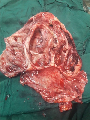
Figure 1 Macroscopic view of the left kidney after nephrectomy was performed.
Consent
Written informed consent was obtained from the patient for publication of this case report.
Discussion
The initial symptom of our patient was left flank pain which gradually got worse and was associated with urinary frequency of around 5–6 times/day, high-grade intermittent fever, chills, rigors and loss of appetite. Literature also supported that the most common symptoms of pyonephrosis are fever, chills and flank pain.Citation10–Citation14
We did not find any stones in the affected kidney, and UPJO might be the possible cause of pyonephrosis in our case. On the other hand, different authors reported that 75% of pyonephrosis cases are due to nephrolithiasis,Citation4 71% of the cases are due to urinary lithiasisCitation15 and 17 out of 23 cases are due to nephrolithiasis.Citation6 This may imply that pyonephrosis is a multifactorial disease, and a further study should be conducted to clearly present its risk factors and causes.
Our diagnosis was based on ultrasound. Different literature also confirmed the importance and even the indispensability of ultrasound and computed tomography in the diagnosis of pyonephrosis.Citation2,Citation10,Citation12,Citation13,Citation16,Citation17 The sensitivity of renal ultrasonography for differentiating hydronephrosis from pyonephrosis is 90%, and the specificity is 97%.Citation17
The ultrasound finding in this patient indicated that the left kidney was replaced by an abscess-containing sac and a huge intraperitoneal loculated abscess with internal septation, associated free inter-loop and pelvic echo debris abscess. Literature also supported that pyonephrosis might be differentiated from hydronephrosis on ultrasonography by the presence of debris, fluid–fluid levels and internal echoes in the collecting system.Citation18
To treat the patient, left-sided nephrectomy and abdominal lavage were performed. The patient had no postoperative complications. Literature also supported that radical nephrectomy can be the preferred treatment for a kidney that has lost most of its function if the contralateral kidney is normal. On top of this, nephrectomy has been found to have fewer complications compared to other treatments.Citation9,Citation19
Conclusion
This case report highlights the importance of recognizing the possibility of underlying kidney rupture in a patient with generalized peritonitis. UPJO might be the possible cause of pyonephrosis in our case. Regarding its treatment, nephrectomy is a preferable option when the affected kidney is not fully functional and the contralateral kidney is normal.
Disclosure
The authors report no conflicts of interest in this work.
References
- EroğluMKandıralıEAkut Pyelonefrit ve pyonefrozTurk Klin J Surg Med Sci20073202428
- ErolAÇobanSTekinAA giant case of pyonephrosis resulting from nephrolithiasisCase Rep Urol2014201413
- BalasPSeqditsasTAntonopoulosDPeritonitis after spontaneous rupture of pyonephrotic kidney into a peritoneal cavityAm J Surg197112156126135557773
- ChenYLWangMCHuangJZPyonephrosisKidney Dial2008201418
- EroğluMKandıralEAkut Pyelonefrit ve pyonefrozTurk Klin J Surg Med Sci20073202428
- LezinMSHofmannRStollerMLPyonephrosis: diagnosis and treatmentBr J Urol19927043603631450841
- MokhmaljiHBraunPMPortilloFJMPercutaneous nephrostomy versus ureteral stents for diversion of hydronephrosis caused by stones: a prospective, randomized clinical trialJ Urol200116541088109211257644
- JimenezJFPaciosMALLlamazaresGConejeroJSole-BalcellsFTreatment of pyonephrosis: a comparative studyJ Urol19781203287289682243
- AndroulakakisPAPyonephrosis: a critical review of 131 casesBr J Urol198254289927082947
- ScarneciuIConstantinaAGrigorescuDMaximLPyonephrosis: diagnosis and treatment: report of 65 casesJ Mol Biol20152122125
- TaheriAAEl AzizSChadliAAcute pyelonephritis in diabetic patientsAnn D’Endocrinol2015764558
- SowYFallBSarrAThiamADiaoBFallPAPyonephrosis: 44 cases in senegalMed Trop (Mars)201171549549822235626
- MohamedAMohammedRManagement of pyonephrosis: our experienceWebmed Cent Urol201235WMC003420
- HasigovAEngbangJPNFidarovFGiant pyonephrosis due to urolithiasis and diabetes: case reportOpen J Urol201667122125
- RabiiRJoualARaisetalHPyonephrosis: diagnosis and treatment: a review of 14 casesAnn D’Urol2000343161164
- SchaefferAJSchaefferEMInfections of the urinary tractWeinAJKavoussiLRCampbell-Walsh Urol9th edPhiladelphia, PASaunders Elsevier200719731985
- Van NieuwkoopCHoppeBPBontenTNPredicting the need for radiologic imaging in adults with febrile urinary tract infectionClin Infect Dis201051111266127221034195
- SchaefferAJSchaefferEMInfected hydronephrosis and pyonephrosisWeinAJKavoussiLRNovickACPartinAWPetersCACampbell-Walsh Urology10 edPhiladelphia, PASaunders Elsevier201235603574
- JimenezJFLopez PaciosMALlamazaresGConejeroJSole-BalcellsFTreatment of pyonephrosis: a comparative studyJ Urol19781203287289682243