
Abstract
Background
Superior lumbar hernia is a rare posterolateral abdominal wall defect and herniation of abdominal contents through the superior lumbar triangle. A lumbar hernia is an unusual defect and only 300 cases of primary lumbar hernias have been reported since the first case report in 1731. To date, most clinicians are usually unfamiliar with the presentation of lumbar hernias, and the diagnosis is often done in a wrong way.
Case report
A 55-year-old female patient presented with complaints of right flank pain and swelling on the right lumbar region. On abdominal examination, there was round protruding swelling immediately beneath the 12th rib of the right upper flank area. The swelling was palpable and bowel sound was heard over it. The swelling was easily reducible and protruded when the patient was coughing or straining. The opposite side of the lumbar region was normal, and chest examination was clear and normal. The hematological and urine analysis laboratory findings were normal.
Discussion
The ultrasound finding revealed the bowel contents within the hernial sac. With the diagnosis of primary acquired superior lumbar hernia, the patient underwent open surgery. The defect was reduced back and repaired using a retro-muscular or sublay prolene mesh (15 cm by 7.5 cm). The patient was discharged at post-operative day four and followed-up for four months.
Conclusion
In conclusion, the diagnosis of lumbar hernias should be considered in all patients presented with complaints of flank pain and swelling in the flank area. In the absence of obvious swelling in the flank region, ultrasound investigation might not establish the diagnosis of lumbar hernias. Hence, ultrasound would be the option to confirm the diagnosis when the swelling is palpable.
Keywords:
Background
The lumbar region is bordered superiorly by the 12th rib, inferiorly by the iliac crest, medially by the erector spinal muscles, and laterally by the external oblique muscle. The superior lumbar is defined medially by the erector spine muscle group, laterally by the internal oblique muscle, and superiorly by the 12th rib.Citation1,Citation2 Lumbar hernias are divided into two types according to the anatomical location of the defect: superior lumbar hernia and inferior lumbar hernia.Citation3 Superior lumbar hernia (a Grynfeltt–Lesshaft hernia) is a rare posterolateral abdominal wall defect and a herniation of abdominal contents through the superior lumbar triangle.Citation4
A lumbar hernia is an unusual defect and only 300 cases of primary lumbar hernias have been reported since the first case report in 1731.Citation3,Citation4 The causes of herniation could be congenital, primary, and secondary hernias. Congenital hernias are those that are discovered in infancy and comprise 20% of all lumbar hernias. These hernias are associated with rib and vertebral anomalies.Citation5 Primary, or spontaneous, lumbar hernias comprise about 54% of the reported cases and are associated with age, rapid weight loss, chronic disease, muscular atrophy, and strenuous physical labor.Citation5,Citation6 Secondary, or acquired, lumbar hernias comprise 25% and are associated with trauma (blunt, penetrating, or crushing) surgery, and infections in pelvic bones.Citation7 Lumbar hernias may contain fat, kidney, and intraperitoneal structures. It can be a rare cause of bowel obstruction due to the strangulation of bowel loops within the hernial sac.Citation8
Clinically, lumbar hernias present as bulges in the posterior region of the abdomen and disappear with the prone position. Most patients complain of discomfort or pain in the lumbar region.Citation1 The area of weakness can be detected by palpation, and occasionally bowel sounds on auscultation and percussion can be heard.Citation1 To date, most clinicians are usually unfamiliar with the presentation of lumbar hernias, and the diagnosis is often done in a wrong way.
Case report
A 55-year-old female patient from Southeast Ethiopia, presented to Goba referral hospital with complaints of right flank pain and swelling on the right lumbar region. The pain was a dull ache and not associated with urinary complaints like dysuria, frequency, urgency or hematuria. She had no cough and chest pain.
The patient reported that she had been visiting more than three hospitals with complaints of right flank pain for the last eight years and had been taking an ultrasound, magnetic resonance imaging, renal function test, and urine analysis. However, the patient reported that the results of these investigations were normal, and she was only treated with different types of anti-pain medications. Four years later, the patient noticed that swelling on the right lumbar region, and the swelling had increased gradually and was associated with pain. She had no history of flank trauma, surgery, chronic constipation, and other abdominal infections. In addition, the patient had no history of chronic cough, difficulty of urination, and history of diabetes mellitus.
On admission, the patient was clinically active and had normal vital signs. On abdominal examination, there was round protruding swelling immediately beneath the 12th rib of the right upper flank region (). The swelling was palpable and bowel sound was heard in the region. The swelling was easily reducible and protruded when the patient was coughing or straining. The opposite side lumbar region was normal, and chest examination was clear and normal. The hematological and urine analysis laboratory findings were normal. The ultrasound picture revealed the bowel contents within the hernial sac. Otherwise, all visceral organs were normal.
Figure 1 (A, B) Swelling or bulge on the right upper flank area (white circle).
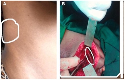
With the diagnosis of primary acquired superior lumbar hernia, the patient underwent open surgery. The open approach was designed due to the absence of laparoscopy services in the hospital. The defect was reduced back and repaired using a retro-muscular or sublay prolene mesh (15 cm by 7.5 cm) as this can maintain the maximum overlap of healthy tissue with the implanted mesh material. The patient was discharged at postoperative day four and followed up for four months. The patient had no recurrence of swelling at four months of follow-up.
Discussion
The confirmation of lumbar hernias was made in 1731. It is a rare case and accounts for less than 2% of all hernias in the abdominal wall.Citation1 Superior lumbar hernia is more common than the inferior lumbar hernia.Citation9 Regarding the clinical manifestation, the patients may present with a variety of signs and symptoms including mass/swelling in the flank area, lower back pain, and symptoms of bowel obstruction.Citation3,Citation10,Citation14,Citation15 Signs and symptoms of intestinal obstruction might also be present if a bowel loop has been incarcerated.Citation10 The patient might complain about a backache, nausea, feeling of weight, or dragging sensation in the lumbar region.Citation11 Sometimes patients might be presented with nonspecific abdominal pain or back pain which could represent sciatica.Citation15 Likewise, the current case also presented with the complaints of right flank pain and had round protruding swelling immediately beneath the 12th rib of the right upper flank area. The swelling was palpable and bowel sound was heard over it.
When there is no obvious bulge in the flank region, ultrasound and magnetic resonance imaging may not establish the diagnosis of lumbar hernias. Ultrasound may confirm the diagnosis when the swelling is palpable.Citation12 Computed tomography scan played the vital role in defining hernia contents accurately.Citation14,Citation16,Citation17 This coincides with our case report in which the ultrasound and magnetic resonance imaging had not confirmed the lumbar hernia. Ultrasound confirmed the diagnosis of superior lumbar hernia when the swelling was noticed by the patient and palpable during physical examination.
Surgery is typically recommended to repair the defect and prevent complications.Citation12 Either an open or laparoscopic procedure can be performed with equivalent success.Citation13 However, the choice of the operative technique depends on the experience of the surgeon and the diameter of the hernia size.Citation1 For primary spontaneous lumbar hernias, minimally invasive techniques are preferred.Citation5 In the current case report, the patient underwent open surgery and the defect was repaired using an underlay prolene mesh. A similar technique has been reported by Sharma and BhatiCitation17 and Ankush et alCitation13 Even though the patient stayed for four days in the hospital and was followed-up for four months as an outpatient, finally, a good result was achieved. Therefore, an appropriate surgical treatment which shortens the length of hospital stay, lower consumption of analgesics, earlier return to normal activity, and less chance of wound infection should be planned.
Conclusion
In conclusion, the diagnosis of lumbar hernias should be considered in all patients that present with complaints of flank pain and swelling in the flank area. In the absence of obvious swelling in the flank region, ultrasound investigation might not establish the diagnosis of lumbar hernias. Hence, ultrasound would be the option to confirm the diagnosis when the swelling is palpable.
Ethical statement
We confirm that any aspect of the work covered in this manuscript that has involved experimental human patient has been conducted with the ethical approval of all relevant bodies and that such approvals are acknowledged within the manuscript. Written informed consent was obtained from the patient for publication of this case report and any accompanying images. Institutional approval was not required for this manuscript.
Acknowledgments
We would like to thank our study subject for her participation in this case report. We also thank Mr Fetene Kassahun and Dr Seid Ahmed (English language instructor at Debre Berhan University, Ethiopia) for their English language editing support.
Disclosure
The authors report no conflicts of interest in this work.
References
- SandersJVCavalcanteJBPereiraIRClinical and surgical anatomy of lumbar hernia: a reviewInt Arch Med20171110
- Moreno-EgeaAGirelaEParlorioEAguayo-AlbasiniJLControversies in the current management of traumatic abdominal wall herniasCirugia Espanola200782526026718021624
- AlvesGRde Andrade SilvaRVCorrêaJRHaygertCJGrynfeltt-Lesshaft a herniaAnn Gastroenterol20122516424714098
- PetersenKSnikerisJHallTSBleichner’s hernia – lumbar herniaAm J Case Rep201314262923569557
- RamkaranCSinghB“Primary Grynfeltt’s-lumbar hernia” – a rare caseIndian J Clin Anatomy Physiol201633382384
- OrcuttTWHernia of the superior lumbar triangleAnn Surg197117322942975541485
- StupalkowskaWPowell-BrettSFKrijgsmanBGrynfeltt-Lesshaft a lumbar hernia: a rare cause of bowel obstruction misdiagnosed as a lipomaJ Surg Case Rep2017913
- HideIGPikeEEUberoiRLumbar hernia: a rare cause of large bowel obstructionPostgrad Med J19997588223123310715766
- DumluETraumatic abdominal wall hernia after a blunt trauma; a case reportJ Clin Anal Med20145144342344
- HsuSDShenKLLiuHDChenTWYuCLumbar hernia: clinical analysis of cases and review of the literatureVisceral Med2008243221224
- RadhakrishnaVTangaSMBilateral superior lumbar hernias: a case report and review of the literatureInt Surg J20174414721474
- SedikAMFathiAMaaliMHusseinAElhoushySA traumatic lumbar hernia: case report and review of the literatureSaudi Surg J2017528486
- SarwalASharmaAKhullarRSoniVBaijalMChowbeyPPrimary lumbar hernia: a rare case report and a review of the literatureAsian J Endosc Surg20181117585902
- LightDGopinathBBanerjeeARatnasinghamKIncarcerated lumbar hernia: a rare presentationAnn R Coll Surg Engl2010923e13e14
- MismarAAl-ArdahMAlbsoulNYounesNUnderlay mesh repair for spontaneous lumbar herniaInt J Surg Case Rep20134653453623608513
- GoyalSSinglaSNakipuriaDRA spontaneous inferior lumbar hernia: rare case report and reviewMOJ Surg2018926061
- SharmaBBhatiTA rare case of lumbar hernia – a case reportJ Dent Med Sci2017161158