
Abstract
Brugada syndrome is a genetic condition that predisposes to an increased risk of ventricular fibrillation and sudden cardiac death in a structurally normal heart. The Brugada type 1 electrocardiogram (ECG) pattern may occur independently of the actual syndrome, and this clinical phenomenon is often referred to as Brugada phenocopy. There are several other factors which have been known to induce this electrocardiographic pattern, and currently, there is a paucity of literature with respect to the pattern that is observed in patients with electrolyte disturbances, specifically hyponatremia. This case report highlights a suspected hyponatremia-induced Brugada type 1 ECG pattern, which subsequently normalized following resolution of the electrolyte derangement.
Introduction
Brugada syndrome (BrS), first described as a clinical entity in 1992, is a genetic condition having a diagnostic electrocardiogram (ECG) pattern which is associated with a heightened risk of ventricular arrhythmias (VAs) and sudden cardiac death (SCD).Citation1 It is considered a primary electrical condition without structural heart disease.Citation2 The ECG pattern consists of ST-segment elevation >2 mm in one or more leads from V1 to V3 with “coved type” descending to inverted T-wave or “saddle back” morphology consistent with Brugada type 1 and type 2 patterns, respectively.Citation3,Citation4 The prevalence varies geographically and ethnically with a male preponderance. The risk stratification and management of patients remains challenging.Citation2
The Brugada type 1 ECG pattern may occur independently of the actual syndrome, and this clinical phenomenon is often referred to as Brugada phenocopy (BrP).Citation2 It is typically recommended that these patients are further risk stratified to determine those having true BrS with potentially fatal arrhythmias vs those with Brugada’s phenocopies as seen in a myriad of clinical situations.Citation5,Citation6
Currently, there is a paucity of literature with respect to the Brugada-type ECG pattern that is observed in patients with electrolyte disturbances, specifically hyponatremia.Citation7–Citation9 This case highlights a highly suspicious severe hyponatremia-induced Brugada type 1 ECG phenocopic pattern, which subsequently normalized following resolution of the electrolyte derangement.
Case report
A 49-year-old Caribbean black gentleman with medical history of hypertension on hydrochlorothiazide and angiotensin-converting enzyme inhibitors presented to the emergency department with acute delirium. There was no pertinent history of syncope, presyncope palpitations, or family history of SCD according to relatives upon admission. The physical examination revealed an obtunded patient with tenuous vital signs with systolic blood pressures of 106 mmHg, tachycardia 112 beats per minute, and pulse oximetry of 96% on room air. Initial laboratory results indicated a serum sodium level of 108 mg/dL (normal 135–145 mg/dL), with other electrolytes being within their respective normal ranges. His serum osmolality was 266 mOsm/kg (normal 275–295 mOsm/kg) and urinary osmolality 586 mOsm/kg (normal 300–900 mOsm/kg). A random urinary sodium was <20 mEq/L.
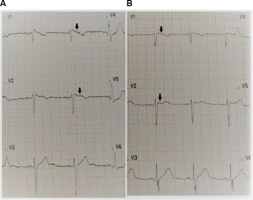
The tentative diagnosis was antihypertensive-induced hypovolemic hyponatremia secondary to the thiazide-like diuretic and angiotensin-converting enzyme inhibitor. The nephrology team was consulted for the management of the severe hyponatremia for which they initiated a 1,000 mL 0.9% normal saline bolus, followed thereafter by a maintenance infusion of 75 mL per hour for 3 days, ensuring an appropriate correction rate of the serum sodium level with surveillance electrolyte testing. On admission, the patient’s ECG displayed a coved ST-segment elevation in V1 and V2, with elevated J points suggestive of a Brugada type 1 pattern (). During his 5-day hospitalization, he underwent extensive investigations which included endocrinologic, infectious, and rheumatologic, all of which were all normal. A chest radiograph was unremarkable and transthoracic echocardiogram displayed mild diastolic dysfunction with left atrial enlargement. Gradually, his electrolyte derangements (hyponatremia and hyposmolality) resolved over the ensuing hospitalization and serial daily ECGs revealed subsequent normalization of the ECG abnormalities (). His neurological status also steadily improved, returning to baseline.
Figure 1 There is the characteristic ST-segment elevation ≥2 mm in ≥1 right precordial lead (V1 to V3), followed by an r'-wave and a straight ST-segment.
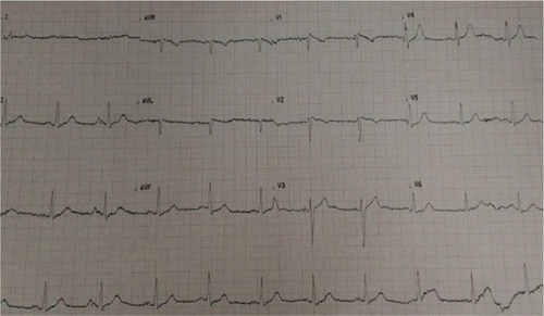
Figure 2 (A) Admission ECG upon presentation indicating a coved ST-segment elevation (black arrows) in V1 and V2, with elevated J points suggestive of a Brugada type 1 pattern. The patient’s serum sodium was 108 mg/dL (normal 135–145 mg/dL). (B) Day 5 ECG indicating resolution of the Brugada type 1 pattern (black arrows) with normalization of serum sodium. The patient’s serum sodium was 135 mg/dL (normal 135–145 mg/dL).
He was subsequently discharged with a modified antihypertensive regimen and early outpatient clinic appointments to both nephrology and cardiology for which he returned to normal, pre-morbid functioning with complete resolution of his hyponatremia. Of note, an outpatient sodium channel blocker test was not performed due to patient declining the procedure during the informed consent discussion amid his concerns of precipitating lethal arrhythmias,Citation10 nor could the patient afford the genetic testing for SCN5A.Citation11,Citation12 The patient and his family had been extensively counseled with respect to accessing online resources and his case submission to the Brugada Phenocopy international registry and online educational portal for further insight into his condition.Citation13
Discussion
It is challenging to ascertain the true burden of BrS due to the unknown prevalence and dynamic variability of ECGs; however, the incidence of the BrS electrocardiographic pattern has ranged from 0.12% to 0.8% in several studies and has accounted for ~4%–12% of all sudden deaths.Citation16 The mainstay of therapy is an implantable cardioverter-defibrillator, but radiofrequency catheter ablation is also considered a novel management strategy.Citation2 Since its first description in 1992, there has been steady progress with respect to the electrophysiological and genetic mechanisms underlying the disease.Citation2
An ECG displaying a BrS type 1 pattern that is triggered by other factors has been called Brugada ECG phenocopy.Citation17 It is imperative to ascertain the distinction between BrS and BrP as it may completely alter a patient’s prognosis and management.Citation13 Several studies provide robust evidence that BrP and BrS ECG patterns are visually identical and indistinguishable, and consequently, an erroneous diagnosis may have a negative impact on patient morbidity and mortality.Citation18–Citation20
There are a multitude of conditions that display BrP with both type 1 and type 2 patterns such as right ventricular ischemia, pulmonary embolism, left anterior descending artery occlusion, right bundle branch block, pectus excavatum, and arrhythmogenic cardiomyopathy.Citation21 Additionally, there are dynamic factors which disrupt transmembrane ionic currents, such as bradycardia and drugs, and can either unmask or exacerbate a BrP ECG pattern.Citation5,Citation22–Citation25 This induction is described as an “acquired form of BrS”. It remains unknown whether acquired BrS is due to individual susceptibility resulting from an increase in latent ion channel dysfunction.Citation6
The literature is not replete with describing the manifestation of BrP ECG pattern in the presence of severe hyponatremia, and currently there are only a few case reports and series that discuss this possible associationCitation5,Citation7,Citation8,Citation26 Mechanistically, it is suspected that severe hyponatremia has a similar potential to diminish the electrochemical ionic gradient, by decreasing the inward sodium current and leaving the transient outward current unopposed. This may result in the loss of the action potential dome in the right ventricular epicardium producing a Brugada phenocopy.Citation27 However, whether induction of the Brugada pattern by severe hyponatremia is associated with increased susceptibility to VAs is currently uncertain.Citation21 This is likely a transient and reversible phenomenon as displayed by the resolution of the electrocardiographic abnormalities when both the serum sodium and transmembrane gradient are normalized over the ensuing hospitalization.
In a study by Xu et al, two cases (7%) displayed BrP with hyponatremia also with a myriad of electrolyte derangements such as acidosis, hyperkalemia, hypocalcemia, hyperphosphatemia, and hyperglycemia. In seven cases (26%), provocative testing using sodium channel blockers was performed, and all failed to reproduce a BrS ECG pattern (BrP class A).Citation7 Additionally, no SCD or malignant VAs were detected.Citation7,Citation28
Our patient’s ECG was mostly consistent with a Brugada “coved type 1” phenocopy for several reasons, although the presence of a negative p-wave component in V2 could possibly suggest electrode malposition.Citation2,Citation4 There is the characteristic ST-segment elevation ≥2 mm in ≥1 right precordial lead (V1 to V3), followed by an rʹ-wave and a straight ST-segment. Additionally, the descending ST-segment crosses the isoelectric line and is followed by a negative and symmetric T-wave. At 40 ms of high takeoff, the decrease in amplitude of ST is ≤4 mm, the duration of QRS is longer than in a right bundle branch block, and there is a mismatch between V1 and V6.Citation3 No high-pass filters were applied to attenuate low-frequency noise.Citation14,Citation15
Our patient was considered to have a low pretest probability of BrS as there was no pertinent medical or familial history of syncope, palpitations, or witnessed nocturnal agonal respiration. He did not undergo provocative testing due to concerns of inducing lethal arrhythmias and SCD.Citation29
As aforementioned, Xu alluded to the fact that all provocative testing failed to replicate a BrP class A patient in similar clinical scenarios. The patient could not afford genetic testing for mutations of the SCN5A gene; however, this is not deemed mandatory since the SCN5A mutation is identifiable in only 20%–30% of probands affected by true BrS.Citation30
As per Anselm et al’s BrP systematic diagnostic criteria, it is likely that our patient did display a type 1, class B, hyponatremia-induced BrP (as drug challenge was not performed) which subsequently resolved after normalization of the electrolyte derangement.Citation31,Citation32 It is imperative to note that while BrS and BrP have identical patterns in precordial leads V1 to V3, patients with BrP have an identifiable underlying condition that elicits these patterns which normalize upon resolution. Furthermore, BrS possesses a high clinical pretest probability of true congenital BrS, whereas conversely, BrP has a low pretest probability based on symptomatology and history. Significantly, patients with BrP have a negative provocative challenge with procainamide, flecainide, or ajmaline, while patients with true congenital BrS have a positive provocative challenge.Citation31
Differentiating between BrS and BrP is important because patients with BrS are at risk of SCD and may require an implantable cardioverter-defibrillator and should refrain from drugs with sodium channel-blocking properties.
Conclusion
In conclusion, it appears likely that severe hyponatremia may induce a suspected Brugada-type ECG phenocopy that reverts to the pre-existing baseline state upon normalization of this electrolyte derangement as evidenced in this case report. Further in-depth research is required to elucidate the mechanistic effects and to determine any prognostic clinical implications.
Copyright permission
The patient has provided written informed consent to have the details of his case published.
Institutional approval
Institutional approval was not required for publication of this case report.
Compliance with ethical standards
All procedures performed in studies involving human participants were in accordance with the ethical standards of the institutional and/or national research committee and with the 1964 Helsinki declaration and its later amendments or comparable ethical standards.
Author contributions
All authors contributed to data analysis, drafting or revising the article, gave final approval of the version to be published, and agree to be accountable for all aspects of the work.
Disclosure
The authors report no conflicts of interest in this work.
References
- BrugadaPBrugadaJRight bundle branch block, persistent ST segment elevation and sudden cardiac death: a distinct clinical and electrocardiographic syndrome. A multicenter reportJ Am Coll Cardiol1992206139113961309182
- BrugadaJCampuzanoOArbeloESarquella-BrugadaGBrugadaRPresent status of Brugada syndrome: JACC state-of-the-art reviewJ Am Coll Cardiol20187291046105930139433
- Bayés de LunaABrugadaJBaranchukACurrent electrocardiographic criteria for diagnosis of Brugada pattern: a consensus reportJ Electrocardiol201245543344222920782
- de LunaABGarcia-NieblaJBaranchukANew electrocardiographic features in Brugada syndromeCurr Cardiol Rev201410317518024827804
- YapYGBehrERCammAJDrug-induced Brugada syndromeEuropace200911898999419482855
- ShimizuWAcquired forms of the Brugada syndromeJ Electrocardiol2005384 Suppl222516226070
- XuGGottschalkBHAnselmDDRelation of the Brugada phenocopy to hyperkalemia (from the International registry on Brugada phenocopy)Am J Cardiol2018121671571729397883
- AlvarezPAVázquez BlancoMLermanJBrugada type 1 electrocardiographic pattern induced by severe hyponatremiaCardiology201111829710021540589
- TameneASattirajuSWangKBendittDGBrugada-like electrocardiography pattern induced by severe hyponatraemiaEuropace201012690590720185483
- UeokaAMoritaHWatanabeAPrognostic significance of the sodium channel blocker test in patients with Brugada syndromeJ Am Heart Assoc2018710e00861710
- Sarquella-BrugadaGCampuzanoOArbeloEBrugadaJBrugadaRBrugada syndrome: clinical and genetic findingsGenet Med201618131225905440
- AntzelevitchCPatocskaiBBrugada syndrome: clinical, genetic, molecular, cellular, and ionic aspectsCurr Probl Cardiol201641175726671757
- GottschalkBHAnselmDDBaranchukABrugada phenocopy international registry and online educational portal2014 Available from: http://www.brugadaphenocopy.comAccessed November 30, 2018
- García-NieblaJLlontop-GarcíaPValle-RaceroJISerra-AutonellGBatchvarovVNde LunaABTechnical mistakes during the acquisition of the electrocardiogramAnn Noninvasive Electrocardiol200914438940319804517
- KnightBPPelosiFMichaudGFStrickbergerSAMoradyFClinical consequences of electrocardiographic artifact mimicking ventricular tachycardiaN Engl J Med1999341171270127410528037
- QuanXQLiSLiuRZhengKWuXFTangQA meta-analytic review of prevalence for Brugada ECG patterns and the risk for deathMedicine (Baltimore)20169550e564327977610
- BaranchukANguyenTRyuMHBrugada phenocopy: new terminology and proposed classificationAnn Noninvasive Electrocardiol201217429931423094876
- GottschalkBHAnselmDDBrugadaJExpert cardiologists cannot distinguish between Brugada phenocopy and Brugada syndrome electrocardiogram patternsEuropace20161871095110026498159
- GottschalkBHBaranchukADifferentiation between Brugada syndrome and Brugada phenocopy ECG patterns: is it possible?Brugada Phenocopy20181119124
- Al-KhatibSMStevensonWGAckermanMJ2017 AHA/ACC/ HRS guideline for management of patients with ventricular arrhythmias and the prevention of sudden cardiac death: Executive summary: a report of the American College of Cardiology/American Heart Association Task Force on clinical practice guidelines and the heart rhythm SocietyHeart Rhythm20181510e190e25229097320
- Moncayo-ArlandiJBrugadaRUnmasking the molecular link between arrhythmogenic cardiomyopathy and Brugada syndromeNat Rev Cardiol2017141274475628703223
- PostemaPGNevilleJde JongJSRomeroKWildeAAWoosleyRLSafe drug use in long QT syndrome and Brugada syndrome: comparison of website statisticsEuropace20131571042104923533266
- DumaineRTowbinJABrugadaPIonic mechanisms responsible for the electrocardiographic phenotype of the Brugada syndrome are temperature dependentCirc Res199985980380910532948
- YanGXAntzelevitchCCellular basis for the Brugada syndrome and other mechanisms of arrhythmogenesis associated with ST-segment elevationCirculation1999100151660166610517739
- JunttilaMJGonzalezMLizotteEInduced Brugada-type electrocardiogram, a sign for imminent malignant arrhythmiasCirculation2008117141890189318391123
- AgrawalYAggarwalSKalavakuntaJKGuptaVAll that looks like “Brugada” is not “Brugada”: Case series of Brugada phenocopy caused by hyponatremiaJ Saudi Heart Assoc201628427427727688678
- MaheshwariASreenivasanSBendittDBrugada Phenocopy: The Art of Recognizing the Brugada ECG Pattern1st edElsevier20188792 Available from: https://market.android.com/details?id=book-kyk0D-wAAQBAJAccessed November 30, 2018
- Abu ShamaRBayes de LunaABaranchukATachycardia-dependent Brugada phenocopy due to hyperkalemiaJ Cardiovasc Electrophysiol20172891084108528603943
- AntzelevitchCBrugadaPBorggrefeMBrugada syndrome: report of the second consensus conference: endorsed by the heart rhythm society and the European heart rhythm associationCirculation2005111565967015655131
- ProbstVWildeAABarcJSCN5A mutations and the role of genetic background in the pathophysiology of Brugada syndromeCirc Cardiovasc Genet20092655255720031634
- AnselmDDBaranchukATerminological clarification of Brugada phenocopy, Brugada syndrome, and the Brugada ECG pattern: Re. early repolarization pattern in patients with provocable Brugada phenocopy: a marker of additional arrhythmogenic cardiomyopathyInt J Cardiol2014171228824360162
- AnselmDDGottschalkBHBaranchukABrugada phenocopies: consideration of morphologic criteria and early findings from an international registryCan J Cardiol201430121511151525418217