
Abstract
Background
Asthma and bronchiectasis are different conditions that frequently coexist. The prevalence of bronchiectasis rises considerably in subjects with severe asthma (25%–51%).
Objective
We evaluated the clinical and biological efficacy of mepolizumab on our pilot population of severe uncontrolled asthmatics with bronchiectasis not related to other pathologies.
Patients and methods
Four patients with severe uncontrolled asthma and diagnosed as bronchiectasis were recruited and started biological treatment with mepolizumab. Standard investigations were performed in all four patients at baseline (T0), after 3 months (T1) and after 1 year (T2) of treatment.
Results
After 1 year (T2) of therapy with mepolizumab, patients showed a significant increment of asthma control test value (12±1.1 vs 24.5±0.3, P<0.01), a reduction of the number of exacerbations/year (5±0.7 vs 0.75±0.75, P<0.01), an increase of pre-bronchodilator FEV1 (1,680±500 vs 1,860±550 mL, P<0.01) and a reduction of eosinophils in blood (0.75±0.14 vs 0.12±0.02 cells/µL, P<0.01), in the sputum (9.6%±2.1% vs 5.6%±2.7%, P<0.05) and in nasal cytology (++ vs +).
Conclusion
The efficacy of mepolizumab in terms of reduction of inflammation and increase of control that we observed in our patients might suggest that targeting the IL-5 in severe eosinophilic asthma with bronchiectasis may be a good therapeutic strategy.
Introduction
Severe asthma is reported, to date, in 5%–15% of the total asthmatic population and is estimated to account for >60% of the total costs of the disease, primarily required for medications and asthma exacerbations. Severe asthma is frequently accompanied by a difficult and often unachievable control that requires critical management. However, today, the availability of biological drugs allows for a more modern therapy tailored to the patient. Although severe asthma still remains a challenge for specialists, the target therapy gave interesting clinical results when prescribed for the selected patients. Today, the phenotypic characterization of severe asthmatic patients has, therefore, become crucial in view of a different management and therapeutic approach.Citation1 The extreme clinical, biological and inflammatory heterogeneity of severe asthma has required the application of cluster analyses that, considering multiple comorbidities and risk factors, divide accordingly severe asthmatic patients into homogenous groups. The result is the identification of several phenotypes of severe asthma as those associated to obesity, exercise, smoking habit, aspirin, allergens, nasal polyposis, menopause, infections and others.Citation2 The investigation and treatment of comorbidities is of major importance in patients with severe asthma, as they contribute to poor disease control.
Severe uncontrolled asthma associated with bronchiectasis is an emerging phenotype. Recent epidemiological studies showed an unexpected prevalence of severe asthma with bronchiectasis that ranges from 2.2%Citation3 to 77%.Citation4 Almost one-third of severe asthmatic patients showed the presence of bronchiectasis on the chest computed tomography (CT) scan. Some groups have described bronchiectasis as a comorbidity associated to asthma,Citation5 while others have identified asthma as being one of many etiologies of bronchiectasis.Citation6
This phenotype has been described in the Type 2 (T2) endotype and particularly in patients with severe, late-onset eosinophilic asthma. It has been mainly observed in women and the elderly, in non-smokers, and it is often associated with an impaired lung function. Identification of bronchiectasis in asthmatic subjects could lead significantly to modifications to both therapy and prognosis.Citation7,Citation8
At the moment, to our knowledge, no study exists suggesting one specific therapeutic approach in these patients, leaving the choice to the specialist, absolutely personal. These patients usually have a severe and uncontrolled asthma that does not respond to the maximum pharmacological therapy as indicated by steps 4 and 5 of Global Initiative for Asthma (GINA) guidelines; thus, a step up with biologics could be appropriately proposed.Citation9
In this study, we focused on our small population of patients with severe asthma and bronchiectasis that we started on biological therapy with the anti-IL5, mepolizumab.
Mepolizumab is a humanized monoclonal antibody used for the treatment of severe eosinophilic asthma (SEA), which recognizes and blocks IL-5, a signaling protein of the immune system.
The aim of this study was to evaluate the clinical and biological efficacy of mepolizumab in patients with severe uncontrolled asthma and bronchiectasis not related to other pathologies.
Patients and methods
Patients
This prospective pilot study included four consecutive patients with severe asthma and diagnosed as having bronchiectasis by means of chest CT scan and a compatible clinical history consistent with chronic sputum production and/or frequent respiratory infections, who were followed up at the accredited outpatient pulmonary clinic for severe asthma of the Institute of Respiratory Diseases of the University of Foggia, Italy during a period of 1 year. Written informed consent was obtained from all subjects, and the Institutional Ethics Committee of the University of Foggia approved the study, which was carried out according to the Declaration of Helsinki. Asthmatics were classified and treated according to GINA guidelines.Citation9 Severe asthmatics were diagnosed according to the European Respiratory Society/American Thoracic Society taskforce criteriaCitation10 on severe asthma. All four patients of the study were treated with high-dose inhaled corticosteroids (ICS) with long-acting β-agonists (LABAs) and long-acting muscarinic receptor antagonists (LAMAs) and started biological treatment with mepolizumab at enrollment, respecting the prescribing criteria. Once the diagnosis was confirmed, additional standard investigations were performed on all four patients with severe asthma (familiar, personal and pathological anamnesis, lung function tests, prick tests, serum eosinophils count, total serum IgE, induced sputum and nasal cytology analysis, fractional exhaled NO measurement). We used the asthma control test (ACT) questionnaire to evaluate the control of symptoms. All measurements were done at baseline (T0), after 3 months (T1) and after 1 year of treatment with mepolizumab (T2).
All subjects enrolled underwent all tests foreseen by the guidelinesCitation8 to identify the etiology of bronchiectasis, including high-resolution chest CT (HRCT): α1-antitripsin deficiency, allergic bronchopulmonary aspergillosis, cystic fibrosis (CF), immunodeficiency and systemic disease. Patients with these conditions, smokers and former smokers were excluded because in this research, we were looking for the presence of bronchiectasis related to asthma and not associated to other pathologies.
Asthmatics enrolled were assessed during a period of stability and at least 4 weeks after an upper respiratory tract infection. No patient at enrollment was treated with oral corticosteroids.
At the first visit, a complete baseline questionnaire requesting information on medical history was administered to all subjects, who were then given a physical examination, atopy assessment and spirometry with bronchodilator reversibility test. During the second visit, subjects underwent blood analysis for eosinophil count and total IgE levels, fractional exhaled nitric oxide (FeNO) measurement, ACT and, finally, sputum induction and nasal cytology.
Atopic status
The skin prick test (SPT) was performed for a panel of inhalant allergens as previously described for common aero-allergens (Lofarma, Milan, Italy). SPT results were considered positive when eliciting a wheal diameter ≥3 mm, using negative (saline) and positive (histamine 10 mg/mL) controls for interpretation.Citation11
Lung function
Pulmonary function tests were performed. FEV1 and FVC were measured using a spirometer (Sensormedics, Milan, Italy). The best value of three maneuvers was expressed as a percentage of the predicted normal value. After baseline evaluation, spirometry was repeated 15 minutes after the subjects had inhaled 400 µg of salbutamol. Reversibility of airway obstruction was expressed in terms of the percentage changes from baseline of the FEV1.Citation12
Induced sputum collection and processing
According to the method described by Toungoussova et al,Citation13 sputum was induced through inhalation of hypertonic saline solution (4.5%) with an ultrasonic nebulizer (DeVilbiss 65; DeVilbiss Corporation, Somerset, PA, USA) and analyzed after selection of mucus plugs. In two subjects, we used the spontaneous sputum.
All subjects enrolled were able to produce adequate sputum samples (defined as containing at least 500 nonsquamous cells). The sputum (spontaneous or induced) was used for cytological analysis.
Measurement of FeNO
Measurement of FeNO was performed according to recent guidelines.Citation14 The Medisoft FeNO device (HYPAIR; Medisoft, Dinant, Belgium), which is semi-portable for repeatable multi-flow measurement of exhaled NO with off-line measurement, was used. It has a software package that provides step-by-step online quality control. The measurement range is 0–600 ppb. Exhaled NO (FeNO) was measured using a previously described restricted breath technique, which employed expiratory resistance and positive mouth pressure to close the velum and exclude nasal NO, and a constant expiratory flow of 50 and 350 mL/s. Repeated exhalations were performed until three plateaus agreed within 5% of inter-observation difference.
Nasal cytology
A scraping of the nasal mucosa on the middle third of the inferior turbinate was performed. After sampling, the material was laid on a microscope slide, air dried and stained using the May–Grunwald–Giemsa method. The smear was observed under a common light microscope equipped with 1,000× objective. In order to analyze the rhino-cytogram, we performed a reading for fields (not <50), in order to observe the cellular elements that composed the nasal mucosa (eosinophils, mast cells, neutrophils, lymphocytes, bacteria, spores, biofilms and so on). To calculate the percentage of each cellular element, semi-quantitative grading counts of each cell type, according to the previously published studies, were performed (grade 1+: occasional groups, grade 2+: moderate number, grade 3+: easily visible, grade 4+: elevated number).Citation15,Citation16
Statistical analysis
Descriptive statistics (ie, means, SDs, percentages) were applied to summarize the continuous and categorical variables. In order to assess the difference between T0 and T2 for all variables analyzed, we performed a Wilcoxon matched-pairs signed rank test, considering a P-value of <0.05 statistically significant. Data were analyzed using GraphPad for MAC.
Results
Demographic, clinical, functional and biological data are shown in . The patients enrolled were four females, with late-onset severe asthma and diagnosed as bronchiectasis (). At enrollment (T0), the FENO50 was 29.2±7.5 ppb, FENO350 was 11.2±4.3 ppb, blood eosinophil count was 750±141.6 cells/mm3, sputum eosinophils were 9.6%±2.1%, FEV1 was 1,680±500 mL and the ACT questionnaire was 12±1.1. The nasal cytology showed a moderate degree of eosinophils (++) and the absence of nasal polyps or rhinosinusitis.
Figure 1 Chest CT scan before treatment (T0).
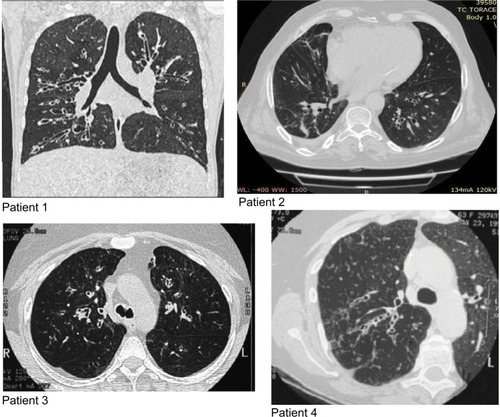
Table 1 Anthropometric and clinical data of patients at baseline (T0) and after 1 year (T2) of treatment with mepolizumab
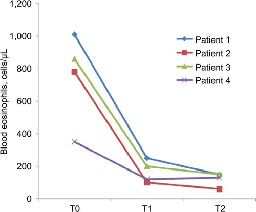
After 1 year (T2) of mepolizumab treatment, patients showed a significant increment of ACT (12±1.1 vs 24.5±0.3, P<0.01; ), a reduction of number of exacerbations/year (5±0.7 vs 0.75±0.75, P<0.01; ), an increase of pre-bronchodilator FEV1 (1,680±500 vs 1,860±550 mL, P<0.01; ) and a reduction of eosinophils in blood (0.75±0.14 vs 0.12±0.02 cells/µL, P<0.01; ), in the sputum (9.6%±2.1% vs 5.6%±2.7%, P<0.05; ) and in nasal cytology (++ vs+; ).
Figure 2 ACT value before treatment (T0), after 3 months (T1) and after 1 year (T2) of treatment with mepolizumab.
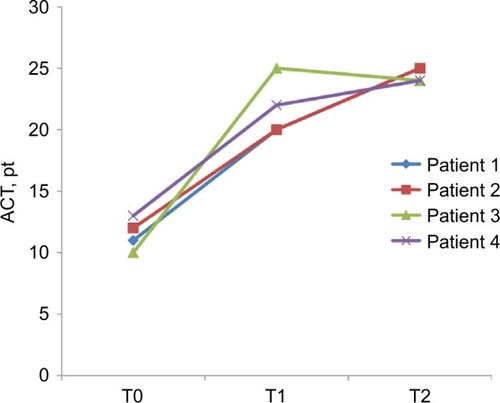
Figure 3 Number of exacerbations/year of asthma before treatment (T0), after 3 months (T1) and after 1 year (T2) of treatment with mepolizumab.
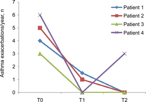
Figure 4 ΔFEV1 preBD vs baseline (mL, %), before treatment (T0), after 3 months (T1) and after 1 year (T2) of treatment with mepolizumab.
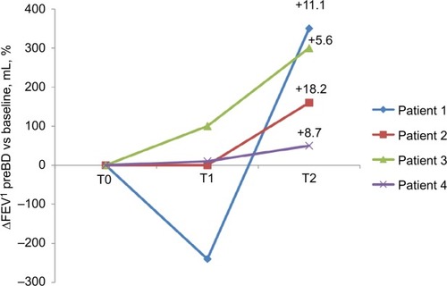
Figure 5 Eosinophils in the sputum (%) before treatment (T0), after 3 months (T1) and after 1 year (T2) of treatment with mepolizumab.
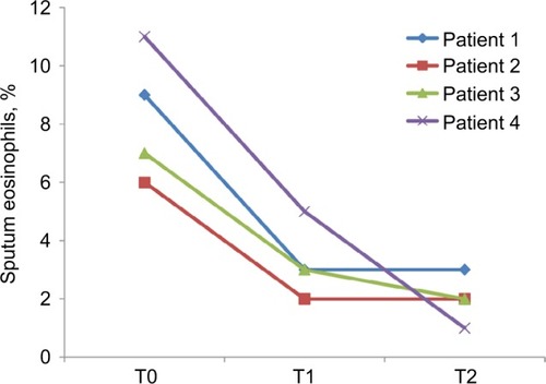
Figure 6 Eosinophils in blood (cells/μL) before treatment (T0), after 3 months (T1) and after 1 year (T2) of treatment with mepolizumab.
All patients, during treatment with mepolizumab, underwent a step down of inhalation therapy, suspending LAMA (100%) and reducing ICS to lower dose (70%).
Discussion
In this pilot study, we demonstrated the efficacy of mepolizumab in an emerging phenotype of patients with severe uncontrolled asthma and diagnosed bronchiectasis not related to other pathologies in terms of reduction of eosinophils and increasing control of the disease.
As previously reported, we confirmed prevalence of female gender in this phenotype as all subjects enrolled were women. Girón et alCitation17 demonstrated that bronchiectasis of unknown etiology and secondary to asthma was more common in women. Gender differences in bronchiectasis are reported in CF disease: females with CF have a more severe form of the disease, poorer lung function and earlier Pseudomonas aeruginosa colonization and conversion to its more aggressive mucoid form. Chotirmall et alCitation18 showed that immune responses in the CF airway are further compromised by estrogen which supresses the protective acute inflammatory burst necessary to clear bacterial infection and prevent exacerbation. As non-CF bronchiectasis usually presents in females who have undergone menopause, the role of estrone, the major estrogen of menopause, should be investigated in future studies as currently no evidence exists to link hormones to the severity of non-CF bronchiectasis in affected females.Citation19
We further confirmed that the coexistence of severe asthma and bronchiectasis is typical of elderly patients with late-onset non-atopic severe uncontrolled eosinophilic asthma.Citation20 Accordingly to Olveira et al,Citation21 the prevalence of bronchiectasis rises considerably in subjects with more severe asthma, as evident from the poorer lung function and lower control of the disease of the four subjects enrolled in our study. As in this study, Coman et alCitation20 further proved the higher incidence of exacerbations in patients with this phenotype, reporting more hospitalizations in severe asthmatics with bronchiectasis. This suggests that the coexistence of both pathologies worsens outcomes.
While early studies documented a relationship between asthma, bronchiectasis and atopy,Citation22 many have not,Citation23 even when specifically looking at subjects with severe asthma, as in our study. Furthermore, our patients did not have rhinosinusitis and nasal polyps and we could explain it with the hypothesis of Shteinberg et alCitation24 that the involvement of the upper airway in patients with bronchiectasis is associated with an early-onset and atopic asthma.
All patients enrolled showed an eosinophilic inflammatory pattern. Although bronchiectasis is usually associated with neutrophilic inflammation,Citation25,Citation26 we know that when there is a coexistence of asthma, the inflammation might be eosinophilic or mixed (neutrophilic–eosinophilic).
The presence of bronchiectasis in patients with an eosinophilic profile has also been described in patients without asthma with a prevalence of 17.5%.Citation27 Those patients with eosinophilic bronchiectasis also showed higher sputum IL-13, further proving the T2 endotype pattern, where this interleukin plays a key role. In their study, Tsikrika et alCitation27 further described low concentration of FENO in bronchiectasis patients, although they had T2 endotype with increased eosinophils in airways and blood. We, therefore, were not surprised to find exactly the same normal level of FENO in our population despite an increased eosinophilic profile. Our explanation agrees with the hypothesis that eosinophilic inflammation in asthma with bronchiectasis is not primarily Th2 driven and possibly another T2-high pathway through Group 2 Innate Lymphoid Cells (ILC2s) plays a role in eosinophilic inflammation.Citation28
As known, environmental stimuli, including pollutants, viruses, helminths and bacterial infections, such as the ones that characterized the history of bronchiectasis, trigger ILC2s-mediated T2 airways inflammation related to airway inflammation. These stimuli induce airway epithelial cell death and production of IL-25, IL-33 and thymic stromal lymphopoietin. These cytokines promote massive expansion of ILC2s. ILC2s produce Th2 cytokines, including IL-5, IL-9 and IL-13 that are involved in the activation/proliferation of eosinophils and mucus hypersecretion/tissue remodeling, respectively.Citation29
The recent expansion in our understanding of the context of IL-5 and IL-5-producing ILC2s in eosinophil activation and the pathogenesis of eosinophil-dependent inflammatory diseases has led to advances in therapeutic options.
Mepolizumab was the first therapeutic humanized monoclonal antibody to human IL-5 to be approved for any indication globally. Mepolizumab acts by preventing IL-5 from binding to IL-5Ra complex expressed on the eosinophil cell surface. This action inhibits IL-5 signaling and the overexpressing functions of peripheral blood and tissue eosinophils.
For the first time, to our knowledge, we demonstrated the efficacy of mepolizumab in patients not only with SEA but also with diagnosed bronchiectasis, in terms of reduction of eosinophils and increasing control of the disease. We could explain these results with the hypothesis that the pathogenesis of this phenotype of patients is driven by ILC2s related to airway inflammation that respond well to the biological therapy with mepolizumab, and it is not influenced by other inflammatory cells such as neutrophils.
It is difficult to determine if the severity of asthma in subjects with coexistent bronchiectasis is due to the presence of bronchiectasis, or if the presence of bronchiectasis is the cause of the severity of asthma itself. As we observed higher levels of peripheral eosinophils in our patients with coexistent bronchiectasis, we suggest that the latter hypothesis is the most probable.Citation20
Our findings suggest that the presence of bronchiectasis could be another criterion to identify an emerging phenotype of SEA that has been attracting increased interest for the unexpected high prevalence observed in asthmatic population and that we need more attention with a large dedicated study designed to better explore it.
Bronchiectasis non-CF is treated in clinical practice with therapy based on very little evidence.Citation30 In our opinion, development of new treatments is urgently needed. Identifying different phenotypes of bronchiectasis may help us in directing therapy toward specific target with better results compared to that actually expected by traditional therapy. The efficacy of mepolizumab that we observed in patients with SEA and bronchiectasis might suggest that targeting the IL-5 in eosinophilic bronchiectasis may be a good therapeutic strategy and may support further larger studies in this direction.
The limitations of our study are mainly related to the small number of cases and to the absence of a comparison with a control group of patients with SEA without bronchiectasis and a group of male subjects. The bronchiectasis documented by HRCT scan was furthermore not classified or scored according to severity and was all included in the analysis, clinically significant or not.
According to Coman et al,Citation20 we believe the extent of the immune–pathological roles of the eosinophils in SEA has yet to be fully understood, and prospective studies on routine CT scan and active treatment of bronchiectasis in subjects with severe asthma are needed to help guide management in this population.
Author contributions
GEC, GS and GC designed the study. GS and DL contributed to the clinical and laboratory work for the study. GEC, GS, GC and MPFB drafted the article and revised it critically for important intellectual content. GEC, GS, GC and DL contributed to final approval of the version to be published. All authors contributed to data analysis, drafting and revising the article, gave final approval of the version to be published, and agree to be accountable for all aspects of the work.
Acknowledgments
Giovanna E Carpagnano and Giulia Scioscia are co-first authors for this work.
Disclosure
GC is a full-time employee for GlaxoSmithKline, Italy. The authors report no other conflicts of interest in this work.
References
- KupczykMWenzelSU.S. and European severe asthma cohorts: what can they teach us about severe asthma?J Intern Med2012272212113222630041
- ChungKFAdcockIMHow variability in clinical phenotypes should guide research into disease mechanisms in asthmaAnn Am Thorac Soc201310 SupplS109S11724313760
- KangHRChoiG-SParkSJThe effects of bronchiectasis on asthma exacerbationTuberc Respir Dis (Seoul)201477520921425473408
- LynchDANewellJDTschomperBACinkTMNewmanLSBethelRUncomplicated asthma in adults: comparison of CT appearance of the lungs in asthmatic and healthy subjectsRadiology199318838298338351357
- BisaccioniCAunMVCajuelaEKalilJAgondiRCGiavina-BianchiPComorbidities in severe asthma: frequency of rhinitis, nasal polyposis, gastroesophageal reflux disease, vocal cord dysfunction and bronchiectasisClinics200964876977319690661
- DimakouKTriantafillidouCToumbisMTsikritsakiKMalagariKBakakosPNon CF-bronchiectasis: aetiologic approach, clinical, radiological, microbiological and functional profile in 277 patientsRespir Med20161161727296814
- PatelISSeemungalTARWilksMLloyd-OwenSJDonaldsonGCWedzichaJARelationship between bacterial colonisation and the frequency, character, and severity of COPD exacerbationsThorax200257975976412200518
- Martínez-GarcíaM-Ade La Rosa CarrilloDSoler-CataluñaJ-JPrognostic value of bronchiectasis in patients with moderate-to-severe chronic obstructive pulmonary diseaseAm J Respir Crit Care Med2013187882383123392438
- (*NEW) 2018 GINA Report: Global Strategy for Asthma Management and Prevention | Global Initiative for Asthma – GINA Available from: https://ginasthma.org/gina-reports/Accessed February 19, 2019
- ChungKFWenzelSEBrozekJLInternational ERS/ATS guidelines on definition, evaluation and treatment of severe asthmaEur Respir J201443234337324337046
- BousquetJHeinzerlingLBachertCPractical guide to skin prick tests in allergy to aeroallergensAllergy2012671182422050279
- Standardization of Spirometry, 1994 UpdateAmerican Thoracic SocietyAm J Respir Crit Care Med19951523110711367663792
- ToungoussovaOMiglioriGBFoschino BarbaroMPChanges in sputum composition during 15 min of sputum induction in healthy subjects and patients with asthma and chronic obstructive pulmonary diseaseRespir Med200710171543154817258444
- American Thoracic Society/European Respiratory SocietyATS/ERS recommendations for standardized procedures for the online and offline measurement of exhaled lower respiratory nitric oxide and nasal nitric oxide, 2005Am J Respir Crit Care Med2005171891293015817806
- GelardiMFiorellaMLLeoGIncorvaiaCCytology in the diagnosis of rhinosinusitisPediatr Allergy Immunol200718s18505217767609
- MeltzerEOEvaluating rhinitis: clinical, rhinomanometric, and cytologic assessmentsJ Allergy Clin Immunol1988825 Pt 29009083057042
- GirónRMde Gracia RoldánJOlveiraCSex bias in diagnostic delay in bronchiectasis: an analysis of the Spanish historical registry of bronchiectasisChron Respir Dis201714436036928393532
- ChotirmallSHGreeneCMOglesbyIK17beta-estradiol inhibits IL-8 in cystic fibrosis by up-regulating secretory leucoprotease inhibitorAm J Respir Crit Care Med20101821627220378727
- VidaillacCYongVFLJaggiTKSohM-MChotirmallSHGender differences in bronchiectasis: a real issue?Breathe201814210812129875830
- ComanIPola-BibiánBBarrancoPBronchiectasis in severe asthmaAnn Allergy Asthma Immunol2018120440941329496464
- OlveiraCPadillaAMiguel-ÁngelMartínez-GarcíaEtiología de las bronquiectasias en Una cohorte de 2.047 pacientes. Análisis del registro histórico español. [Etiology of bronchiectasis in a cohort of 2,047 patients. An analysis of the spanish historical bronchiectasis registry]Arch Bronconeumol201753736637428118936
- SäynäjäkangasOKeistinenTTuuponenTKiveläSLLinks between hospital diagnoses of bronchiectasis and asthmaAllergy19975211112011229404566
- OguzulgenIKKervanFOzisTTurktasHThe impact of bronchiectasis in clinical presentation of asthmaSouth Med J2007100546847117534081
- ShteinbergMNassrallahNJrbashyanJUriNSteinNAdirYUpper airway involvement in bronchiectasis is marked by early onset and allergic featuresERJ Open Res20184100115201729362708
- JatakanonALimSKharitonovSAChungKFBarnesPJCorrelation between exhaled nitric oxide, sputum eosinophils, and methacholine responsiveness in patients with mild asthmaThorax199853291959624291
- ColePJInflammation: a two-edged sword – the model of bronchiectasisEur J Respir Dis Suppl19861476153533593
- TsikrikaSDimakouKPapaioannouAIThe role of non-invasive modalities for assessing inflammation in patients with non-cystic fibrosis bronchiectasisCytokine20179928128628863927
- Padilla-GaloAOlveiraCFernández de Rota-GarciaLFactors associated with bronchiectasis in patients with uncontrolled asthma; the NOPES score: a study in 398 patientsRespir Res20181914329548297
- YanagibashiTSatohMNagaiYKoikeMTakatsuKAllergic diseases: from bench to clinic – contribution of the discovery of interleukin-5Cytokine201798597028863833
- ChalmersJDChotirmallSHBronchiectasis: new therapies and new perspectivesLancet Respir Med20186971572629478908