
Abstract
Introduction
Maternal red cell alloimmunization to Rh antigen in pregnant women occurs when the immune system is sensitized by foreign red blood cell surface antigens, in this case fetal red blood cells, inducing an immune response. Various antigens of blood group systems may cause alloimmunization, especially the Rh, Kel, Fy, JK, and MNS systems. This study aimed to determine alloimmunization to the different frequencies of Rh antigen among pregnant women in South Western Uganda.
Methods
A total of 1369 pregnant women consented and were recruited into a cross-sectional study during their regular antenatal visits during the period August 2020 to July 2021. Samples (4 mL) of anticoagulated and coagulated blood were obtained, and Rh blood grouping including Rh antigen and the indirect antiglobulin test (IAT) was carried out using the agglutination technology of the LISS ID-Card technique in the Ortho Biovue ID-Micro Typing System.
Results
Out of 1369 participants recruited to the study, 78 (5.7%) were D−, 1291 were D+, and 134 (9.8%) had alloantibodies. Among those with alloantibodies, 115 (85.8%) were D+ and 19 (14.2%) D−. The percentage alloimmunization according to the Rh antigens was highest in e (9.72%), c (2.48%), C (2.34%) and E (0.94%) antigens. With the ABO system, alloimmunization was highest in blood group B (10.7%), followed by A (10.6%), O (9.2%) and then AB (7.1%). Alloimmunization was more prevalent in D− (24%) than in D+ participants (8.9%). Rhesus antigen e was the most prevalent antigen (99.8%), followed by c. The alloimmunization rate of 9.8% among these participants is high, and appears in both D+ and D− women. The other Rhesus antigens are seen to cause alloimmunization, with antigen e causing the highest prevalence. In conclusion, there is a need to identify antibodies and study the outcome for clinical significance, especially in D+ women, to facilitate proper pregnancy management.
Introduction
Antibodies of blood groups are immunoglobulins that are produced to react with corresponding antigens, mainly on the surface of red blood cells. These may be acquired naturally or through immunization in response to antigens present on red cells that are foreign for the individual.Citation1 The ABO blood group system was first described by Karl Landsteiner in 1900; since then, more than 60 different red blood cell antigens have been discovered and linked to eliciting an antibody response, which is commonly referred to as alloimmunization, with anti-D being the most commonly reported antigen.Citation2
Alloimmunization is known to result from the genetic difference in red blood cell antigens between the fetus an the mother, or in patients with multiple transfusions. This alloimmunization occurs due to exposure of foreign red cell antigens inherited by the fetus from father to the antenatal immune system and subsequent sensitization. The Rhesus (Rh) antigen is the most common cause of this antibody response (alloimmunization), followed by ABO, while the Kidd, Kell, and Duffy systems are less common causes.Citation3,Citation4 Exposure during gestation occurs as a result of the transplacental crossing of fetal red blood cells during delivery, miscarriage, or ectopic pregnancy, or when an invasive procedure such as amniocentesis is carried out.Citation1 Age, gravidity/parity, and poor obstetric/gynecological history are other factors that are linked to alloimmunization in pregnant women.
Rh alloimmunization and hemolytic disease of the fetus and newborn are closely linked; this was first described by Levine and later by Louis.Citation5 The antigens that are commonly linked with perinatal hemolytic disease of the fetus and newborn are mainly of the Rh system, and include D, C, E, c, e, f, and Cw, among others. ABO antibodies are mainly of the IgM type and do not commonly cause hemolytic disease of the fetus and newborn, as they cannot cross the placenta, but ABO-related hemolytic disease of the fetus and newborn has been reported to occur in group O mothers with high titers of naturally occurring IgA anti-A or B. Hemolytic disease of the fetus and newborn commonly follows antigenic exposure by transfusion, or fetomaternal hemorrhage or leakage.Citation1,Citation6
In South Western Uganda, the distribution of the ABO and Rh antigens has been determined, with O (49.4%) being the highest in the ABO system and e (99.8%) antigen the most common in the Rh antigen system, but the prevalence of Rh alloimmunization is not known.Citation7 The prevalence of alloantibodies and related hemolytic disease of the fetus and newborn varies from population to population, and there are very limited data on this subject in this part of the country. Determination of the prevalence of maternal alloimmunization to Rh antigens in both the D+ and the D− populations of pregnant women during the antenatal period is key in the control of hemolytic disease of the newborn.
During antenatal visits, Rh alloimmunization is not routinely screened for and antigen typing is restricted to D phenotype screening only. Young women of child-bearing age with recurrent pregnancies in South Western Uganda are at higher risk of developing hemolytic disease of the newborn as a result of antibodies against non-screened Rh antigens, which can be fatal.Citation8 This study aimed to obtain information on the prevalence of alloimmunization and how it is distributed within the different Rh blood phenotypes among pregnant women, to provide a baseline for policy information and future planning towards pregnancy management, as a way of preventing hemolytic disease of the newborn.Citation9–11
Materials and Methods
Between August 2020 and July 2021, a cross-sectional, descriptive study was conducted in which a total of 1396 pregnant women attending an antenatal clinic were recruited after providing their written informed consent. Samples (4 mL) of anticoagulated and coagulated blood were collected. Screening for Rh antigen and detection of the presence of alloantibodies was carried out using the direct antigen grouping and indirect antiglobulin test (IAT), both employing the agglutination technique, in which glass beads and reagents are contained in a column of gel to capture agglutinates in a semi-solid medium. These tests were performed using the Ortho Biovue ID-Micro Typing System (Ortho Clinical Diagnostics, Raritan, NJ, USA).
Internal quality control was achieved by including both known positive and negative controls, in accordance with the relevant guidelines for quality assurance. The study identification number and individual initials were used to identify ID-Cards. The aluminum foil was removed, then 50 µL cell suspension of 0.8% concentration was added, followed by 40 µL of the participant’s sample, to the respective microtubes, and incubated at 37°C for 10 minutes. ID-Cards were then centrifuged for 15 minutes and visually inspected for agglutination.
Results
In total, 1369 pregnant women fulfilling the eligibility criteria were recruited into the study. The maternal age ranged from 16 to 45 years, with a mean age of 26 years. Most of the study population (74.7%) were less than 30 years of age. There were 592 primigravida (G0) women (43.8%) and the rest were multigravida (G2 to G5+) ().
Table 1 Demographic and Clinical Characteristics of the Participants (N=1369)
Out of the 1369 pregnant women enrolled in the study, 1291 (94.6%) were D+, of whom 387 (29.8%) were group A individuals, 214 (16.5%) group B, 54 (4.1%) group AB, and 664 (49.6%) group O ().
Table 2 Distribution of D Antigens by Blood Group (N=1369)
Of the total number enrolled, 134 (9.8%) pregnant women had Rh alloantibodies. The distribution of alloimmunization was 115 (8.9%) among the D+ pregnant women, as shown in . Among the D− pregnant women, 19 (24.4%) had the alloantibodies (). Regarding the Rh antigen distribution, alloimmunization was highest in the C, 32 (12.2%), and lowest in the E antigens, 13 (6.1%) ().
Table 3 Distribution of IAT-Positive Results by Rh Blood Group (N=1369)
Table 4 Rh Antigen and IAT Distribution (N=1369)
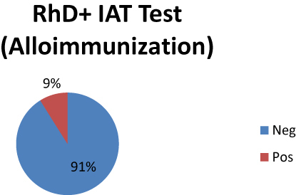
Figure 1 Percentage distribution of Rh alloimmunization among the Rh D+ pregnant women.
Of the five major Rh antigens, c (99.3%) and e (99.8%) antigens were found to be the most common antigens, with antigen e (9.72%) having the highest alloimmunization, followed by D (8.77%), while E (0.94%) was the lowest (). The distribution of alloimmunization among ABO blood groups showed the highest level in blood group B, 25 (10.7%), and the lowest in AB, 4 (7.1%) ().
Table 5 Distribution of IAT-Positive Results by ABO Blood Group (N=1369)
Discussion
The distribution of maternal alloimmunization varies in different populations in different countries. The alloimmunization rate in this study was 9.8%, with that among the D− participants being 24.4%, which is much higher than reported by Das et alCitation4 (2.27%), Sahoo et alCitation12 (3.6%), and KaharCitation13 (1.02%) in studies conducted in India and Europe, where the prevalence was in the range of 0.5–2.57%. The higher rate of alloimmunization in this study could be due to the variations in the population ethnicity. This rate is much lower than that reported by LiCitation14 (45.5%) in China, and this could be due to the recruitment criteria, which emphasized alloimmunized participants.
The alloimmunization prevalence is higher in this population when compared with developed countries such as the Netherlands, as reported by Koelewijn et alCitation15 (1.2%), and Sweden (0.5%), studied by Filbey et al.Citation16 It is also found to be higher in comparison with other developing countries, such as a frequency value of 4.8% in Nigeria in the study by Jeremiah et al.Citation10 The overall alloimmunization rate was also high compared with studies conducted in India by Mangwana et alCitation17 (1.3%) in New Delhi and by Dholakiya et alCitation18 (1.5%) in South India. The reduced rates of alloimmunization in developed countries could mainly be due to well-developed antenatal services and anti-D prophylaxis programs,Citation19–21 but in developing countries such as India and in African countries, there is a significant risk due to a lack of anti-D prophylaxis.Citation18,Citation22 The variations may also be due to differences in race, environment, and the socioeconomic status of a given area.
In this study, 115 (8.9%) of the D+ pregnancies were alloimmunized, which indicates the presence of alloantibodies in the D+ participants, as seen in other studies such as those by Das et alCitation4 (2.27%) and Ghesquière et al.Citation4,Citation18,Citation23 This prevalence has also been reported by Lurie et al,Citation24 Adeniji et al,Citation25 Mbalibulha et al,Citation26 and Pahuja et alCitation27 as 0.2%, 0.15%, 10.3%, and 0.12%, respectively. This requires special follow-up and intervention for the management of hemolytic disease of the fetus and newborn. This will require the formulation of guidelines for universal antibody screening for pregnant women in this area.
Rhesus antigen e was observed to be the most prevalent antigen, at 99.8%, but with less alloimmunization stimulation (9.72%); this is similar to a study conducted in Ivory Coast (99.9%), but different from results obtained in Nigeria by Gwaram et al (96.1%)Citation28 and Jeremiah et al (95.6%)Citation10 This disparity may be due to ethnic variations originating from the heterogeneous nature of the different populations. Antigens of the Rhesus blood group have been recorded to vary among races. All of the Rh antigens (D, C, E, e, and c) have been found to cause hemolytic reactions, although with different capabilities, and particularly delayed reactions resulting in hemolytic disease of the newborn.
Rh D alloimmunization is high in this population, and the study clearly shows that alloimmunization is also present among D+ women, at a level of 8.9%. This suggests that irregular antibodies were detected in both the D− and D+ pregnant women. There is need to determine the titer of these antibodies and to establish the clinical significance at that certain critical level, so that the clinical course be followed up to enable proper management. It is important to note that universal antenatal screening of all pregnant women is key, since in D+ women, just as in D− women, alloantibodies can be formed. There is a need to consider universal antenatal screening and to formulate pregnancy management guidelines accordingly.
Ethical Approval
All the participants were taken through the consent procedure and gave their written informed consent to join the study, and parental written informed consent (assent) was obtained for participants under the age of 18 years. The participant protection procedures were followed in conformity with the Declaration of Helsinki. Ethical clearance was sought and obtained from the ethical review board of the School of Medicine as part of a large study at the College of Health Science, Makerere University (REC Ref No 2019-114), and was registered under number HS508ES by the Uganda National Council for Science and Technology.
Disclosure
All authors declare no competing conflicts of interest relevant to the work presented in this article.
Additional information
Funding
References
- Rudman SV. Textbook of Blood Banking and Transfusion Medicine. 2nd ed. Elsevier Saunders; 2005.
- Reid ME, Lomas-Francis C, Olsson ML. RH - Rh Blood Group System, in the Blood Group Antigen FactsBook. 3rd. Boston: Academic Press; 2012:147–262.
- Gupta GK, Balbuena-Merle R, Hendrickson JE, et al. Immunohematologic aspects of alloimmunization and alloantibody detection: a focus on pregnancy and hemolytic disease of the fetus and newborn. Transfus Apher Sci. 2020;59(5):102946. doi:10.1016/j.transci.2020.102946
- Das S, Shastry S, Rai L, et al. Frequency and clinical significance of red cell antibodies in pregnancy – a prospective study from India. Indian J Pathol Microbiol. 2020;63(2):241–246. doi:10.4103/IJPM.IJPM_737_19
- Habiba U, Munir A, Waris S, et al. Frequency and types of red cell alloantibodies in pregnant females. In Proc. 2020;34:21–25.
- Makroo RN, Bhatia A, Gupta R, et al. Prevalence of Rh, Duffy, Kell, Kidd & MNSs blood group antigens in the Indian blood donor population. Indian J Med Res. 2013;137(3):521–526.
- Mbalibulha Y, Natukunda B, Livex OA, et al. ABO and rh antigen distribution among pregnant women in South Western Uganda. J Blood Med. 2022;13:351–355. doi:10.2147/JBM.S360769
- Hoffbrand AV, Daniel C, Edward GD. Post Graduate Hematology. John Wiley & Sons; 2005:271.
- Ainley L, Jardim JR, Tan J, et al. Prevalence of maternal alloantibodies in a large teaching hospital and their impact on outcomes of fetuses/neonates. Transfus Med. 2017;27(3):228–230. doi:10.1111/tme.12399
- Jeremiah ZA, Mordi A, Buseri FI, Adias TC. Frequencies of maternal red blood cell alloantibodies in Port Harcourt, Nigeria. Immunohematology. 2011;5:39–41.
- Bondagji NS. Rhesus alloimmunization in pregnancy. A tertiary care center - Experience in the Western region of Saudi Arabia. Saudi Med J. 2012;33:688.
- Sahoo BB, Mishra MS, Mishra D, Panigrahy R, Parida P. Prevalence of red cell alloantibodies in pregnant women. Haematol Int J. 2020;4(1):1–9.
- Kahar M. Frequency of red cell alloantibodies in pregnant females of navsari district: an experience that favours inclusion of screening for irregular erythrocyte antibody in routine antenatal testing profile. J Obstet Gynaecol India. 2018;68(4):300–305. doi:10.1007/s13224-017-0984-5
- Li S. Distribution of maternal red cell antibodies and the risk of severe alloimmune haemolytic disease of the foetus in a Chinese population: a cohort study on prenatal management. BMC Pregnancy Childbirth. 2020;20(1):539. doi:10.1186/s12884-020-03235-w
- Koelewijn JM, Slootweg YM, Folman C, et al. Diagnostic value of laboratory monitoring to predict severe hemolytic disease of the fetus and newborn in non-D and non-K-alloimmunized pregnancies. Transfusion. 2020;60(2):391–399. doi:10.1111/trf.15631
- Filbey D, Hanson U, Wesström G. The prevalence of red cell antibodies in pregnancy correlated to the outcome of the newborn: a 12 year study in central Sweden. Acta Obstet Gynecol Scand. 1995;74(9):687–692. doi:10.3109/00016349509021175
- Mangwana S, Simon N, Sangwan L. RH phenotype, ABO and kell antigens, alleles and haplotypes frequencies in North Indian blood donor population. Glob J Transfus Med. 2021;6(1):81.
- Dholakiya SK, Bharadva S, Vachhani J, et al. Red cell alloimmunization among antenatal women attending tertiary care center in Jamnagar, Gujarat, India. Asian J Transfus Sci. 2021;15(1):52. doi:10.4103/ajts.AJTS_72_17
- Mayer B, Hinkson L, Hillebrand W, et al. Efficacy of antenatal intravenous immunoglobulin treatment in pregnancies at high risk due to alloimmunization to red blood cells. Transfus Med Hemotherapy. 2018;45(6):429–436. doi:10.1159/000490154
- Moinuddin I, Millward FC, Millward P. Prevalence and specificity of clinically significant red cell alloantibodies in pregnant women - a study from a tertiary care hospital in Southeast Michigan. J Blood Med. 2019;2019(10):283–289. doi:10.2147/JBM.S214118
- Moise K. Management of pregnancy complicated by Rhesus (D) alloimmunization; 2016:3.
- Mukhtar I, Abdulkadir A. Frequencies of ABO and Rhesus (D) blood group phenotypes among pregnant women attending antenatal clinic at Murtala Muhammad Specialist Hospital, Kano, Nigeria. J Med Trop. 2019;21(1):31–36. doi:10.4103/jomt.jomt_4_19
- Ghesquière L, Garabedian C, Coulon C, et al. Management of red blood cell alloimmunization in pregnancy. J Gynecol Obstet Hum. 2018;47(5):197–204. doi:10.1016/j.jogoh.2018.02.001
- Lurie S, Eliezer E, Piper I, et al. Is antibody screening in Rh (D)-positive pregnant women necessary? J Matern Fetal Neonatal Med. 2003;14(6):404–406. doi:10.1080/14767050412331312260
- Adeniji AA, Fuller I, Dale T, et al. Should we continue screening rhesus D positive women for the development of atypical antibodies in late pregnancy? J Matern Fetal Neonatal Med. 2007;20(1):59–61. doi:10.1080/14767050601123317
- Mbalibulha Y, Natukunda B, Mugyenyi G, et al. Occurrence of anti-D alloantibodies among pregnant mothers in Kasese District, Western Uganda. J Blood Med. 2015;6:125–129. doi:10.2147/JBM.S80977
- Pahuja S, Gupta SK, Pujani M, et al. The prevalence of irregular erythrocyte antibodies among antenatal women in Delhi. Blood Transfus. 2011;9(4):388–393. doi:10.2450/2011.0050-10
- Etura JE, Amaechi RA, Akpotuzor JO, et al. Demographics of rhesus phenotype of blood donors in Calabar: a case study of University of Calabar Teaching Hospital, Calabar, Cross River State, Nigeria. Adv Hematol. 2020;2020:2659398. doi:10.1155/2020/2659398