 ?Mathematical formulae have been encoded as MathML and are displayed in this HTML version using MathJax in order to improve their display. Uncheck the box to turn MathJax off. This feature requires Javascript. Click on a formula to zoom.
Abstract
The genus Calea is reported for many biological activities such as antiinflammatory, antiplasmodial, antifungal, antimicrobial, and cytotoxic activities. Most of the pharmacological activities are credited to germacranolides, a sesquiterpene lactone common to this genus. Dried aerial parts of Calea pinnatifida Banks were extracted with dichloromethane, which generated the dichloromethane crude extract (DCE). The main purpose of this study was to evaluate the anticancer activity of DCE performed in sulforhodamine B cytotoxicity in vitro assay against human cancer cell lines and in vivo Ehrlich models. The DCE showed a high potency and selectivity for the melanoma and kidney cell line. Two in vivo assays were also conducted in the Ehrlich ascites tumor and Ehrlich solid tumor. In the Ehrlich ascites tumor assay, the treatment with DCE increased survival rates at the highest dose (200 mg/kg). Interestingly, in the Ehrlich solid tumor, by increasing the number of treatments from one to three times a week, the tumor growth was inhibited by a lower dose (100 mg/kg). These results encouraged follow-up studies with C. pinnatifida in order to identify the active principles and to determine the anticancer mechanism of action.
Introduction
Plants have served as a rich source of therapeutic agents for many centuries, being used themselves or as the basis for synthetic drugs. Despite the great developments in organic synthesis, 55% of recent chemotherapeutic drugs are derived from or based upon natural products.Citation1 The use of plants as food and in folk and traditional medicine has led to their being one of the main agents in the research and development of cancer chemopreventive drugs.Citation2,Citation3
Calea pinnatifida Banks is used in folk medicine in the treatment of stomach ache, giardiasis, and amoebiasis.Citation4 Belonging to the Asteraceae family, extracts from the leaves of this plant are composed chiefly of fatty esters, 4-glycosyloxybenzoic acid, anisic acid, sitosterol, stigmasterol, polyacetilene, and germacranolides.Citation5
The genus Calea has approximately 110 species. It is found in tropical and subtropical regions, and contains germacranolides as a common chemical component.Citation4–Citation6 Many biological activities, including antiinflammatory, antiplasmodial, antileishmanial, acaricidal, antifungal, antimicrobial, and cytotoxic, have been reported for this genus.Citation6–Citation12 Germacranolides have also been reported to have other properties such as the ability to induce apoptosis, cytotoxicity, antifungal activity, and inhibitory activity against NF-κB.Citation6,Citation10–Citation14
Arucanolide, a germacranolide isolated from C. urticifolia, induced apoptosis in HL60 cells by dissipating the mitochondrial membrane potential, and triggering apoptosis-inducing factor.Citation15 Among the compounds identified in C. pinnatifida, the germacranolides seem to be the most relevant substances, as they promote many biological activities in the potential development of anticancer agents.Citation10–Citation14
Since previous literature has not examined C. pinnatifida and its anticancer-related influences, the aim of the present study is to examine the antitumor activity of the crude extract taken from this species in both in vitro and in vivo assays.
Materials and methods
Plant material
The aerial components of C. pinnatifida Banks were collected in wildcrafting and identified at the Multidisciplinary Center for Chemical, Biological, and Agricultural Research (CPQBA, University of Campinas, São Paulo, Brazil). A voucher specimen, which was positively identified by MSc Katia Calago Althoff, botanist, was deposited at the CPQBA Herbarium (Number 107).
Obtaining dichlomethane and ethanolic crude extract (DCE and ECE)
Dried aerial parts of C. pinnatifida were ground prior to use in a Stephen mill (model UM 40), and an aliquot (300 g) was extracted by soxhlet (1000 mL; 2 × 48 hours) with dichloromethane (Merck® KGaA, Darmstadt, Germany). This provided the dichloromethane crude extract (DCE) after solvent evaporation. The plant residue was extracted with ethanol 96°GL (Merck®) in the same conditions cited above. The solvent was evaporated at 45°C under vacuum, and it was subsequently lyophilized, resulting in ethanolic crude extract (ECE).
In vitro anticancer activity assay
Human tumor cell lines, UACC-62 (melanoma), MCF-7 (breast), NCI-ADR/RES (resistant ovary), 786-O (kidney), NCI-H460 (lung, non-small cell), PC-3 (prostate), OVCAR-3 (ovary), HT29 (colon), and K-562 (leukemia), were kindly provided by the US National Cancer Institute. Stock cultures were grown in medium containing 5 mL RPMI 1640 (Gibco®–BRL; Life Technologies, Carlsbad, CA) supplemented with 5% fetal bovine serum. Gentamicine (50 µg/mL) was added to the experimental cultures. Cells in 96-well plates (plates: 100 µL cells/well; inoculation density ranging from 4 to 7 × 10⁴ cells/mL) were exposed to sample concentrations in DMSO/RPMI (0.25, 2.5, 25, and 250 µg/mL) at 37°C, with 5% CO2 in air for 48 hours. The final DMSO concentration did not affect cell viability. Following this preparation, cells were fixed with 50% trichloroacetic acid, and cell proliferation was determined by spectrophotometric quantification (540 nm) of cellular protein content using sulforhodamine B assay.Citation16,Citation17 Doxorubicin (doxorubicin chloridrate, 50 mg; 11% purity; Eurofarma, São Paulo, Brazil) was adopted as a positive control. Using the concentration response curve for each cell line, total growth inhibition (TGI) was determined through nonlinear regression analysis () using ORIGIN 8.0 software (OriginLab Corporation, Northampton, MA).Citation18–Citation20
Table 1 Comparative TGI (µg/mL) in human tumor cell lines
In vivo assay – acute toxicity
BALB/c mice were treated intraperitoneally (IP) with DCE at doses of 100, 300, 500, and 1000 mg/kg. Groups were observed during 4 hours and then daily for 14 days. The following general toxicity parameters were evaluated: body weight loss, locomotion, behavior (agitation, lethargy), respiration, salivation, tearing eyes, cyanosis, and mortality.Citation21,Citation22 The lethal dose was calculated by linear regression, as described by Litchfield and Wilcoxon.Citation22
In vivo assay – Ehrlich tumor
DCE was evaluated in vivo on an Ehrlich ascitic tumor (EAT) and Ehrlich solid tumor (EAT) assay. Ehrlich tumor cells were maintained in the ascites form by peritoneal passages in mice via weekly transplantation of 5 × 10⁵ tumor cells. Male BALB/c mice aged 8–10 weeks and weighing 25–30 g were used for Ehrlich tumor experiments. The animals were obtained from the Multidisciplinary Center for Biological Investigation on Laboratory Animal Science-UNICAMP, and were maintained under controlled temperature conditions (22°C–24°C), light (12 hours of light, 12 hours of dark) and humidity (45%–65%), with food and water ad libitum. All procedures were conducted in accordance with the principles and guidelines adopted by the Institutional Committee for Ethics in Animal Research at the University of Campinas (CEEA, UNICAMP, protocol 1421-1, December 19, 2007).
Ehrlich ascites assay
Ehrlich tumor cells (1.0 × 10⁴) were implanted IP, and the animals (n = 10) were treated with the samples, IP, once a week over the course of 21 days; the animals were followed for a total of 90 days. The animals were divided into three groups: negative control group (vehicle); positive control group (doxorubicin chloridrate, 50 mg; 11% purity) at a dose of 3.0 mg/kg; and DCE treated groups. DCE was administered at doses of 50, 100, and 200 mg/kg. Anticancer activity was assessed by the survival rates of the groups.Citation3,Citation23
Ehrlich solid assay
Ehrlich tumor cells (1.0 × 10⁵) were implanted subcutaneously in the region of the back, and the animals (n = 8) were treated with the samples, IP, every 48 hours over the course of 15 days. The animals were divided into groups: negative control group (vehicle); positive control group (doxorubicin chloridrate, 50 mg; 11% purity) at a dose of 3.0 mg/kg; and DCE treated groups. DCE was administered at doses of 50 and 100 mg/kg. The tumor volume was measured using a pachymeter (height × width × length). Anticancer activity was assessed by comparing the relative tumor weight (tumor weight/animal weight) and tumor volume using the equation:
(1)
Citation3
Statistical analyses
Results are expressed as the mean ± standard deviation (SD) per group. Statistical evaluation was done using analysis of variance, followed by Duncan’s new multiple range test using StatSoft® software (StatSoft, Inc, Tulsa, OK). Graphics were designed using the ORIGIN® software. Differences were considered significant at P ≤ 0.05 and are represented by asterisks (*P < 0.05, **P < 0.01, ***P < 0.001).
Results
The evaluation of DCE in the in vitro sulforhodamine B assay demonstrated that this extract has a strong relationship between its concentration and the effect on and selectivity for the melanoma (UACC-62) and kidney (786-O) cell lines.
The TGI shown in demonstrated the activity of DCE, ECE, and doxorubicin. DCE is very potent and kills all cell lines; meanwhile, the ECE did not show any antiproliferative effects. Doxorubicin shows high cytotoxic activity in almost all cell lines except for NCI-ADR/RES, which expresses a multidrug resistance phenotype.
Subsequently, the DCE was selected for in vivo studies. Through IP administration, DCE showed a 50% lethal dose of 500 mg/kg in an acute toxicity test. The IP route was chosen because the absorption barriers are similar to the intravenous route, which more closely resembles current clinical applications for chemotherapy treatment. The results of this toxicity study established the doses used for in vivo assays.Citation22
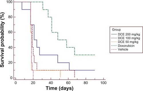
The in vivo EAT anticancer activity was determined by the survival life span by comparing the mortality rate of the treatment groups versus the control group (). Only the highest dose (200 mg/kg) increased the survival rate when compared with the negative control group (P = 0.009). In spite of the antitumor activity, the 200 mg/kg dose caused the death of one experimental animal before the negative control group; therefore, the dose had no relationship with tumor development. Both the 50 mg/kg and the 100 mg/kg doses did not show any anticancer activity in this assay using once a week treatment, and the positive control (doxorubicin) increased the overall survival time, demonstrating antitumor activity, as expected (P < 0.001).
Figure 1 Kaplan–Meier survival curves of mice treated with Calea pinnatifida DCE.
Abbreviation: DCE, dichloromethane crude extract.
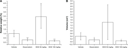
In vivo EST anticancer activity was determined by the reduction of the relative tumor weight and tumor volume. In this assay, 100 mg/kg of DCE showed anticancer activity in a treatment that was administered every 48 hours. This dose inhibited 66.7% of the relative tumor weight (P = 0.0002) and 57.6% of tumor volume (P = 0.012), whereas the 50 mg/kg dose increased the tumor weight (P = 0.004), but did not show any differences in the volume from negative control (P = 0.06). The positive control (doxorubicin) inhibited 71.6% of the tumor weight (P = 0.0007) and 66.5% of tumor volume (P = 0.032) ().
Figure 2 Ehrlich solid relative tumor weight and tumor volume of mice treated with Calea pinnatifida DCE. Ehrlich solid relative tumor weight (A) and tumor volume (B) of mice treated with C. pinnatifida DCE.
Abbreviation: DCE, dichloromethane crude extract.
Discussion
Because of the high number of cancer cases, the interest in alternative therapies using natural products are increasing, especially those derived from plants.Citation24,Citation25 In order to look for new sources of therapeutic anticancer agents, many plant extracts and active principles have been studied in in vitro and in vivo cancer models, and the correlation of both studies became one of the key steps for the success of this type of research.Citation1
Since the knowledge that different cell lines display varying sensitivities towards cytotoxic compounds, the use of more than one cell line is considered necessary for the detection of anticancer compounds.Citation21 Bearing this in mind, cell lines of different histological origins were used in the present study. An in vivo model could potentially broaden these results, but more data needs to be compiled prior to this being a viable option to overpass the limitations of the in vitro models.Citation26
Previous studies demonstrated that other Calea species have many different biological properties, such as cytotoxicity and antiinflammatory activities.Citation6,Citation7 However, the present study showed, for the first time, the antitumor activity of C. pinnatifida crude extract, both in in vitro and in vivo experimental models. The DCE inhibited cell growth at low concentrations ranging from 8.0 to 70.0 µg/mL (TGI value) with highest selectivity noted for kidney (786-O) and melanoma (UACC-62) cell lines with TGI of 8.41 and 13.77 µg/mL, respectively (). These data were used as the determining point to study the activity of DCE in a murine cancer model.
In the EAT model, the C. pinnatifida DCE antitumoral activity was confirmed by the prolongation of the life-span in once-a-week treated groups at the dose of 200 mg/kg. These results are important since the EAT is a very aggressive tumor,Citation27 killing the negative control group after 20 days of tumor cells inoculation. The EAT implantation induced a local inflammatory reaction, increasing vascular permeability and resulting in cellular migration and progressive ascites fluid formation.Citation23 The ascites fluid formation is essential for tumor growth, as this constitutes the direct nutritional source for tumor cells.Citation3,Citation28 Since the extract increased animal survival, these findings suggest either a direct cytotoxic effect on tumor cells, as previously observed in vitro, or an indirect local effect provided by an antiinflammatory activity as described by other species of the genus Calea.Citation3,Citation5,Citation29
To investigate whether the inhibitory effect of C. pinnatifida on Ehrlich tumors would be local or systemic, we evaluated the effect of IP treatment in another cancer model, the EST. In order to avoid the acute toxicity observed in the EAT model at a dose of 200 mg/kg, we increased the frequency of dosing and reduced the maximum dose administrated. The smaller interval between treatments proved to yield better anticancer activity since 100 mg/kg was able to diminish the tumor size in EST, whereas the same dose did not increase the survival rate in the EAT model. The possible explanation for these results may be the maintenance of DCE blood levels in an every-two-day schedule rather than a once-a-week treatment schedule, when using the same dose level. However, further pharmacokinetics studies are required to determine the half-life of DCE.
Despite the anticancer activity of the DCE at 100 mg/kg, the dose of 50 mg/kg increased tumor weight, demonstrating that the use of the DCE in cancer treatment should be carefully reviewed with more assays to understand the molecular mechanisms involved in this activity, as well as to identify the active principles with proinflammatory and protumorigenic activity. Although there are not enough data to draw strong conclusions, these results present a systemic effect of the DCE by both reducing the solid subcutaneous tumor, as well as increasing the survival rate in the ascitic model.
All the substances involved in the antitumor effect of C. pinnatifida DCE are unknown, even though some compounds found in the extract such as the germacranolides could explain these results.Citation4 These compounds have been described as potent apoptosis inductors, as well as inhibitors of NF-κB, an important immune and inflammatory response mediator.Citation11,Citation12,Citation15 The induction of apoptosis and immunomodulatory activity are important mechanisms for some cancer treatments.Citation30
Both in vitro and in vivo anticancer activity observed in the present study stimulate future research using the DCE obtained from C. pinnatifida as a chemotherapeutic agent. Additional phytochemical studies are in progress to identify the active principles involved in this antitumor activity.
Acknowledgments
The authors are grateful for the technical support offered by Benício Pereira from the Division of Agrotecnology – CPQBA/UNICAMP and to Adilson Sartoratto from the Division of Organic and Pharmaceutical Chemistry – CPQBA/UNICAMP. The authors also thank FAPESP (2008/53652-7), CNPq, and Capes for their financial support.
Disclosure
The authors report no conflicts of interest in this work.
References
- NewmanDJCraggGMNatural products as sources of new drugs over the 30 years from 1981 to 2010J Nat Prod201275331133522316239
- CraggGMNewmanDJNature: a vital source of leads for anticancer drug developmentPhytochem Rev200982313331
- NascimentoFRCruzGVPereiraPVAscitic and solid Ehrlich tumor inhibition by Chenopodium ambrosioides L. treatmentLife Sci200678222650265316307762
- PruskiJFUrbatschLEFive new species of Calea (Compositae: Heliantheae) from planaltine BrazilBrittonia1988404341356
- FerreiraZSRoqueNFGottliebOROliveiraFGottliebHEStructural clarification of germacranolides from Calea speciesPhytochemistry198019714811484
- YamadaMMatsuuraNSuzukiHGermacranolides from Calea urticifoliaPhytochemistry200465233107311115541738
- Venegas-FloresHSegura-CobosDVázquez-CruzBAntiinflamatory activity of the aqueous extract of Calea zacatechichiProc West Pharmacol Soc20024511011112434549
- do NascimentoAMSalvadorMJCandidoRCItoIYde OliveiraDCAntimicrobial activity of extracts and some compounds from Calea platylepisFitoterapia200475551451915261392
- do NascimentoAMSalvadorMJCandidoRCde AlbuquerqueSde OliveiraDCTrypanocidal and antifungal activities of p-hydroxyacetophenone derivatives from Calea uniflora (Heliantheae, Asteraceae)J Pharm Pharmacol200456566366915142345
- MatsuuraNYamadaMSuzukiHInhibition of preadipocyte differentiation by germacranolides from Calea urticifolia in 3T3-L1 cellsBiosci Biotechnol Biochem200569122470247416377913
- RibeiroVLdos SantosJCMartinsJRAcaricidal properties of the essential oil and precocene II obtained from Calea serrata (Asteraceae) on the cattle tick Rhipicephalus (Boophilus) microplus (Acari: Ixodidae)Vet Parasitol20111791–319519821402447
- WuHFronczekFRBurandtCLJrZjawionyJKAntileishmanial Germacranolides from Calea zacatechichiPlanta Med201177774975321128202
- BorkPMSchmitzMLKuhntMEscherCHeinrichMSesquiterpene lactone containing Mexican Indian medicinal plants and pure sesquiterpene lactones as potent inhibitors of transcription factor NF-kappaBFEBS Lett1997402185909013864
- RiveroAQuintanaJEiroaJLPotent induction of apoptosis by germacranolide sesquiterpene lactones on human myeloid leukemia cellsEur J Pharmacol20034821–3778414660007
- NakagawaYIinumaMMatsuuraNA potent apoptosis-inducing activity of a sesquiterpene lactone, arucanolide, in HL60 cells: a crucial role of apoptosis-inducing factorJ Pharmacol Sci200597224225215699578
- SkehanPStorengRScudieroDNew colorimetric cytotoxicity assay for anticancer-drug screeningJ Natl Cancer Inst19908213110711122359136
- HolbeckSLUpdate on NCI in vitro drug screen utilitiesEur J Cancer200440678579315120034
- ShoemakerRHThe NCI60 human tumour cell line anticancer drug screenNat Rev Cancer200661081382316990858
- FoucheGCraggGMPillayPKolesnikovaNMaharajVJSenabeJIn vitro anticancer screening of South African plantsJ Ethnopharmacol2008119345546118678239
- LongatoGBRizzoLYSousaIMIn vitro and in vivo anticancer activity of extracts, fractions, and eupomatenoid-5 obtained from Piper regnellii leavesPlanta Med201177131482148821391177
- LapaAJSouccarCLima-LandmanMTRCastroMSALimaTCMMétodos de Avaliação da Atividade Farmacológica de Plantas MedicinaisPorto AlegreMetrópole2003 Portuguese
- LitchfieldJTJrWilcoxsonFA simplified method of evaluating dose-effect experimentsJ Pharmacol Exp Ther19499629911318152921
- SacomanJLMonteiroKMPossentiAFigueiraGMFoglioMACarvalhoJECytotoxicity and antitumoral activity of dichloromethane extract and its fractions from Pothomorphe umbellataBraz J Med Biol Res200841541141518545814
- RatesSMPlants as source of drugsToxicon200139560361311072038
- JemalASiegelRWardEHaoYXuJThunMJCancer Statistics, 2009CA Cancer J Clin200959422524919474385
- SmithJANgoHMartinMCWolfJKAn evaluation of cytotoxicity of the taxane and platinum agents combination treatment in a panel of human ovarian carcinoma cell linesGynecol Oncol200598114114515963813
- LoboCRuiz-BellidoMAAledoJCMárquezJNúñezDeCastroIAlonsoFJInhibition of glutaminase expression by antisense mRNA decreases growth and tumourigenicity of tumour cellsBiochem J2000348Pt 225726110816417
- FecchioDSiroisPRussoMJancarSStudies on inflammatory response induced by Ehrlich tumor in mice peritoneal cavityInflammation19901411251312323805
- Vendramini-CostaDBde CastroIBRuizALMarquissoloCPilliRAde CarvalhoJEEffect of goniothalamin on the development of Ehrlich solid tumor in miceBioorg Med Chem201018186742674720729093
- HanahanDWeinbergRAHallmarks of cancer: the next generationCell2011144564667421376230