
Abstract
Glypican-3 (GPC3), a member of heparan sulfate proteoglycans, attaches to the cell membrane and is frequently observed to be elevated in hepatocellular carcinoma (HCC). However, GPC3 is not detected in normal liver tissues and benign liver lesions. Consequently, GPC3 is currently being used as a diagnostic biomarker and HCC-specific positron emission computed tomography probe to identify HCCs in normal liver tissues and benign liver lesions. The overexpression of GPC-3 in serum or liver tissue also predicts poor prognosis for HCC patients. In addition, GPC3 promotes HCC growth and metastasis by activating the canonical Wnt and other signaling pathways. Targeting of GPC3, including GC33, HN3 and YP7, might offer new immunotherapeutic tools for HCC treatment.
Introduction
Hepatocellular carcinoma (HCC) is one of the most common malignant tumors and the third leading cause of cancer-related death in the world.Citation1 Although a great progress has been made in HCC treatment, including curative surgery and nonsurgical treatment, the HCC prognosis remains poor.Citation1 The result of treatment depends on the HCC stage at the time of diagnosis. HCC can be cured if diagnosed at an early stage. However, most HCCs are diagnosed at an advanced stage when patients visit physicians with symptoms.
Glypican-3 (GPC3), a member of the heparan sulfate (HS) proteoglycan family, is attached to the cell surface by a glycosyl-phosphatidylinositol (GPI) anchor and can be cleaved off the cell surface. The soluble GPC3 can be detected in serum (sGPC3).Citation2,Citation3 GPC3 is widely expressed in human embryos and plays a significant role in morphogenesis and growth, by mechanisms involving insulin-like growth factor, bone morphogenetic protein (BMP), fibroblast growth factor (FGF) or hedgehog (Hh) signaling pathway.Citation4–Citation6 GPC3 can be detected in the fetal liver from embryonic weeks 18 to 30, but cannot be identified in any normal adult hepatic tissue.Citation4–Citation7 In recent years, extensive research has been carried out on the role of GPC3 in diagnosis, progression and treatment of HCC in vivo and in vitro.Citation8,Citation9 Here, we summarize current evidences for the use of GPC3 as a diagnostic biomarker, its oncogenic function and as a immunotherapeutic target for HCC patients.
GPC3 as a biomarker for diagnosis and prognosis of HCC
In 1997, Hsu et alCitation7 first reported that mRNA and protein levels of GPC3 were upregulated to a greater extent in most HCCs than in normal liver, cholangiocarcinoma and metastatic carcinomas of the liver. Since then, the diagnostic value of GPC3 in HCC has been studied extensively, and increasing studies have confirmed that GPC3 would be a useful serological and immunohistochemical biomarker for HCC. By immunolabeling GPC3 with a monoclonal antibody, Capurro et alCitation8 revealed that 72% of HCCs were GPC3-positive; however, GPC3 was undetectable in normal liver tissues, cirrhosis or benign lesions. Other studies conducted on GPC3 across the world also revealed similar results.Citation9–Citation13 Besides, the membrane-bound GPC3 can also be cleaved off by lipase from the GPI anchor.Citation14 Thus, the diagnostic value of sGPC3 was evaluated later by several studies. Our study also verified that GPC3 was a sensitive and specific biomarker for diagnosis of early HCC due to its high expression in HCC tissue.Citation9 Moreover, we found that sGPC3 was detectable in 48.8% of patients with negative serum α-fetoprotein (AFP), which further confirmed that GPC3 might be a serum marker for HCC and indicated that GPC3 could be used to distinguish AFP-negative HCC from cirrhotic nodules.Citation11 Qiao et al analyzed the serum levels of GPC3, AFP and human cervical cancer oncogene (HCCR) in 189 cases (101 HCC, 40 cirrhosis, 18 hepatitis and 30 control healthy donors).Citation10 They concluded that GPC3 was the best sensitive biomarker of the three biomarkers mentioned earlier, and the combination of these three biomarkers, with a sensitivity of 80.2%, was much higher than AFP alone.Citation11 In recent years, in vivo and in vitro studies displayed the use of Zr-α GPC3, a HCC-specific positron emission computed tomography (PET) probe, in HCC imaging as well as the detection of GPC3 levels by small-animal PET. Those results suggested that the identification of small liver lesions with a HCC-specific PET probe would help physicians to make the differential diagnosis between HCCs and benign lesions in cirrhotic patients so that patient management could be significantly altered.Citation13,Citation14 In addition, extensive studies to determine the prognostic value of GPC3 were carried out in HCC patients.Citation15–Citation19 Increasing evidence revealed that high GPC3 expression was a prominent prognostic factor that predicted a poor outcome for HCCs. Clinicopathological studies on GPC3-Immunohistochemistry stainings revealed that high GPC3 expression was associated with poor postoperative disease-free survival (DFS) and overall survival (OS) and that it also served as an independent risk factor.Citation15–Citation17 Patients with overexpressed GPC3 in HCC tissues showed notably shorter OS and DFS than those with underexpressed GPC3. Several meta-analytic studies also supported the fact that a high GPC3-IHC score was one of the prognostic factors in HCC, as there was a significantly negative correlation between GPC3 expression and OS or DFS of HCC patients.Citation18,Citation19
GPC3-mediated signaling pathway in HCC progression
In addition to being a reliable indicator for the diagnosis and prognosis of HCC, GPC3 has a significant role in the progression of HCC (). GPC3 functions as a coreceptor/storage site for some ligands, e.g. Wnt and FGF, via its HS side chains, and facilitates ligand and/or its receptors to stimulate the signaling pathways involved in HCC growth and invasion. Several studies showed that GPC3 could promote the growth of hepatoma cells in vivo and in vitro.Citation20–Citation23 Capurro et al suggested that GPC3 promoted the growth of hepatoma cells by stimulating canonical Wnt/β-catenin signaling pathway activity in vivo and in vitro.Citation20 Recently, this GPC3-mediated activation of Wnt signaling pathway in HCC cells has been confirmed by other reports.Citation21,Citation22 Li et al showed that ectopic GPC3 could increase the c-Myc expression, a typical target of the canonical Wnt signaling pathway; c-Myc can also transcriptionally activate GPC3 directly in HCC cells.Citation21 Zittermann et alCitation22 reported that soluble GPC3, which is cleaved off the cell membrane at the GPI anchor domain, could inhibit the in vivo growth of HCC cells by blocking the canonical Wnt signaling pathway in tumors generated by Huh6 and Huh7, and Akt and ERK signaling activation in HepG2- and Huh7-derived tumors, respectively. In other words, this activity can be observed only when GPC3 is attached to the cell membrane. On the other hand, it is possible that GPC3 promotes the growth of HCC cells by mediating other signaling pathways. For example, Sun et al proved that suppression of GPC3 inhibited cell proliferation and enhanced apoptosis via upregulation of TGF-β2 in vivo and in vitro.Citation23 In addition, it has been demonstrated that several FGF members (FGF8, FGF17 and FGF18) were upregulated in the great majority of HCC samples.Citation24
Figure 1 The diagram of possible GPC3-mediated signaling pathway in HCC progression.
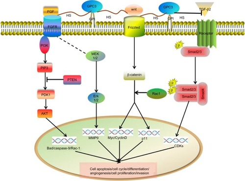
Metastasis is an important aspect of HCC progression, and epithelial–mesenchymal transition (EMT) is considered as the first step in the metastatic cascade.Citation25 Recently, we have found that the expression of GPC3 in HCC tissue was upregulated during HCC progression from Barcelona Clinic Liver Cancer stage A or B to stage C. The increased expression of GPC3 in tumor tissues was closely related to the level of EMT markers, as well as to the cancer vascular invasion. HepG2 cells, expressing a higher level of GPC3, possessed stronger ability of invasion and exhibited more EMT-like changes than those of HCC cell lines that expressed lower levels of GPC3 (Hep3B and Huh7). Our studies suggested that GPC3 promoted HCC progression and metastasis by inducing EMT in tumor cells, and the ERK signaling pathway is involved in this GPC3-induced process.Citation26 In addition, Ruan et al also reported that GPC3 promoted the metastasis of HCC in vitro and in vivo.Citation27
GPC3 is a new therapeutic target for HCC
Based on the HCC-specific expression of GPC3 in liver, it is an emerging target for liver cancer therapy. A number of studies demonstrated that GPC3 is a potential therapeutic target for HCC.Citation28–Citation30 Currently, antibodies targeting GPC3, including human antibody HN3 and humanized mouse antibodies YP7 and GC33, are in different stages of preclinical or clinical development. GC33, a humanized mouse antibody recognizing a C-terminal domain of GPC3, showed notable cytotoxic activity against GPC3-positive hepatoma cells in vivo by complement-dependent cytotoxicity and/or antibody-dependent cell cytotoxicity.Citation31–Citation34 GC33 exhibited marked effect against metastatic or advanced HCC in a phase I trial,Citation35 and the patients well tolerated a dose escalation of GC33 (2.5–20 mg/kg).Citation36 Currently, more clinical trials for GC33 alone (phase II clinical trials) and GC33 combined with sorafenib, a chemodrug, are recruiting volunteers (phase I clinical trials). A randomized phase II clinical trial on GC33 was conducted in 185 patients with HCC metastasis. In this clinical trial, the dose of GC33 was set at 1,600 mg (intravenous) on days 1 and 8, and then every 2 weeks thereafter. There was no significant change in mean progression-free survival (PFS) between GC33 and placebo groups (2.6 mo vs 1.5 mo, HR = 0.97, P = 0.87).Citation37 However, higher exposure of GC33 with FcgR3A-158V polymorphism or CD16 expression intensity may prolong PFS, and therefore, further studies to analyze the GPC3-positive HCC immuno-microenvironment are necessary.Citation37 YP7, a new humanized mouse anti-GPC3 antibody, has high affinity and recognizes the C-terminal epitope that overlaps the GC33-binding site and displays the capacity of suppressing tumor activity in vivo. HN3, a human single-domain antibody, can also inhibit HCC cell lines and growth of xenograft tumors by binding to the GPC3 N-terminal and C-terminal domains.Citation38 Currently, the GAO study group found that treatment with GPC3 monoclonal antibodies, such as HN3 and YP7, could suppress the growth of HepG2- and Hep3B-generated tumor xenografts.Citation39 Treatment with HN3 showed higher antitumor activity than YP7.Citation40 Both HN3 and YP7 exhibit antitumor activity in vitro and in vivo, but their efficacies in HCC patients need to be determined and require further clinical trials.
GPC3 cannot play a dominant role in the apoptosis of HCC cells, and the fact that the GC33 or HN3 antibody could completely eliminate HCC cells needs further trials for confirmation. In combination with chemotherapy, armed antibodies, such as antibody–drug conjugates, bispecific antibodies and chimeric antigen receptor T-cell adoptive therapy may be better potential ways for HCC therapy.Citation39
Conclusion
GPC3 is overexpressed in most of the HCC tumors, and was also used as an indicator for HCC diagnosis and prognosis. It is used as a potential target for developing therapeutic antibodies for HCC treatment. However, the relationship between GPC3 structure and function remains unclear. In addition, in order to develop GPC3-targeted therapies in HCC treatment, the expression and regulation of GPC3 in HCC need further confirmation.
Acknowledgments
The authors would like to thank Ms Shan Shan Wang for her support in providing a summary of possible signaling networks for GPC3. This study was supported by the Capital Science and Technology Development Fund (2014-1-2181), Beijing Municipal Administration of Hospitals Clinical Medicine Development of Special Funding (ZYLX201610) and Beijing Municipal Administration of Hospitals’ Ascent Plan (DFL20151602).
Disclosure
The authors report no conflicts of interest in this work.
References
- ZhuRXSetoWKLaiCLYuenMFEpidemiology of hepatocellular carcinoma in the Asia-Pacific regionGut Liver201610333233927114433
- FilmusJCapurroMRastJGlypicansGenome Biol20089522418505598
- TraisterAShiWFilmusJMammalian Notum induces the release of glypicans and other GPI anchored proteins from the cell surfaceBiochem J2008410350351117967162
- YamauchiNWatanabeAHishinumaMThe glypican 3 oncofetal protein is a promising diagnostic marker for hepatocellular carcinomaMod Pathol200518121591159815920546
- CapurroMIXuPShiWLiFJiaAFilmusJGlypican-3 inhibits Hedgehog signaling during development by competing with patched for Hedgehog bindingDev Cell200814570071118477453
- ZhangLLiuHSunLLiNDingHZhengJGlypican-3 as a potential differential diagnosis marker for hepatocellular carcinoma: a tissue microarray-based studyActa Histochem2012114654755222119409
- HsuHCChengWLaiPLCloning and expression of a developmentally regulated transcript MXR7 in hepatocellular carcinoma: biological significance and temporospatial distributionCancer Res19975722517951849371521
- CapurroMWanlessIRShermanMGlypican-3: a novel serum and histochemical marker for hepatocellular carcinomaGastroenterology20031251899712851874
- LiuHLiPZhaiYDiagnostic value of glypican-3 in serum and liver for primary hepatocellular carcinomaWorld J Gastroenterol201016354410441520845507
- QiaoSSCuiZQGongLSimultaneous measurements of serum AFP, GPC-3 and HCCR for diagnosing hepatocellular carcinomaHepatogastroenterology201158110–1111718172421940340
- LiBLiuHShangHWLiPLiNDingHGDiagnostic value of glypican-3 in alpha fetoprotein negative hepatocellular carcinoma patientsAfr Health Sci201313370370924250310
- LiuJWZuoXLWangSDiagnosis accuracy of serum glypican-3 level in patients with hepatocellular carcinoma and liver cirrhosis: a meta-analysisEur Rev Med Pharmacol Sci201519193655367326502856
- ShamJGKievitFMGriersonJRGlypican-3-targeted Zr PET imaging of hepatocellular carcinomaJ Nucl Med201455579980424627434
- ZhuDQinYWangJNovel Glypican-3-binding peptide for in vivo hepatocellular carcinoma fluorescent imagingBioconjug Chem201627383183926850086
- FuSJQiCYXiaoWKLiSQPengBGLiangLJGlypican-3 is a potential prognostic biomarker for hepatocellular carcinoma after curative resectionSurgery2013154353654423601901
- YoritaKTakahashiNTakaiHPrognostic significance of circumferential cell surface immunoreactivity of glypican-3 in hepatocellular carcinomaLiver Int201131112013120964802
- ShirakawaHSuzukiHShimomuraMGlypican-3 expression is correlated with poor prognosis in hepatocellular carcinomaCancer Sci200910081403140719496787
- LiJGaoJZDuJLWeiLXPrognostic and clinicopathological significance of glypican-3 overexpression in hepatocellular carcinoma: a meta-analysisWorld J Gastroenterol201420206336634424876756
- XiaoWKQiCYChenDPrognostic significance of glypican-3 in hepatocellular carcinoma: a meta-analysisBMC Cancer20141410424548704
- CapurroMIXiangYYLobeCFilmusJGlypican-3 promotes the growth of hepatocellular carcinoma by stimulating canonical Wnt signalingCancer Res200565146245625416024626
- LiLJinRZhangXOncogenic activation of GPC3 by c-Myc in human hepatocellular carcinomaHepatology20125641380139022706665
- ZittermannSICapurroMIShiWFilmusJSoluble glypican 3 inhibits the growth of hepatocellular carcinoma in vitro and in vivoInt J Cancer201012661291130119816934
- SunCKChuaMSHeJSoSKSuppression of glypican 3 inhibits growth of hepatocellular carcinoma cells through up-regulation of TGF-β2Neoplasia201113873574721847365
- GauglhoferCSagmeisterSSchrottmaierWUp-regulation of the fibroblast growth factor 8 subfamily in human hepatocellular carcinoma for cell survival and neoangiogenesisHepatology201153385486421319186
- AcloqueHAdamsMSFishwickKBronner-FraserMNietoMAEpithelial–mesenchymal transitions: the importance of changing cell state in development and diseaseJ Clin Invest200911961438144919487820
- WuYLiuHWengHGlypican-3 promotes epithelial–mesenchymal transition of hepatocellular carcinoma cells through ERK signaling pathwayInt J Oncol20154631275128525572615
- RuanJLiuFChenXInhibition of glypican-3 expression via RNA interference influences the growth and invasive ability of the MHCC97-H human hepatocellular carcinoma cell lineInt J Mol Med201128449750321617840
- BaumhoerDTornilloLStadlmannSRoncalliMDiamantisEKTerraccianoLMGlypican 3 expression in human nonneoplastic, preneoplastic, and neoplastic tissues: a tissue microarray analysis of 4,387 tissue samplesAm J Clin Pathol2008129689990618480006
- LlovetJMChenYWurmbachEA molecular signature to discriminate dysplastic nodules from early hepatocellular carcinoma in HCV cirrhosisGastroenterology200613161758176717087938
- ZhuZWFriessHWangLEnhanced glypican-3 expression differentiates the majority of hepatocellular carcinomas from benign hepatic disordersGut200148455856411247902
- HoMKimHGlypican-3: a new target for cancer immunotherapyEur J Cancer201147333333821112773
- FilmusJCapurroMGlypican-3: a marker and a therapeutic target in hepatocellular carcinomaFEBS J2013280102471247623305321
- NakanoKIshiguroTKonishiHGeneration of a humanized anti-glypican 3 antibody by CDR grafting and stability optimizationAnticancer Drugs2010211090791620847643
- NakanoKOritaTNezuJAnti-glypican 3 antibodies cause ADCC against human hepatocellular carcinoma cellsBiochem Biophys Res Commun2009378227928419022220
- IshiguroTSugimotoMKinoshitaYAnti-glypican 3 antibody as a potential antitumor agent for human liver cancerCancer Res200868239832983819047163
- ZhuAXGoldPJEl-KhoueiryABFirst-in-man phase I study of GC33, a novel recombinant humanized antibody against glypican-3, in patients with advanced hepatocellular carcinomaClin Cancer Res201319492092823362325
- YenCJDanieleBKudoMRandomized phase II trial of intravenous RO5137382/GC33 at 1600 mg every other week and placebo in previously treated patients with unresectable advanced hepatocellular carcinomaJ Clin Oncol201432Suppl 5 abstract 4102
- FengMHoMGlypican-3 antibodies: a new therapeutic target for liver cancerFEBS Lett2014588237738224140348
- GaoWTangZZhangYFImmunotoxin targeting glypican-3 regresses liver cancer via dual inhibition of Wnt signalling and protein synthesisNat Commun20151166536
- GaoHLiKTuHDevelopment of T cells redirected to glypican-3 for the treatment of hepatocellular carcinomaClin Cancer Res201420246418642825320357