
Abstract
Purpose
Carotid artery stenosis (CAS) is a leading cause of cerebral infarction, its early diagnosis and intervention are necessary. In light of the important role of microRNAs (miRNAs) in cerebrovascular disease, this study aimed to investigate the expression pattern and clinical significance of serum miR-455-5p in the onset and development of CAS, as well as its underlying mechanism.
Patients and Methods
Seventy patients with asymptomatic CAS were recruited, and the development of cerebral ischemia events (CIEs) was recorded during the five-years follow-up. qRT-PCR was performed for the serum miR-455-5p detection. ROC curve was applied for the diagnostic ability evaluation. By constructing multivariable logistic or cox regression model, odds ratio (OR) or hazard ratio (HR) were calculated to assess the impact of each risk factor on independent variables. Human aortic endothelial cells (HAECs) were treated with ox-LDL to induce endothelial cell damage. The role of miR-455-5p in the cell viability, apoptosis, oxidative stress and inflammatory response was detected.
Results
Serum miR-455-5p showed low expression in cases with CAS, and had an independent influence on the degree of CAS. The diagnostic ability of serum miR-455-5p to diagnose CAS was determined via ROC curve, with the AUC of 0.927. During follow-up, patients with low miR-455-5p expression showed high incidence of CIEs. In multivariable cox regression model, degree of CAS and miR-455-5p were significant risk factors for the development of CIEs in the CAS patients. In vitro, miR-455-5p was at a low expression in HAECs cell models and can prevent cells from ox-LDL induced cell apoptosis, oxidative stress and inflammatory response. SOCS3 was a target gene of miR-455-5p and upregulated in ox-LDL treated cells.
Conclusion
Down-regulated expression of serum miR-455-5p is hopeful to be used as a biomarker for the early diagnosis of CAS. MiR-455-5p is an independent risk factor for the degree of CAS, and has a certain predictive value for the development of CIEs. That might be associated with the protective role of miR-455-5p against ox-LDL-induced endothelial cell injury via targeting SOCS3.
Introduction
Carotid artery stenosis (CAS) is a common atherosclerotic disease, which commonly occurs in the elderly.Citation1 CAS can be caused by carotid atherosclerosis, fibromuscular dysplasia, and hematologic diseases.Citation2 Thrombosis at the site of carotid artery stenosis or rupture of unstable plaque can lead to distal arterial embolism, which can cause cerebral infarction and endanger the life of patients.Citation3 It is reported that 30–60% of cerebral infarction patients are caused by CAS.Citation4 With the changes in environment, diet and living and working habits caused by social modernization, the incidence of cardiovascular and cerebrovascular diseases has increased significantly. Studies have shown that the rate of intracranial artery stenosis in elderly people over 60 years old in China is 5.9%-6.9%.Citation5 The early clinical symptoms of CAS patients are not obvious, and the main manifestations are headache, insomnia, dizziness, and so on. Once the acute attack, it may cause life-threatening stroke, and increase the disease morbidity and mortality. Therefore, early diagnosis and intervention of CAS patients are particularly important.
MicroRNA (miRNA) is a kind of non-coding single-stranded RNA molecule with 18 to 25 nucleotides in length. It can regulate the expression of relevant coding proteins by promoting degradation or inhibiting transcription via binding to the 3’-untranslated regions (UTR) of corresponding target message RNA (mRNA).Citation6 As a conserved regulator, miRNAs can regulate various biological processes, including cell proliferation and differentiation, angiogenesis, cholesterol metabolism, tumor formation, and apoptosis.Citation7 As previously reported, miR-455-5p can participate in neurological diseases via playing anti-inflammatory and neuroprotective effects. In middle cerebral artery occlusion (MCAO) rat models, miR-455-5p is detected to be downregulated, and miR-455-5p overexpression can attenuate cerebral ischemic stroke-induced neuronal apoptosis and oxidative injury.Citation8 Based on the bioinformatic analysis, miR-455-5p is identified to be suppressed in patients with ischemic stroke.Citation9 In vitro, miR-455-5p can inhibit the proliferation and migration of vascular smooth muscle cells (VSMCs), which is the main pathogenesis of CAS.Citation10 In addition, oxidative stress, endothelial dysfunction, and low-grade chronic inflammation also play an essential role in the pathogenesis of CAS.Citation11 Moreover, miR-455-5p is reported to exert a powerful mediator for oxidative stress, endothelial dysfunction, and inflammation in different diseases.Citation12–14 The previous findings demonstrate the potential role of miR-455-5p in CAS. However, the clinical values of miR-455-5p in CAS have not been elucidated, and it attracts our concern.
According to clinical characteristics, asymptomatic CAS cases are often neglected due to a lack of clinical manifestations, which is harmful to the control of disease progression. Herein, asymptomatic CAS cases were included in the current study. The expression pattern of miR-455-5p in the CAS patients and healthy controls were compared and its diagnostic value was analyzed. In addition, we followed up with the patients and recorded the occurrence of cerebral ischemia events (CIEs) events. Furthermore, the clinical predictors that can influence the occurrence of CIEs were also analyzed.
Materials and Methods
Study Subjects
70 cases who were diagnosed with asymptomatic CAS and 65 healthy controls were recruited in the present study. The sample size estimation was based on preliminary data, and calculated by using a two independent proportions power analysis with alpha = 0.05 and a power of 90% (beta=0.1). And results indicated 60 individuals are needed for each group. All cases underwent Doppler duplex sonography at Renhe Hospital, and the asymptomatic CAS was diagnosed when the internal carotid artery was greater than 50%.Citation15 Moderate carotid stenosis was defined as the degree of carotid artery stenosis between 50 and 69%, and severe carotid stenosis was defined as the degree of carotid stenosis ≥70%. The exclusion criteria were as follows: (1) Had a history of cerebrovascular disease; (2) Transient ischemic attack in recent one year; (3) Patients with intracranial vascular stenosis; (4) Malignant tumors and severe cardiac or renal insufficiency or thyroid dysfunction. Individuals in the control group were those who get a regular medical checkup in the same hospital. All participants underwent the Doppler ultrasonography and were asked if there was a history of neurological disease. All controls had normal Doppler ultrasound or a less than 20% in the internal carotid artery, and individuals who had a history of cerebrovascular disease were excluded. This study was performed in line with the principles of the Declaration of Helsinki. Approval was granted by the Ethics Committee of Renhe Hospital. All patients were informed of the purpose of the study and signed informed consent forms.
Serum Sample Collection
After fasting for 12 hours, 10 mL of peripheral venous blood were collected from each subject. After being centrifuged at 3000 r/min for 15 min, the blood samples were centrifuged at 15,000 r/min for 15 min for the serum sample collection. Half-blood samples were used for biochemical analysis, and the rest were used for the following qRT-PCR.
Demographic and Clinical Indexes
Demographic characteristics such as age, sex, body mass index (BMI), and smoking history were collected. The patient’s history of hypertension and hyperglycemia was recorded by asking for medical history. Blood lipid parameters, including total cholesterol (TC), triglyceride (TG), high-density lipoprotein (HDL), low-density lipoprotein (LDL), and the fasting blood glucose (FBG) levels were measured in an automated biochemical analyzer (Olympus AU2700; Toshiba, Tokyo, Japan) through using commercial kits based on the manufacturer instruction.
qRT-PCR
The total RNA was extracted using the TRIzol method and reverse transcribed into cDNA using miRcute Plus miRNA first-strand cDNA Kit (Tiangen, Beijing, China). The reaction conditions were as follows: 16 °C for 30 min, 42 °C for 30 min, 85°C for 5 min. Then qRT-PCR was done using the miRcute Plus miRNA qPCR Kit (Tiangen, Beijing, China) for the miRNA level detection. The conditions were as follows: 95 °C for 10 min, 95 °C for 15s, 60 °C for 1 min, 40 cycles. The expression levels of miR-455-5p were calculated based on the 2−ΔΔCt method,Citation16 and U6 was applied for the reference gene. The primers used were as follows: miR-455-5p forward, 5’-CGGTATGTGCCTTTGGACT-3’ and reverse, 5’-GTCGTATCCAGTGCAGGG-3’; and U6 forward, 5’-CTCGCTTCGGCAGCACA-3’ and reverse, 5’-AACGCTTCACGAATTTGCGT-3’.
Follow Up
All CAS cases completed five years of follow-up between October 2016 and October 2021. The cerebral ischemia events (CIEs) were recorded by telephone or during outpatient follow-up visits. The end events of follow-up observation were the occurrence of transient ischemic attack (TIA), stroke, or sudden death.
Cell Culture and Treatment
Human aortic endothelial cells (HAECs) were gained from ATCC, and incubated under the environment of 37°C with 5% CO2. Different concentrations of oxidized low-density lipoprotein (ox-LDL, Union-Biology Company, Beijing, China) were added to the culture medium of HAECs to induce the endothelial cell damage. Sequences of miR-455-5p mimic and its negative control (miR-NC) were synthesized by Sangon (Shanghai, China). Sequences were transfected into HAECs using Lipofectamine 3000 (Invitrogen, Grand Island, NY, USA) to regulate the miR-455-5p levels in vitro.
CCK-8 Assay
Cells in each group were collected and inoculated into 96-well plates at the concentration of 5×10³ cells/well. After 0h, 24 h, 48 h, 72 h culture, 10 μL CCK-8 solution was added to each well, and the incubator continued to incubate for 2.5 h. The absorbance value of each well at 450 nm was detected by a microplate reader (BioTek China, Beijing, China).
Flow Cytometry Assay
Cells in each group were collected, and 100 μL cell suspension was obtained with 1×10⁸ cells/L buffer. Annexin V-FITC and PI were added according to Annexin V-FITC/ PI kit instruction book, respectively. The cells were incubated in a dark room at room temperature for 15 min, and the apoptosis was detected by FACS flow cytometry (Jiyuan, Guangzhou, China).
ELISA Assay
The enzyme-linked immunosorbent assay (ELISA) kit was used for the measurement of protein levels. Levels of inflammatory factors (sICAM-1, IL-1β, and IL-6) and oxidative stress-related factors (SOD, ROS and MDA) in the supernatant of cells were detected using an ELISA assay.
Luciferase Reporter Assay
TargetScan Release 7.0 (http://targetscan.org/) was applied for the target gene prediction of miR-455-5p, and then the luciferase reporter assay was performed. The cells were co-transfected with miR-455-5p mimic or inhibitor, and the wide-type (WT) or mutant seed region (MUT) of miR-455-5p in the 3’-UTR of SOCS3. Lipofectamine 2000 (Invitrogen, USA) was used for cell transfection. The relative luciferase activity was measured by Dual-Luciferase Reporter System (Promega Corporation, USA) according to the instructions of the manufacturer. Renilla luciferase was used for normalization. Each sample was repeated three times.
Statistical Analysis
All data were analyzed using SPSS 23.0 software package. The cell experiments were repeated at least three times. Measurement data were expressed as mean ± standard deviation (SD), and student’s t test was used for comparison between groups. Categorical variables were expressed as number, and compared between groups using chi-square test. The diagnostic ability of the indicators was evaluated by drawing receiver operating characteristic (ROC) curve. By constructing a multivariable logistic or cox regression model, odds ratio (OR) or hazard ratio (HR) were calculated to assess the impact of each risk factor on independent variables. K-M plots were plotted for predictive accuracy evaluation. Indexes with P value less than 0.05 in the univariable regression analysis were included in multivariable regression models. P < 0.05 was considered to be statistically significant.
Results
Demographic and Clinical Indexes
The demographic and clinical index of the study population was recorded in . The comparative analysis between the two groups suggested that there was no statistical difference in the general information between the control and CAS groups, including age, gender, BMI and smoking history (, P > 0.05). In addition, the biochemical indexes also showed no significant difference between the two groups, including FBG, TC, TG, HDL, and LDL (, P > 0.05). But a higher proportion of patients in the CAS group had hypertension than in the control group (P < 0.05).
Table 1 Comparison of General Data Between the Disease Group and the Control Group
Comparison of Serum miR-455-5p Levels Between the Two Groups
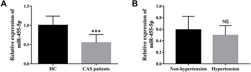
Serum levels of miR-455-5p were measured in the two study groups using qRT-PCR assay. As shown in , serum miR-455-5p was at the low expression in the CAS group compared with the control group, and the student’s t test suggested a significant difference (P < 0.001). In addition, serum miR-455-5p levels were also compared between CAS cases with hypertension and without ones. A decreased expression of serum miR-455-5p was detected in cases with hypertension, but the difference did not reach a significant level (, P > 0.05).
Figure 1 Levels of serum miR-455-5p between different groups. (A) Comparison of serum miR-455-5p levels between CAS group and healthy controls (HC). (B) A decreased expression of serum miR-455-5p was detected in cases with hypertension compared with the non-hypertension group, but the difference did not reach a significant level. ***Means P < 0.001 when compared with the HC group.
Analysis of Clinical Factors Influencing the Degree of Carotid Artery Stenosis
To analyze the influence of various clinical parameters and miR-455-5p on the degree of CAS, the univariable and multivariable regression analyses were applied. The degree of CAS was taken as the dependent variable, and all clinical indicators and miR-455-5p levels were taken as independent variables. Univariate regression analysis was performed first, and the results indicated that FBG, hypertension, TC, TG, HDL, LDL, and miR-455-5p had a significant influence on the degree of carotid artery stenosis in CAS cases (, all P < 0.05). Then the independent variables with P value less than 0.05 in the univariable regression analysis were further included in the multivariate regression model. The analysis results demonstrated that LDL (OR = 4.380, 95% CI=1.101–17.419, P = 0.036), miR-455-5p (OR = 0.154, 95% CI= 0.039–0.606, P = 0.007) were independent influence factors for the degree of carotid artery stenosis ().
Table 2 Influence of Different Clinical Variables and miR-455-5p on the Degree of Carotid Stenosis in CAS Patients
Analysis of the Clinical Values of miR-455-5p in CAS and the Development of Cerebral Ischemia Events (CIEs)
In light of the significant influence of miR-455-5p on the degree of carotid artery stenosis, its diagnostic ability for CAS was detected. Based on the serum miR-455-5p levels in the healthy controls and CAS cases, the ROC curve was drawn and the sensitivity and specificity were calculated. As shown in , serum miR-455-5p showed the diagnostic potential to distinguish CAS from healthy individuals with the area under the curve (AUC) of 0.927, the diagnostic sensitivity and specificity were 82.9% and 90.8%, respectively.
Figure 2 Clinical values of serum miR-455-5p in CAS and the development of CIEs. (A) Analysis of the clinical diagnostic ability of miR-455-5p in CAS. Serum miR-455-5p showed the diagnostic potential to identify CAS from healthy individuals with the area under the curve (AUC) of 0.927, the diagnostic sensitivity and specificity were 82.9% and 90.8%, respectively. (B) The Kaplan-Meier (K-M) curve that drawn based on the follow-up results. The incidence of CIEs was higher in people carrying low miR-455-5p levels, the Log rank test indicated a significant difference between the low and high miR-455-5p expression group (P = 0.002).
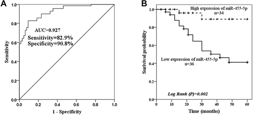
In the current study, all CAS cases completed five years of follow-up, and the CIEs were recorded. According to records, a total of 19 cases developed CIEs, in which 13 TIAs and 6 strokes. Among the 19 CIE cases, 17 individuals were in the low miR-455-5p expression group and 2 cases were in the high miR-455-5p expression group. Then the Kaplan-Meier (K-M) curve was drawn based on the follow-up results, and the even-free survival rates were compared using the Log rank test. It can be seen from that, low miR-455-5p expression was positively related to the occurrence of CIEs (log Rank P = 0.002). Furthermore, the Cox regression model was further used to evaluate the contribution of clinical indicators to CIEs. As shown in , four variables showed significant influence on the occurrence of CIEs in the univariable cox regression analysis, including hypertension, TG, degree of carotid stenosis and miR-455-5p (all P < 0.05). Then the four variables were included in the multivariable Cox regression model, and the results revealed that degree of CAS (HR = 3.241, 95% CI = 1.294–9.045, P = 0.013) and miR-455-5p (HR = 0.128, 95% CI = 0.029–0.573, P = 0.007) were significant risk factors for the development of CIEs in the CAS patients.
Table 3 Multivariable Cox Regression Analysis of the Independent Factors Affecting the Occurrence of Cerebral Ischemia Events
MiR-455-5p Overexpression Can Protect Against ox-LDL Induced Cell Apoptosis
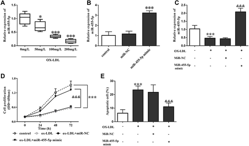
HAECs were treated with different concentrations of ox-LDL to induce endothelial cell damage. As shown in , levels of miR-455-5p decreased gradually with increased ox-LDL concentration. At the concentration of 100 mg/L, the decreasing trend reached an extremely significant level (P < 0.001), so 100 mg/L of ox-LDL was applied in the following experiments. Then the rescue experiments were done by regulating miR-455-5p levels. After miR-455-5p mimic transfection, the levels of miR-455-5p were increased ( and ). In addition, cell proliferation limitation and apoptosis were also observed in cells treated with ox-LDL, but these influences were reversed by miR-455-5p overexpression ( and ).
Figure 3 MiR-455-5p overexpression can protect against ox-LDL-induced cell apoptosis. (A) Levels of miR-455-5p decreased gradually with increased ox-LDL concentration. (B) After miR-455-5p mimic transfection, the levels of miR-455-5p were increased. (C) In ox-LDL treated HAECs, miR-455-5p mimic transfection also increased the levels of miR-455-5p. (D) Ox-LDL led to the inhibition of HAECs viability, which was reversed by miR-455-5p overexpression. (E) miR-455-5p overexpression inhibited ox-LDL-induced cell apoptosis. *Means P< 0.05, ***Means P < 0.001 when compared with the HC group; &&&Means P < 0.001 when compared with the ox-LDL group.
MiR-455-5p Overexpression Can Reverse the Influence of ox-LDL on Cell Oxidative Stress and Inflammatory Response
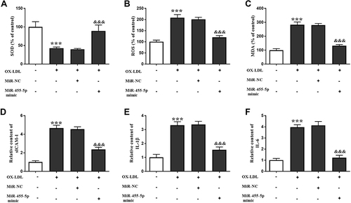
As shown in , the ox-LDL treatment led to the oxidative stress of HAECs, which was reflected by the decrease of SOD and increase of ROS and MDA. But after miR-455-5p mimic transfection, these trends were reversed significantly (). In addition, levels of inflammatory factors were also elevated after ox-LDL treatment, and miR-455-5p played the opposite role by inhibiting the release of sICAM-1, IL-1β and IL-6 ().
Figure 4 MiR-455-5p overexpression can reverse the influence of ox-LDL on cell oxidative stress and inflammatory response. (A–C) ox-LDL treatment led to the decreasing of SOD and increasing in ROS and MDA, which was reversed by miR-455-5p overexpression. (D–F) Levels of inflammatory factors were also elevated after ox-LDL treatment, and miR-455-5p played the opposite role by inhibiting the release of sICAM-1, IL-1β, and IL-6. ***Means P < 0.001 when compared with the HC group; &&&Means P < 0.001 when compared with the ox-LDL group.
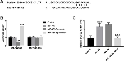
SOCS3 is a Target Gene of miR-455-5p
Bioinformatics analysis showed that miR-455-5p contains binding sites for SOCS3 (). Furthermore, the luciferase reporter assay results demonstrated that transfection of miR-455-5p mimic decreased the luciferase activity in cells transfected with wild type 3’-UTR of SOCS3, whereas the luciferase activity was increased by miR-455-5p inhibitor transfection significantly (). However, mutation in the miR-455-5p binding sites in the 3’-UTR of SOCS3 abolished the effect on the luciferase activity (). The results indicated that SOCS3 is a direct target gene of miR-455-5p. In addition, an increased expression of SOCS3 was detected in ox-LDL-treated cells, which was reversed by miR-455-5p overexpression ().
Figure 5 SOCS3 is a target gene of miR-455-5p. (A) The binding sites between miR-455-5p and SOCS3. (B) Luciferase activity of cells in different groups. (C) Levels of SOCS3 in different cell groups. ***Means P < 0.001 when compared with the HC group; &&&Means P < 0.001 when compared with the ox-LDL group.
Discussion
According to clinical characteristics, asymptomatic CAS cases are often neglected due to a lack of clinical manifestations, which is harmful to the control of disease progression. In recent years, some new markers have been widely reported and may be used as auxiliary tools for CAS diagnosis, such as miRNAs. In the current study, abnormal expression of miR-455-5p was detected in the serum of asymptomatic CAS, and it showed a close association with the occurrence of CIEs. Therefore, serum miR-455-5p was identified to be a promising biomarker for the diagnosis and prognosis of CAS.
At present, there have many studies on the risk factors of CAS, and the recognized risk factors include age, male sex, smoking, elevated cholesterol level, hypertension, and so on. In the present study, the demographic and clinical indexes of the study groups were compared. It was found that in the CAS group, more cases suffered from hypertension. Besides, the influence of hypertension on the degree of CAS was also determined by univariable regression analysis. The finding is consistent with the previous findings on the contributing role of hypertension in CAS.Citation17 In addition, the univariable regression analysis also indicated TC, TG, HDL, and LDL to be the influence factors for the degree of CAS. These factors may promote the occurrence of atherosclerosis and the formation of plaque, and thus participate in the progress of CAS.Citation18,Citation19 Dyslipidemia is one of the important risk factors for CAS. LDL binds to free cholesterol in the blood and can be oxidized to oxidized LDL (ox-LDL), which can promote macrophages to become foam cells, leading to the formation of atherosclerosis.Citation20 In the current study, after adjusting for other clinical indexes, LDL was identified to be an independent factor for the degree of CAS.
As previous studies reported, the role of miRNAs in cerebrovascular diseases has been widely reported. Several miRNAs have been detected to influence the stability of atherosclerosis and thus participate in the CAS process, such as miR-128-3p, and miR-223-3p.Citation21,Citation22 MiR-455-5p is a neuron-specific miRNA, it plays an important role in central nervous function regulation and nerve growth and development.Citation23,Citation24 Current studies have shown that miR-455-5p is under-expressed in ischemic stroke mice, and overexpression of miR-455-5p can reduce neuronal apoptosis and alleviate cerebral ischemic reperfusion injury.Citation24,Citation25 In the present study, miR-455-5p was determined to be at a low expression in the serum of CAS cases. And cases accompanied with hypertension owned lower levels of serum miR-455-5p than non- hypertension ones, but the difference did not reach a significant level. We deduced that miR-455-5p downregulation might have certain association with hypertension, but this is not the main reason for the relationship between miR-455-5p and the development of CAS. In ox-LDL induced human vascular endothelial cells (HUVECs), miR-455-5p is identified to be involved in the protective role of HOXA-AS3 silencing against atherosclerosis.Citation26 In addition, miR-455-5p also can inhibit the proliferation and migration of vascular smooth muscle cells (VSMCs) in vitro, which is the main pathogenesis of CAS.Citation10 Clinically, miR-455-5p is downregulated in patients with atherosclerosis.Citation10 The findings infer that miR-455-5p might be involved in the progression of CAS via regulating the process of atherosclerosis. As expected, the current results demonstrated the independent influence of miR-455-5p on the degree of carotid artery stenosis. Furthermore, its diagnostic potential for CAS was also determined by the ROC curve. These findings forced us to conclude that serum miR-455-5p had the potential for the early identification of CAS, and is related to the severity of the disease. It is known that miRNAs are produced by all cell types, and the same miRNA may be derived from a variety of cell sources, such as endothelial cells, monocytes and macrophages, vascular smooth cells, and platelets, and eventually are secreted in blood. The present study demonstrated the abnormal expression of miR-455-5p in the serum of CAS cases, but its origin is not elucidated, which can be taken into consideration in future studies.
CAS is recognized as a major risk factor for cerebral ischemia events (CIEs).Citation27 Transient ischemic attack (TIA) occurs when atherosclerotic plaques in carotid artery stenosis are shed. And the transient ischemic attack is characterized by short duration and repeated attacks. Therefore, plaque stability is closely related to the occurrence of CIEs.Citation28 It is known that macrophage inflammation affects atherosclerotic plaque stability and thus contributes to the development of cerebrovascular diseases.Citation28 Moreover, miR-455-5p downregulation has been reported to be associated with macrophage polarization.Citation29 Therefore, the influence of miR-455-5p on CIEs should be investigated. In the present study, CIEs occurred in nearly 30% of asymptomatic CAS patients during our five-year follow-up period. Interestingly, a high proportion of patients with low expression levels showed poor prognosis compared with the high expression group, indicating that serum miR-455-5p might have a close association with the occurrence of CIEs. Moreover, the multivariable cox regression analysis confirmed our hypothesis, and miR-455-5p was an independent influence factor for the development of CIEs in the CAS patients. The findings prompted that serum miR-455-5p can predict the occurrence of CIEs in CAS patients to a certain extent, providing clinical evidence for early intervention. Of course, our study population is relatively small, other studies with huge study samples should be performed to verify the clinical findings.
Endothelial cell injury is one of the most important changes in cerebrovascular events.Citation30 Based on the in vitro experiments, cell viability inhibition and cell apoptosis promotion were observed in ox-LDL treated HAECs, indicating the endothelial cell injury induced by ox-LDL. But miR-455-5p overexpression reversed the bad effects, demonstrating the protective role of miR-455-5p. In addition, ox-LDL-induced oxidative stress and inflammatory response of endothelial cells are the important mechanisms of cerebrovascular events.Citation31 In the current study, miR-455-5p prevented HAECs from ox-LDL-induced oxidative stress and inflammatory response, which might be the protective mechanism of miR-455-5p in CAS and long-term ischemic events. Furthermore, the suppressor of cytokine signaling 3 (SOCS3), the negative regulator of IL-6/JAK/STAT3 signaling, was identified to be a target gene of miR-455-5p. Moreover, upregulated expression of SOCS3 was detected in ox-LDL treated cells. Consistently, the same expression trend was also reported in rabbits with atherosclerosis.Citation32 In another model of Ang II–induced vascular dysfunction, SOCS3 inhibition can protect against systemic Ang II–induced vascular dysfunction and hypertension in mice.Citation33 The evidence supported our conclusion that SOCS3 might be involved in the underlying protective mechanism of miR-455-5p against ox-LDL-induced endothelial cell injury. However, the speculation should be verified in future studies including in vivo studies, and more experiments such as tunnel assay are needed to verify cell phenotypic changes after miR-455-5p treatment.
Conclusion
In conclusion, the down-regulated expression of serum miR-455-5p is hopeful to be used as a biomarker for the early identification of CAS. MiR-455-5p serves as an independent risk factor for the degree of CAS, and has a certain predictive value for the occurrence of CIEs in CAS patients. That might be associated with the protective role of miR-455-5p against ox-LDL-induced endothelial cell injury through targeting SOCS3. The current findings provide a promising biomarker for the early diagnosis and timely therapeutic interference of CAS, and provide a theoretical basis for the mechanism of the disease progression.
Disclosure
The authors report no conflicts of interest in this work.
References
- Yi L, Tang J, Shi C, et al. Pentraxin 3, TNF-alpha, and LDL-C are associated with carotid artery stenosis in patients with ischemic stroke. Front Neurol. 2019;10:1365. doi:10.3389/fneur.2019.01365
- Bai X, Feng Y, Li L, et al. Treatment strategies for asymptomatic carotid artery stenosis in the era of lipid-lowering drugs: protocol for a systematic review and network meta-analysis. BMJ Open. 2020;10(7):e035094. doi:10.1136/bmjopen-2019-035094
- Iko M, Aikawa H, Go Y, et al. Treatment outcomes of carotid artery stenting with two types of distal protection filter device. Springerplus. 2014;3:132. doi:10.1186/2193-1801-3-132
- Taguchi A, Ohba S, Itoh Y, Ohshita J, Yonezawa K. [A case of carotid artery stenosis associated with repetitive stimulation of the hyoid]. No Shinkei Geka. 2018;46(9):811–818. Japanese. doi:10.11477/mf.1436203818
- Jackson DC, Sandoval-Garcia C, Rocque BG, et al. Cognitive deficits in symptomatic and asymptomatic carotid endarterectomy surgical candidates. Arch Clin Neuropsychol. 2016;31(1):1–7. doi:10.1093/arclin/acv082
- Xiao S, Zhu H, Luo J, Wu Z, Xie M. miR4255p is associated with poor prognosis in patients with breast cancer and promotes cancer cell progression by targeting PTEN. Oncol Rep. 2019;42(6):2550–2560. doi:10.3892/or.2019.7371
- Huang W, Wu Y, Cheng D, He Z. Mechanism of epithelialmesenchymal transition inhibited by miR203 in nonsmall cell lung cancer. Oncol Rep. 2020;43(2):437–446. doi:10.3892/or.2019.7433
- Wu R, Yun Q, Zhang J, Bao J. Long non-coding RNA GAS5 retards neural functional recovery in cerebral ischemic stroke through modulation of the microRNA-455-5p/PTEN axis. Brain Res Bull. 2021;167:80–88. doi:10.1016/j.brainresbull.2020.12.002
- He W, Chen S, Chen X, Li S, Chen W. Bioinformatic analysis of potential microRNAs in ischemic stroke. J Stroke Cerebrovasc Dis. 2016;25(7):1753–1759. doi:10.1016/j.jstrokecerebrovasdis.2016.03.023
- Zhang X, Liu Y, Zhao J, Yan T. MiR-455-5p serves as a biomarker of atherosclerosis and inhibits vascular smooth muscle cell proliferation and migration. Per Med. 2021;18(3):213–221. doi:10.2217/pme-2020-0136
- Yasue H, Mizuno Y, Harada E. Coronary artery spasm - Clinical features, pathogenesis and treatment. Proc Jpn Acad Ser B Phys Biol Sci. 2019;95(2):53–66. doi:10.2183/pjab.95.005
- Chen P, Miao Y, Yan P, Wang XJ, Jiang C, Lei Y. MiR-455-5p ameliorates HG-induced apoptosis, oxidative stress and inflammatory via targeting SOCS3 in retinal pigment epithelial cells. J Cell Physiol. 2019;234(12):21915–21924. doi:10.1002/jcp.28755
- Xu X, Wu Z, Qiu H, Wu J. Circular RNA circPHC3 promotes cell death and apoptosis in human BMECs after oxygen glucose deprivation via miR-455-5p/TRAF3 axis in vitro. Neuropsychiatr Dis Treat. 2021;17:147–156. doi:10.2147/NDT.S288669
- Zhang JS, Hou PP, Shao S, et al. microRNA-455-5p alleviates neuroinflammation in cerebral ischemia/reperfusion injury. Neural Regen Res. 2022;17(8):1769–1775. doi:10.4103/1673-5374.332154
- Gurbuz O, Kumtepe G, Ozkan H, Karal IH, Ercan A, Ener S. Red blood cell distribution width predicts long term cardiovascular event after on-pump beating coronary artery bypass grafting. J Cardiothorac Surg. 2016;11:48. doi:10.1186/s13019-016-0465-4
- Livak KJ, Schmittgen TD. Analysis of relative gene expression data using real-time quantitative PCR and the 2(-Delta Delta C(T)) Method. Methods. 2001;25(4):402–408. doi:10.1006/meth.2001.1262
- Chen Y, Liu Y, Luo C, Lu W, Su B. Analysis of multiple factors involved in acute progressive cerebral infarction and extra- and intracranial arterial lesions. Exp Ther Med. 2014;7(6):1495–1505. doi:10.3892/etm.2014.1624
- Li A, Huang W, Yang Q, Peng L, Liu Q. Expression of the C677T polymorphism of the 5, 10-Methylenetetrahydrofolate Reductase (MTHFR) gene in patients with carotid artery atherosclerosis. Med Sci Monit. 2020;26:e920320. doi:10.12659/MSM.920320
- Pac-Kozuchowska E, Krawiec P, Grywalska E. Selected risk factors for atherosclerosis in children and their parents with positive family history of premature cardiovascular diseases: a prospective study. BMC Pediatr. 2018;18(1):123. doi:10.1186/s12887-018-1102-2
- Li C, Chen Q, Zhang M, et al. The correlation between lipoprotein(a) and coronary atherosclerotic lesion is stronger than LDL-C, when LDL-C is less than 104 mg/dL. BMC Cardiovasc Disord. 2021;21(1):41. doi:10.1186/s12872-021-01861-6
- Farina FM, Hall IF, Serio S, et al. miR-128-3p is a novel regulator of vascular smooth muscle cell phenotypic switch and vascular diseases. Circ Res. 2020;126(12):e120–e135. doi:10.1161/CIRCRESAHA.120.316489
- Barbalata T, Moraru OE, Stancu CS, Sima AV, Niculescu LS. MiR-223-3p levels in the plasma and atherosclerotic plaques are increased in aged patients with carotid artery stenosis; association with HDL-related proteins. Mol Biol Rep. 2021. doi:10.1007/s11033-021-06636-y
- Strickland IT, Richards L, Holmes FE, Wynick D, Uney JB, Wong LF. Axotomy-induced miR-21 promotes axon growth in adult dorsal root ganglion neurons. PLoS One. 2011;6(8):e23423. doi:10.1371/journal.pone.0023423
- Yao S, Tang B, Li G, Fan R, Cao F. miR-455 inhibits neuronal cell death by targeting TRAF3 in cerebral ischemic stroke. Neuropsychiatr Dis Treat. 2016;12:3083–3092. doi:10.2147/NDT.S121183
- Chen J, Zhu C, Jia W, Wang J, Gu L. MiR-455-5p attenuates cerebral ischemic reperfusion injury by targeting FLT3. J Cardiovasc Pharmacol. 2020;76(5):627–634. doi:10.1097/FJC.0000000000000898
- Chi K, Zhang J, Sun H, et al. Knockdown of lncRNA HOXA-AS3 suppresses the progression of atherosclerosis via sponging miR-455-5p. Drug Des Devel Ther. 2020;14:3651–3662. doi:10.2147/DDDT.S249830
- Wang C, Lv G, Zang D. Risk factors of carotid plaque and carotid common artery intima-media thickening in a high-stroke-risk population. Brain Behav. 2017;7(11):e00847. doi:10.1002/brb3.847
- Nagata M, Minami M, Yoshida K, et al. Calcium-binding protein S100A4 is upregulated in carotid atherosclerotic plaques and contributes to expansive remodeling. J Am Heart Assoc. 2020;9(18):e016128. doi:10.1161/JAHA.120.016128
- Chi X, Ding B, Zhang L, Zhang J, Wang J, Zhang W. lncRNA GAS5 promotes M1 macrophage polarization via miR-455-5p/SOCS3 pathway in childhood pneumonia. J Cell Physiol. 2019;234(8):13242–13251. doi:10.1002/jcp.27996
- Liu X, Zhang X, Zhang Y, et al. Kernelized k-local hyperplane distance nearest-neighbor model for predicting cerebrovascular disease in patients with end-stage renal disease. Front Neurosci. 2021;15:773208. doi:10.3389/fnins.2021.773208
- Chen S, Zhou H, Zhang B, Hu Q. Exosomal miR-512-3p derived from mesenchymal stem cells inhibits oxidized low-density lipoprotein-induced vascular endothelial cells dysfunction via regulating Keap1. J Biochem Mol Toxicol. 2021;35(6):1–11. doi:10.1002/jbt.22767
- Yang X, Jia J, Yu Z, et al. Inhibition of JAK2/STAT3/SOCS3 signaling attenuates atherosclerosis in rabbit. BMC Cardiovasc Disord. 2020;20(1):133. doi:10.1186/s12872-020-01391-7
- Li Y, Kinzenbaw DA, Modrick ML, Pewe LL, Faraci FM. Context-dependent effects of SOCS3 in angiotensin II-induced vascular dysfunction and hypertension in mice: mechanisms and role of bone marrow-derived cells. Am J Physiol Heart Circ Physiol. 2016;311(1):H146–156. doi:10.1152/ajpheart.00204.2016