
Abstract
Introduction
Alveolar bone residual ridge resorption remains a major challenge for dental implant placement in patients with edentulism. Fenugreek seed extracts have been reported to have potential roles in bone metabolism.
Purpose
This study aimed to evaluate the effects of fenugreek seed ethanolic extract (FSEE) on bone cells, inflammation, hormones, and angiogenesis parameters of alveolar bone tissue following teeth extraction in an ovariectomized (OVX) model.
Methods
A total of 30 adults female Wistar rats were assigned into two major groups. Each group consisted of control, OVX, OVX+FSEE 100 mg/kg BW, OVX+FSEE 200 mg/kg BW, and OVX+FSEE 400 mg/kg BW. The FSEE treatment was applied through the intragastric route for 7 days in the first group and for 30 days in the second group of animals. The first molar tooth of the right maxilla was extracted before the FSEE treatment. The level of 17β-estradiol was measured by the ELISA method. The dissected maxilla alveolar bone processus was sectioned for histological evaluation by hematoxylin–eosin staining and an immunohistochemistry assay.
Results
This study found that FSEE reduced the blood estrogen level and increased estrogen receptor-α (ER-α) expression. FSEE administration modified the number of bone cells, angiogenesis, vascular endothelial growth factor (VEGF), sclerostin, and the osteoprotegerin/receptor activator of nuclear factor kappa-β ligand (OPG/RANKL) ratio. Alterations were seen in the inflammatory markers interleukin-6 (IL-6), transforming growth factor-β1 (TGF-β1), and the macrophage-1/macrophage-2 (M1/M2) ratio.
Conclusion
In this study, inflammation was found to be attenuated by reductions in IL-6 and sclerostin, and an increase in TGF-β1. The maturation of bone osteocytes increased along with the increase in ER-α expression and ratio of OPG/RANKL.
Introduction
Edentulism is defined as the permanent loss of natural teeth in the oral cavity. Both partial and complete edentulism can reduce a person’s quality of life because adequate teeth are required for mastication processes, speech, and aesthetic value. A higher prevalence of edentulism is reported among elderly people and in populations of developing countries. Since the incidence of edentulism is correlated with poor socioeconomic factors, edentulism is of concern as a global public health issue.Citation1,Citation2
Dental implants may be proposed as a way to maintain the dental function of edentulous individuals. Conventional dental implant placement requires the presence of a proper alveolar residual ridge. Alveolar residual ridge disposition may present a challenge for dental implant procedures.Citation3 Alveolar residual ridge resorption results from bone remodeling after teeth extraction. Resorption involves osteoclast activities that induce structural bone loss of the alveolar bone.Citation4,Citation5
Estrogen deficiency, immune activation, and oxidative stress may be involved in the biomechanism of residual ridge resorption.Citation4,Citation6 Inflammation initiates interactions between T cells and osteoclasts. This interaction leads to T-cell activation, which increases the release of receptor activator of nuclear factor kappa-Β ligand (RANKL) and the cytokines tumor necrosis factor-α (TNF-α), interleukin-1β (IL-1β), and interleukin-6 (IL-6). Inflammation promotes osteoclast activity in bone resorption.Citation4 Chronic inflammatory processes lead to an imbalance between free radicals and antioxidant enzymes, resulting in oxidative stress conditions.Citation7 Osteoclast activity is influenced by the state of hormones, especially estrogen. A hypoestrogenic state induces a decline in bone volume, bone density, and bone mineral density.Citation8 These biomechanisms indicate the importance of treatment to inhibit residual ridge resorption.
Fenugreek (Trigonella foenum-gaecum) is widely known as a folk medicine. It acts as a galactagogue by increasingthe prolactin level.Citation9 Bioactive compounds of fenugreek seed have been proposed as potential treatments for hyperglycemia and hypercholesterolemia, and as antimicrobials, antioxidants, etc.Citation10 Studies on the phytoestrogenic roles of fenugreek seed in an ovariectomized model found that it improves brain-derived neurotrophic factor, memory, and cognitive performance.Citation11,Citation12 The binding affinity of fenugreek compound to the estrogen receptor is stronger than that of other phytoestrogenic plants.Citation13 The anti-inflammatory effects of fenugreek seed have been demonstrated in ovariectomized rats supplemented with a high-fat diet.Citation14 The estrogenic potential of FSEE has been reported to be superior to that of fenugreek aqueous extract in improving the weight of reproductive organs and tissue in ovariectomized mice.Citation15 To the authors’ knowledge, limited information is available on the effects of FSEE on bone parameters in the ovariectomized model. Because of the importance of optimal residual ridge in cases of edentulism, and the strong potential for the phytoestrogenic role of FSEE, further exploration is necessary to overcome edentulism. Thus, this study aimed to evaluate the effect of FSEE in inhibiting alveolar bone residual ridge resorption in ovariectomized rats, focusing on estrogen, bone cells, inflammation, and angiogenesis parameters.
Materials and Methods
Plant Materials and Extraction
Fenugreek seeds were obtained from Materia Medika Herbal Laboratories, owned by the East-Java Province Government (Malang, East Java, Indonesia). The air-dried (28±2°C) seeds were powdered before being extracted by maceration. The powder was macerated in 96% ethanol solvent (5 mL/g) for 72 hours at 37°C. The filtrate was evaporated using a rotary vacuum evaporator at 50°C.Citation16 The extracts were kept at 4°C before being applied to the animals.Citation17
Experimental Design
Animals were randomly assigned into two groups of 15. Each group consisted of subgroups, namely, sham surgery as the control, ovariectomized (OVX), OVX+fenugreek 100 mg/kg BW, OVX+fenugreek 200 mg/kg BW, and OVX+fenugreek 400 mg/kg BW. Each subgroup consisted of three animals. The first and second groups of animals differed in the duration of fenugreek treatment, ie, 7 and 30 days, respectively (). Thirty days post-ovariectomy, the animals underwent tooth extraction followed by FSEE administration. The extracts were diluted in distillate water to a volume of 1 mL before being administered to the animals. The extract was applied in a single daily dose via the intragastric route.Citation18
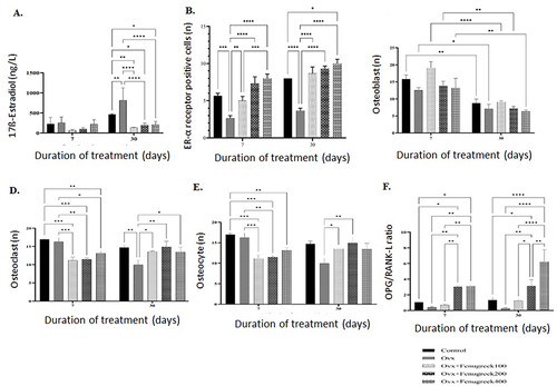
Figure 1 Effect of FSEE on blood estrogen (A), ER-α expression (B), osteoblasts (C), osteoclasts (D), osteocytes (E), and ratio of OPG/RANKL (F). Data are shown as mean ± SEM. *p<0.05, **p<0.001, ***p<0.0002, ****p<0.0001.
Animals
All animal procedures followed the guidelines of the National Center for the Replacement, Refinement and Reduction of Animals in Research (NC3Rs), and were approved in advance by the Health Research Ethic Committee of the Faculty of Medicine, Universitas Brawijaya (no 173/EC/KEPK/06/2022). Female adult Wistar rats, aged 4–6 months, were obtained from Institut Teknologi Bandung (Bandung, West Java, Indonesia). The animals were maintained in a room with controlled temperature and humidity, and a 12–12-hour light–dark cycle. Standard laboratory food and drink were provided ad libitum.Citation19
Ovariectomy Surgery
Ovariectomy procedures were performed under sterile conditions. Animals were anesthetized with ketamine–xylazine via intraperitoneal injection. A single ventral transverse skin incision was carried out to expose the ovaries. Ovaries were ligated and severed using 2/0 silk string. After fat-pad repositioning, muscle-pad skin was sutured in two layers. The sham control animals received a similar incision without removal of the ovaries.Citation20,Citation21
Extraction of Teeth
The teeth were extracted following a modified version of previous protocols. In brief, the animals were sedated and anesthetized using ketamine–xylazine, injected intraperitoneally. Preoperatively, the area surrounding the first molar right maxilla was cleaned using iodine solution. The teeth were completely extracted using sterile forceps. The socket was gently irrigated using saline liquid. Animals were placed individually until complete recuperation from the anesthesia (approximately 24 hours), with water provided ad libitum, followed by regular feeding on the next day.Citation22,Citation23
Collection of Blood Samples and Enzyme-Linked Immunosorbent Assay (ELISA)
Blood was collected under anesthesia via intracardiac puncture. After incubation at room temperature, the clotted blood samples were centrifuged at 3500 rpm for 10 minutes. The supernatants were collected and refrigerated at −20°C until further analysis. The level of 17β-estradiol was measured using a commercial ELISA kit, ie, Rat 17β-estradiol (BT Laboratory, catalogue no E1393Ra). The assay was performed according to the manufacturer’s protocols.Citation24
Tissue Collection and Immunohistochemistry Assay
Dissected bone alveolar processes of the maxilla were fixated in 4% paraformaldehyde overnight at 4°C. After washing in phosphate-buffered saline (PBS), the tissues were decalcified in 10% ethylenediaminetetraacetic acid (EDTA) for 2–4 weeks. The tissue was then dehydrated and embedded in paraffin using standard histological procedures. Then, 5‑µm sections were prepared for hematoxylin and eosin (H&E) staining and immunohistochemical analysis. Deparaffinized sections were immersed in citrate buffer for antigen retrieval. After washing in PBS, the sections were blocked in 3% hydrogen peroxide followed by primary and secondary antibody incubation, and stained with diaminobenzidine buffer as the chromogen.Citation25 The antibodies used for this study were: sclerostin polyclonal antibody (Thermo Fisher Scientific, catalogue no PA5-37943); interleukin-6 (IL-6) antibody (C12-1-hIL-6; Santa Cruz Biotechnology, catalogue no sc-32296); vascular endothelial growth factor (VEGF) expression was analyzed using anti-VEGFA antibody (VG-1) (Abcam, catalogue no ab1316); receptor activator of nuclear factor-κβ ligand (RANKL) was analyzed using RANKL antibody (12A668) (Santa Cruz Biotechnology, catalogue no sc-52950); osteoprotegerin (OPG) was analyzed using osteoprotegerin antibody (E-10) (Santa Cruz Biotechnology, catalogue no sc-377079); transforming growth factorβ1 (TGF-β1) was analyzed using TGF-β1 polyclonal antibody (Elabscience, catalogue no E-AB-33090); macrophage-1 (M1) and macrophage-1 (M2) were analyzed using anti-CD163 and anti-CD163 rabbit monoclonal antibody (Boster Bio, catalogue no M00812).
Statistical Analysis
Data are shown as mean ± SEM. Statistical analysis was carried out using two-way ANOVA to analyze the mean differences between groups, in GraphPad Prism 9.0.0. The significance value was set at p<0.05.Citation17
Results
The level of blood estrogen (17β-estradiol) in the ovariectomized group (257.09±143.38 ng/L) increased by 13% compared to the control group (225.75±160.69 ng/L) after 7 days of follow-up. The increment of blood estrogen dramatically increased (75.06%) at 30 days post-ovariectomy. FSEE at a dose of 100 mg/kg BW had the greatest effect on reducing the blood estrogen level, in both 7 days (65.86±23.68 ng/L) and 30 days (139±3.56 ng/L). Higher doses of FSEE resulted in an increase in blood estrogen levels ().
Alveolar bone tissue among rats in the ovariectomized group showed a lower expression of ER-α than in the control group (2.67±0.58 vs 5.67±0.58 ER-α-positive cells) at 7 days. At the longer follow-up (30 days), a greater reduction (54.17%) in ER-α was seen. FSEE increased the expression of ER-α in a dose-dependent manner, ie, 5±1, 7.33±1.25, and 8±1 ER-α-positive cells for doses of 100, 200, and 400 mg/kg BW, respectively, at 7 days. A similar escalation was demonstrated after 30 days of treatment ().
The number of osteoblasts in alveolar bone tissue after 7 days of treatment was 12.53±1.27, 19.13±3.16, 13.83±2.23, and 13.20±4.85 for ovariectomized, and FSEE doses of 100, 200, and 400 mg/kg BW, respectively. Lower numbers of osteoblasts were observed after 30 days of treatment, ie, 7.17±2.19, 9.13±0.42, 7.23±1.09, and 6.37±0.66 for the respective groups (). In contrast, the numbers of osteoclasts were lower in the ovariectomized group at both 7 and 30 days compared to controls. The increase in osteoclasts was more prominent after 30 days’ administration of FSEE (). The number of osteocytes in ovariectomized mice was not different from those of controls at 7 days, whereas at 30 days, the number of osteocytes decreased in the ovariectomized group (9.97±2.29) compared to controls (14.73±1.12). FSEE increased the number of osteocytes significantly at 30 days of treatment (). Meanwhile, the ratio of OPG/RANKL was drastically reduced in ovariectomized rats ().
FSEE administration did not significantly affect the number of tissue blood vessels in the alveolar bone tissue at 7 days of treatment. The dose of 400 mg/kg BW increased the number of blood vessels significantly compared to 100 and 200 mg/kg BW doses at 30 days of treatment (). Conversely, the number of VEGF-positive cells significantly increased in the FSEE-treated groups at 7 and 30 days of treatment (). The inflammatory cytokine IL-6 sharply increased after ovariectomy. IL-6 significantly reduced after FSEE treatment, with the lowest IL-6 level being observed with the highest dose application (). In contrast, TGF-β1 () and the ratio of M1/M2 decreased after ovariectomy (). Treatment with FSEE increased the level of TGF-β1, the ratio of OPG/RANKL, and the ratio of M1/M2 macrophages. The highest dose of FSEE reduced the expression of sclerostin by about 61.54% and 75.75% compared to the ovariectomized group at 7 days and 30 days, respectively (). The histological and immunohistochemistry staining of alveolar bone tissue sections is shown in .
Figure 2 Effect of FSEE on number of blood vessels (A), VEGF expression (B), IL-6 expression (C), TGF-β1 expression (D), sclerostin expression (E), and ratio of M1/M2 (F). Data are shown as mean ± SEM. *p<0.05, **p<0.001, ***p<0.0002, ****p<0.0001.
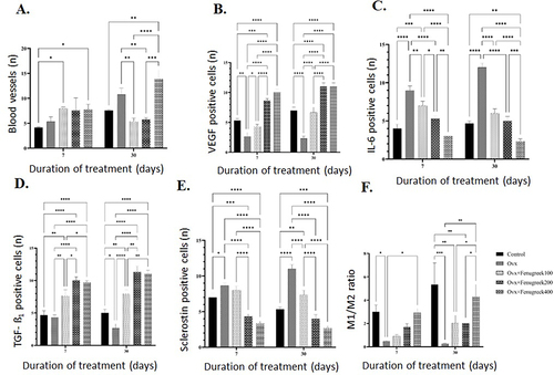
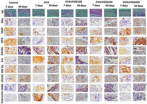
Figure 3 Hematoxylin–eosin staining and immunohistochemical analysis of alveolar tissue sections after different durations of FSEE treatment.
Discussion
Most previous studies have confirmed a decline in estrogen levels after ovariectomy in rats.Citation26,Citation27 In the present study, a rise in the blood estrogen level was shown following ovariectomy surgery. Extragonadal aromatization by peripheral tissues has been suggested as a basic mechanism of the escalation in circulatory estrogen post-ovariectomy. Liver, adrenal, and adipose tissue produce P450 aromatase protein, which is closely related to peripheral estrogen conversion in ovariectomized rats.Citation28
Estrogen is essential for angiogenesis, a process that promotes neovascularization. This effect is mediated through the activation of ER-α and VEGF stimulation.Citation29 Intriguingly, this study showed that blood vessel augmentation is not linked to the rise in VEGF among ovariectomized rats. Despite VEGF stimulation, vascular remodeling can be mediated by the activation of G-protein estrogen receptor (GPER). This receptor modulates the differentiation of endothelial cells via both direct and indirect stimulation.Citation30 Estrogen signaling through ER-α inhibits osteoblast and osteocyte apoptosis, as well as attenuating osteoclast activity, thereby decreasing bone resorption.Citation31 The decline in ER-α in this study may explain the finding of alveolar bone osteoblast, osteoclast, and osteocyte reduction in ovariectomized rats, even as the circulatory estrogen increased.
This study showed that ovariectomy induced IL-6 and reduced TGF-β1 cytokines. The overexpression of IL-6 is linked to the inflammation process.Citation32 Oral inflammation stimulates the release of sclerostin as a bone turnover marker, thereby increasing the activity of osteoclasts.Citation33 The cytokine IL-6 also plays a role as a stimulus for extragonadal aromatization is IL-6 cytokine via activation of CYP19 promoter with glucocorticoid costimulator.Citation34 Hyperestrogen stimulates the generation of reactive oxygen species (ROS) as a marker of early oxidative stress under pathological conditions.Citation35 Oxidative stress presents low antioxidant enzymes and high MDA level reported in edentulous subjects with residual ridge resorption. Both inflammation and oxidative stress were established as molecular pathway of residual ridge resorption.Citation7,Citation36
Fenugreek inhibited inflammation through the reduction of IL-6, as proposed in an earlier study.Citation37 The polarization of macrophages is influenced by fenugreek methanolic extract (FME). M1-type marker cytokines, such as TNF-α and IL-6, were reduced following FME treatment. On the other hand, gene markers for M2 increased significantly after FME.Citation38 The M2 macrophage is a proangiogenic phenotype which plays a role in the expression of VEGF.Citation39 This is supported by a study which found that consuming fenugreek also enhanced the expression of VEGF in people with coronary artery disease; however, this effect was obtained from 8 weeks of fenugreek extract administration.Citation40
The role of fenugreek in estrogen hormone regulation remain inconclusive, depending on the particular case. The reduction of estrogen was reported in a hyperstimulation rat model, with decreases in follicle-stimulating hormone (FSH) and luteinizing hormone (LH). Conversely, FSH stimulation was observed after the administration of fenugreek to subjects with polycystic ovary syndrome.Citation41 The current findings concurred with non-significant results following fenugreek extract administration on serum estradiol, bone resorption, and bone formation markers.Citation18 However, another report revealed that a similar species of Trigonella induced the maturation of osteoblasts through extracellular calcium concentrations.Citation42 Further explanation for the effect of our FSEE in inhibiting alveolar bone residual ridge resorption may be associated with the antioxidant properties of fenugreek. The scavenger activities of fenugreek have been identified as coming from its seed oils, namely, palmitic acid and linoleic acid.Citation43 A previous study revealed the role of fenugreek seed extract in the combination of ovariectomy and metabolic disorder via treatment with a high-fat diet. The role of fenugreek as an inhibitor of cholesterol, inflammation, and histopathological changes in cholesterol-associated tissue has been well elucidated.Citation14 Further exploration is necessary to describe the molecular mechanisms through which fenugreek extract inhibits residual ridge resorption in the ovariectomy model, as well as the metabolic profiles.
Conclusion
FSEE reduced the levels of 17β-estradiol, IL-6, and sclerostin. In contrast, FSEE increased ER-α expression, osteocyte maturation, and TGF-β1 expression.
Disclosure
The authors report no conflicts of interest in this work.
Acknowledgments
The authors would like to thank the Faculty of Medicine, Universitas Brawijaya, for the use of laboratory facilities.
References
- Al‑Rafee MA. The epidemiology of edentulism and the associated factors: a literature Review. J Fam Med Prim Care. 2020;6:169–170. doi:10.4103/jfmpc.jfmpc_1181_19
- Tyrovolas S, Koyanagi A, Panagiotakos DB, et al. Population prevalence of edentulism and its association with depression and self-rated health. Sci Rep. 2016;6:1–9. doi:10.1038/srep37083
- Alshenaiber R, Cowan C, Barclay C, Silikas N. Analysis of residual ridge morphology in a group of edentulous patients seeking nhs dental implant provision—a retrospective observational lateral cephalometric study. Diagnostics. 2021;11:1–8. doi:10.3390/diagnostics11122348
- Kondo T, Kanayama K, Egusa H, Nishimura I. Current perspectives of residual ridge resorption: pathological activation of oral barrier osteoclasts. J Prosthodont Res. 2023;67:12–22. doi:10.2186/jpr.JPR_D_21_00333
- Tan WL, Wong TLT, Wong MCM, Lang NP. A systematic review of post-extractional alveolar hard and soft tissue dimensional changes in humans. Clin Oral Implants Res. 2012;23:1–21. doi:10.1111/j.1600-0501.2011.02375.x
- Gupta S, Singh SV, Arya D. Residual ridge resorption-a review of etiology. Polymorphism. 2019;2:107–113.
- Harish SV, Pudi S, Gade RR, Vudi S, Bn VK, Thota SSB. Assessment of salivary malondialdehyde and superoxide dismutase levels in completely edentulous patients: an in vivo study. Cureus. 2022;14. doi:10.7759/cureus.27949
- Yousefzadeh N, Kashfi K, Jeddi S, et al. Ovariectomized rat model of osteoporosis. EXCLI J. 2020;19:89–107. doi:10.17179/excli2019-1990
- Abdou RM, Fathey M. Evaluation of early postpartum fenugreek supplementation on expressed breast milk volume and prolactin levels variation. Egypt Pediatr Assoc Gaz. 2018;66:57–60 doi:10.1016/j.epag.2018.07.003.
- Visuvanathan T, Than LTL, Stanslas J, Chew SY, Vellasamy S. Revisiting Trigonella foenum-graecum L.: pharmacology and therapeutic potentialities. Plants. 2022;11:1–14. doi:10.3390/plants11111450
- Konuri A, Bhat KMR, Rai KS, Gourishetti K, Phaneendra MYS. Supplementation of fenugreek with choline–docosahexaenoic acid attenuates menopause induced memory loss, BDNF and dendritic arborization in ovariectomized rats. Anat Sci Int. 2021;96:197–211. doi:10.1007/s12565-020-00574-8
- Kruse JL, Congdon E, Olmstead R, et al. Inflammation and improvement of depression following electroconvulsive therapy in treatment-resistant depression. J Clin Psychiatry. 2018;79:1–16. doi:10.4088/JCP.17m11597
- Echeverria V, Echeverria F, Barreto GE, Echeverría J, Mendoza C. Estrogenic plants: to prevent neurodegeneration and memory loss and other symptoms in women after menopause. Front Pharmacol. 2021;12:1–25. doi:10.3389/fphar.2021.644103
- Nagamma T, Konuri A, Bhat KM, Udupa P, Rao G, Nayak Y. Prophylactic effect of Trigonella foenum-graecum L. seed extract on inflammatory markers and histopathological changes in high-fat-fed ovariectomized rats. J Tradit Complement Med. 2022;12:131–140. doi:10.1016/j.jtcme.2021.07.003
- Brogi H, Elbachir H, Amrani NE, Amsaguine S, Radallah D. Fenugreek seeds estrogenic activity in ovariectomized female rats. Curr Issues Pharm Med Sci. 2019;32:138–145. doi:10.2478/cipms-2019-0026
- Lohvina H, Sándor M, Wink M. Effect of ethanol solvents on total phenolic content and antioxidant properties of seed extracts of fenugreek (Trigonella foenum-graecum l.) varieties and determination of phenolic composition by hplc-esi-ms. Diversity. 2022;14. doi:10.3390/d14010007
- Kurnianingsih N, Artamevia D, Winarta AK, Wulandari AP. Modifying effect of anthocyanin from purple sweet potatoes on visceral fat tissue inflammation and liver oxidative stress in psychological stress-induced mice: purple sweet potatoes on psychological stress. J Tropical Life Sci. 2023;13:393–398. doi:10.11594/jtls.13.02.18
- Folwarczna J, Zych M, Nowinska B, Pytlik M. Effects of fenugreek (Trigonella foenumgraecum L.) seed on bone mechanical properties in rats. Eur Rev Med Pharmacol Sci. 2014;18:1937–1947.
- Badi N, Fazelipour S, Naji T, Babaei M, Hessari AK. Histomorphometric and biochemical study of liver and thyroid hormones following administration of MoO3 nanoparticles in female rats. Iran J Vet Med. 2022;16:188–201 doi:10.22059/IJVM.2021.330872.1005196.
- Abedinzade M, Nasri S, Omodi MJ, Ghasemi E, Ghorbani A. Efficacy of Trigonella foenum-graecum seed extract in reducing metabolic and inflammatory alterations associated with menopause. Iran Red Crescent Med J. 2015;17:e26685. doi:10.5812/ircmj.26685
- Sophocleous A, Idris AI. Rodent models of osteoporosis. Bonekey Rep. 2014;3:1–9. doi:10.1038/bonekey.2014.109
- Khoswanto C. A new technique for research on wound healing through extraction of mandibular lower incisors in Wistar rats. Eur J Dent. 2019;13:235–237. doi:10.1055/s-0039-1694312
- Nam OH, Cheon K, Kim MS, Lee HS, Choi SC. Evaluation of the periodontal and pulpal healing of replanted rat molars with doxycycline root conditioning. J Periodontal Implant Sci. 2019;49:148–157. doi:10.5051/jpis.2019.49.3.148
- Widayati A, Mustika D, Karina Riskawati Y, Iskandar A, Kurnianingsih N. The combination of high fat diet and monosodium glutamate altering adipogenesis, brain resistin and serum cortisol level in female rat. MNJ. 2022;8:113–116. doi:10.21776/ub.mnj.2022.008.02.8
- Moreno-Villagrana AP, Gutiérrez-Valdés DH, Flores-Luna MG. Immunohistochemical analysis of alveolar bone preserved with autologous teeth graft. Osteopontin expression and its regulatory functions in preserved alveolar ridge. Int J Odontostomatol. 2021;15:616–625. doi:10.4067/S0718-381X2021000300616
- Hao F, Gu Y, Tan X, et al. Estrogen replacement reduces oxidative stress in the rostral ventrolateral medulla of ovariectomized rats. Oxid Med Cell Longev. 2016;2016. doi:10.1155/2016/2158971
- Khaleghi M, Rajizadeh MA, Bashiri H, et al. Estrogen attenuates physical and psychological stress-induced cognitive impairments in ovariectomized rats. Brain Behav. 2021;11:1–15. doi:10.1002/brb3.2139
- Zhao H, Tian Z, Hao J, Chen B. Extragonadal aromatization increases with time after ovariectomy in rats. Reprod Biol Endocrinol. 2005;3:1–9. doi:10.1186/1477-7827-3-6
- Elsayed DH, Helmy SA, Dessouki AA, El-Nahla AM, Abdelrazek HMA, El-Hak HNG. Influence of genistein and diadizine on regularity of estrous cycle in cyclic female Wistar rat: interaction with estradiol receptors and vascular endothelial growth factor. Open Vet J. 2022;12:639–648. doi:10.5455/OVJ.2022.v12.i5.8
- Bartella V, De Francesco EM, Perri MG, et al. The G protein estrogen receptor (GPER) is regulated by endothelin-1 mediated signaling in cancer cells. Cell Signal. 2016;28:61–71. doi:10.1016/j.cellsig.2015.11.010
- Manolagas SC, O’Brien CA, Almeida M. The role of estrogen and androgen receptors in bone health and disease. Nat Rev Endocrinol. 2013;9:699–712. doi:10.1038/nrendo.2013.179
- Kishimoto T. Interleukin-6: discovery of a pleiotropic cytokine. Arthritis Res Ther. 2006;8. doi:10.1186/ar1916
- Liao C, Liang S, Wang Y, Zhong T, Liu X. Sclerostin is a promising therapeutic target for oral inflammation and regenerative dentistry. J Transl Med. 2022;20:1–13. doi:10.1186/s12967-022-03417-4
- Simpson E, Rubin G, Clyne C, et al. The role of local estrogen biosynthesis in males and females. Trends Endocrinol Metab. 2000;11:184–188. doi:10.1016/S1043-2760(00)00254-X
- Cormio A, Cormio G, Musicco C, Sardanelli AM, Gasparre G, Gadaleta MN. Mitochondrial changes in endometrial carcinoma: possible role in tumor diagnosis and prognosis (Review). Oncol Rep. 2015;33:1011–1018. doi:10.3892/or.2014.3690
- Kumar D, Singhal A, Bansal S, Gupta S. Extraction, isolation and evaluation Trigonella foenum-graecum as mucoadhesive agent for nasal gel drug delivery. J Nepal Pharm Assoc. 2015;27:40–45. doi:10.3126/jnpa.v27i1.12149
- Huang H, Wang X, Yang L, et al. The effects of fenugreek extract on growth performance, serum biochemical indexes, immunity and NF-κB signaling pathway in broiler. Front Vet Sci. 2022;9:1–10. doi:10.3389/fvets.2022.882754
- Hassan N, Withycombe C, Ahluwalia M, Thomas A, Morris K. A methanolic extract of Trigonella foenum-graecum (fenugreek) seeds regulates markers of macrophage polarization. Funct Foods Heal Dis. 2015;5:417–426 doi:10.31989/ffhd.v5i12.216.
- Riabov V, Gudima A, Wang N, Mickley A, Orekhov A, Kzhyshkowska J. Role of tumor associated macrophages in tumor angiogenesis and lymphangiogenesis. Front Physiol. 2014;5:1–13. doi:10.3389/fphys.2014.00075
- Roohbakhsh E, Barari A, Abbaszadeh H. The effect of interval training and consuming fenugreek seed extract on FGF- 21 and VEGF gene expression in patients with coronary artery diseases. Quartery Horiz Med Sci. 2021;27:130–147. doi:10.32598/hms.27.2.3456.1
- Swaroop A, Jaipuriar AS, Gupta SK, et al. Efficacy of a novel fenugreek seed extract (Trigonella foenum-graecum, furocystTM) in polycystic ovary syndrome (PCOS). Int J Med Sci. 2015;12:825–831. doi:10.7150/ijms.13024
- Ibrahim HIM, Darrag HM, Alhajhoj MR, Khalil HE. Biomolecule from Trigonella stellata from Saudi flora to suppress osteoporosis via osteostromal regulations. Plants. 2020;9:1–16. doi:10.3390/plants9111610
- Akbari S, Abdurahman NH, Yunus RM, Alara OR, Abayomi OO. Extraction, characterization and antioxidant activity of fenugreek (Trigonella-Foenum Graecum) seed oil. Mater Sci Energy Technol. 2019;2:349–355 doi:10.1016/j.mset.2018.12.001.