
Abstract
Introduction
Gonial angle is an important craniofacial parameter providing information about symmetry and vertical dimensions of the facial skeleton. It can be measured on panoramic radiographs and lateral cephalograms. Reliable assessment of the gonial angle is challenged by the superimpositions associated with lateral cephalograms. The aim of the current study was to assess the precision of panoramic imaging in measuring the gonial angles compared to lateral cephalograms in adult patients with different mandibular divergence patterns.
Methods
Panoramic radiographs and lateral cephalograms of 448 adults (18–30 years old) were utilized in the study. The gonial angle was determined on the lateral cephalograms using an online AI-driven assessment tool (WebCephTM) and compared to the panoramic measurements among the different gender, malocclusion, and mandibular divergence groups.
Results
Statistically significant differences were recorded between measurements taken on lateral cephalograms or panoramic radiographs (p=0.022). In addition, statistically significant differences were reported in gonial angle measurements on panoramic radiographs among the different mandibular divergence groups (p=0.004) for FMA (p=0.002) for Sn-GoMe.
Conclusion
While cephalometry is considered the gold standard tool for reliable gonial angle assessment, panoramic radiographs were more accurate in detecting the differences between the divergence groups in the current study.
Introduction
A comprehensive patient assessment is essential to the success of orthodontic treatment and dental care. During diagnosis and treatment planning, a thorough assessment of the occlusion, soft tissue relationships, and the skeletal form is necessary. Clinical examination, patient’s photographs, dental casts, and radiographs are typically used for this purpose.Citation1–3 Panoramic radiographs constitute an integral part of the standard of care in dentistry. It is used by dentists and orthodontists alike offering wide panoramic visualization of the maxillofacial region. They serve as invaluable screening tools for detecting abnormalities in the teeth and alveolar bone including diagnosis of cysts or tumours, dental anomalies, tooth eruption paths, bone pathology, and mandibular asymmetry. Its non-invasive nature, reduced radiation exposure, affordability, and ability to showcase the entire dentition, temporomandibular joints, and surrounding anatomy make panoramic radiographs an integral part in modern dental practice. For many clinicians, it is an adequate tool to guide a well-informed decision for a treatment plan.Citation4,Citation5
Similarly, lateral cephalometric radiographs are widely used as a screening tool for orthodontic diagnosis and treatment planning and are considered another cornerstone in the field of diagnostic dentistry. It provides a detailed profile of the skeletal disproportion associated with malocclusions and facilitates the prediction of future growth changes in the craniofacial structures. Lateral cephalometric radiographs show a sagittal view of the skeletal, soft tissues, and dental structures. For lateral cephalometric evaluations, certain anatomical landmarks and points on the skull are used to allow quantitative analyses and measurements.Citation6,Citation7
The gonial angle, a critical metric in craniofacial analysis, is defined as the angle formed by the convergence of two reference lines: one extending from the midpoint of the mandibular ramus to the mandibular body, and the other extending from the mandibular body to the lower border of the mandible. This angular measurement is crucial in assessing mandibular morphology and skeletal relationships. When making cephalometric measurements from radiographs, lateral and anteroposterior projections are typically utilized. However, accurate measurements of an individual’s gonial angle become challenging due to superimpositions on the lateral cephalograms.Citation8–10 The aim of the current study was to assess the precision of panoramic imaging in measuring the gonial angles compared to lateral cephalograms in adult patients with different mandibular divergence patterns.
Materials and Methods
Panoramic radiographs and lateral cephalograms of 448 adults (241 females and 207 males, 18–30 years old) were selected from the orthodontic records archive at the Postgraduate Clinics of Riyadh Elm University, Riyadh, KSA. All radiographs were classified as pre-treatment records of patients currently undergoing orthodontic treatment in the same facility, identified as non-growing subjects, no medical conditions, and no history of surgery or trauma involving the mandible. Radiographs were excluded if the records indicated that subjects had any syndromes, skeletal or facial anomalies, missing teeth (other than the 3rd molars), history of previous orthodontic treatment, or if the radiograph showed any technical or exposure errors or were not taken at the same timepoint. All participants provided written informed consent and the study was approved by the Institutional Review Board of Riyadh Elm University (FRP/226224220).
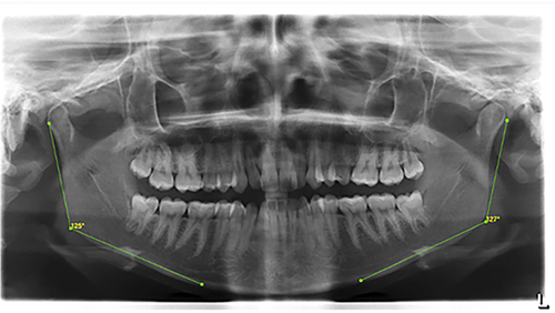
All collected cephalometric radiographs were traced using the WebCephTM program (AssembleCircle Corp., Gyeonggi-do, Republic of Korea) which utilizes AI in tracing. Anatomical landmarks were identified and located by the software and checked manually so that lines and angles could be drawn and measured. Measurements on the lateral cephalograms included, the gonial angle, ANB angle, Frankfort-mandibular plane angle (FMA), and mandibular plane to cranial base (SN-GoMe) (). On the panoramic radiograph, the gonial angle was measured by intersection of the posterior ramal border and the tangent to the inferior border of the mandible. All measurements were taken and reviewed individually by two calibrated examiners ( and ).
Table 1 Anatomical Landmarks, Lines, and Angles Used in the Study
Figure 1 Lateral cephalogram showing the landmarks locations and measurements made using WebCeph.
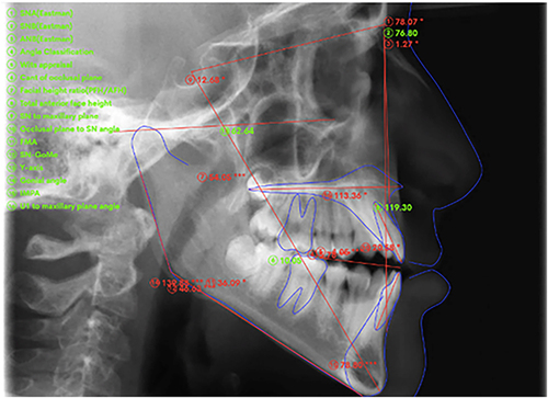
Figure 2 Gonial angle measured on a panoramic radiograph.
Anteroposterior classification of the sample (according to the ANB angle) revealed a distribution as follows: Class I (n=75, 16.74%), Class II (n=282, 62.94%), and Class III (n=91, 20.3%). Vertically, based on the FMA values, the sample was classified as: hypodivergents (n=87, 19.4%), normodivergents (n=161, 35.93%) and hyperdivergents (n=200, 44.6%) cases. However, the percentages changed slightly when considering the Sn-GoMe rendering a classification of: hypodivergents (n=63, 14.06%), normodivergents (n=170, 37.94%) and hyperdivergents (n=215, 47.99%) cases.
Statistical Analysis
The sample size needed for conducting this study was calculated using the G*Power 3.1 software. The measurements were recorded by the primary investigator for 100 cases and repeated after an interval period of 2 weeks by the same investigator to assess intrarater reliability. The same measurements were recorded by another investigator using the same protocol to assess the interrater reliability. The measurement errors were assessed by Dahlberg’s method and intraclass correlation coefficient (ICC) was used to assess reliability. Data was analysed using IBM-SPSS for Windows version 28.0 (SPSS Inc., Chicago, IL). One-way analysis of variance (ANOVA) and regression analysis were used for the comparisons. A p-value of less than 0.05 was considered significant.
Results
The intra- and inter-examiner reliability tests showed no statistically significant differences between readings and excellent reliability for all measurements (ICC ≥ 0.90). Since panoramic measurements of the right and left gonial angles were very high correlated (ICC = 0.968), the average was calculated and compared to the cephalometric gonial angle values.
No statistically significant differences were recorded for the gonial angle measurements taken on lateral cephalograms or panoramic radiographs between both genders (p=0.379 and p=0.116 respectively, ). However, comparison of the gonial angle measurements between lateral cephalograms and panoramic radiographs showed statistically significant difference (p=0.022, ).
Table 2 Descriptive Statistics and Comparison of the Gonial Angle Measurements on Lateral Cephalograms and Panoramic Radiographs Between Both Genders (Significant at P≤0.05)
Table 3 Comparison of the Gonial Angle Measurements on Lateral Cephalograms and Panoramic Radiographs
Statistically significant differences were reported in gonial angle measurements on panoramic radiographs among different mandibular divergence groups (p=0.004) for FMA (p=0.002) for Sn-GoMe ( and ). Nonetheless, gonial angle measurements were non-significantly different when compared between the lateral cephalograms and panoramic radiographs among different malocclusions ().
Table 4 Comparison of Gonial Angle Measurements Between the Lateral Cephalograms and Panoramic Radiographs Among Different Mandibular Divergence Groups (FMA)
Table 5 Comparison of Gonial Angle Measurements Between the Lateral Cephalograms and Panoramic Radiographs Among Different Mandibular Divergence Groups (Sn-GoMe)
Table 6 Comparison of Gonial Angle Measurements Between the Lateral Cephalograms and Panoramic Radiographs Among Different Malocclusions. Significant at P≤0.05
Discussion
The gonial angle radiographic measurement is frequently used in dentistry as an indicator to determine the growth pattern of subjects and to specify the rotation of the mandible. It is also an important parameter for assessing facial asymmetry.Citation11 Gonial angle measurements are also utilized for age prediction in circumstances such as a mass catastrophe, human remains, and missing persons.Citation12 Increased gonial angle indicates downward and backward rotation of the mandible (high angle) while decreased gonial angle indicate upward and forward direction of the mandible (low angle). Therefore, accurate measurement of the gonial angle is crucial for proper treatment planning especially for orthodontics and craniofacial surgical cases.Citation13,Citation14 The aim of the current study was to investigate the accuracy of panoramic imaging in measuring the gonial angles compared to lateral cephalograms in adult patients with different mandibular divergence patterns.
When comparing panoramic and lateral cephalograms, it is crucial to acknowledge the distinctions of their respective digital imaging techniques and the principles of tridimensional geometry that underlie their acquisition. These differences cause potential biases inherent in their interpretation. Even though they are two-dimensional, panoramic photographs depict structures from different perspectives, requiring a grasp of 3D concepts to accurately interpret them. Conversely, lateral cephalograms provide a more direct representation of sagittal relationships but still necessitate an understanding of 3D craniofacial morphology to contextualize their findings. These inherent differences emphasize the importance of considering potential biases when interpreting radiographic findings. Other factors such as patient positioning, anatomical variations, and technical limitations can also contribute to biases in image quality and interpretation. Clinicians must be aware of the impact of all these factors to ensure accurate diagnoses and treatment planning especially for their orthodontics and oral surgery cases.Citation15,Citation16
WebCeph is an online AI-based platform developed to provide cephalometric applications and allow automatic tracings and analysis. Its use has grown significantly in popularity, particularly among orthodontists and orthognathic surgeons. It offers automatic superimposition, treatment simulation, and manual landmark editing while automating measurement computation.Citation17 WebCeph reliability, performance, and accuracy have been assessed previously with conflicting results. While some studies indicated its suitability for clinical uses and research purposes highlighting advantages such as convenience of digital imaging in terms of storage, enhancement, and transmission quality, other reports indicated problems related to inconsistency of measurements.Citation18–20 In the current study, WebCeph was used to perform cephalometric tracing and analysis of the gonial angle.
The results of the study demonstrate that there are statistically significant differences in the values of gonial angle measured on cephalograms and panoramic radiographs. Fisher-Brandies et al,Citation21 in their study, reported a difference of 2.2–3.6 degrees in the gonial angle between panoramic and lateral cephalogram and the difference was statistically significant. It is possible to suggest that using panoramic radiography for measuring the gonial angle should be cautiously considered and that cephalometric analysis serves as the gold standard tool when measuring the gonial angle especially for critical decisions with orthodontics treatment or surgical planning and for forensic purposes requiring comprehensive analysis. Although panoramic radiographs are occasionally a preferred option for measuring gonial angles since they allow clear and independent viewing of the right and left gonial angles, the accuracy of the measurements should always be prioritized.
In the current study, gender had irrelevant influence on gonial angle range. In addition, when gonial angle measurements were compared between the lateral cephalograms and panoramic radiographs among different malocclusions, differences were nonsignificant. However, comparing the values among different mandibular divergence groups, statistically significant differences were reported for the panoramic radiographs’ measurements. The difference could possibly be due to using different anatomical landmarks and points to perform the analysis. In addition, according to a recent report,Citation22 there are several concerns related to the precision of measuring the FMA automatically using AI-based tools such as the WebCeph platform used in the current study. Previous investigations had confirmed the nonagreement in FMA measurements between manual and digital tracing.Citation23,Citation24 In addition, several studies have found that it was hard to locate some landmarks such as the gonion, porion, menton, gnathion, orbitale, and articulare using these tools.Citation25,Citation26
Conclusion
The present study conducted a comprehensive panoramic and cephalometric assessment of gonial angle measurements among the different mandibular divergence groups using an online AI-driven assessment tool. The findings indicated significant differences between both imaging modalities in measuring the gonial angle and variations in the gonial angles across different mandibular divergence patterns. While cephalometry remains to be the gold standard tool for reliable gonial angle assessment, panoramic radiographs were more accurate in detecting the differences between the divergence groups in the current study.
Disclosure
The authors declare no conflicts of interest in this work.
References
- Arnett GW, Bergman RT. Facial keys to orthodontic diagnosis and treatment planning. Part I. Am J Orthod Dentofac Orthop. 1993;103(4):299–312. doi:10.1016/0889-5406(93)70010-L
- Rischen RJ, Breuning KH, Bronkhorst EM, Kuijpers-Jagtman AM, Glogauer M. Records needed for orthodontic diagnosis and treatment planning: a systematic review. PLoS One. 2013;8(11):e74186. doi:10.1371/journal.pone.0074186
- Auconi P, Caldarelli G, Scala A, Ierardo G, Polimeni A. A network approach to orthodontic diagnosis. Orthod Craniofac Res. 2011;14(4):189–197. doi:10.1111/j.1601-6343.2011.01523.x
- Akcam MO, Altiok T, Ozdiler E. Panoramic radiographs: a tool for investigating skeletal pattern. Am J Orthod Dentofac Orthop. 2003;123(2):175–181. doi:10.1067/mod.2003.3
- Sadat-Khonsari R, Fenske C, Behfar L, Bauss O. Panoramic radiography: effects of head alignment on the vertical dimension of the mandibular ramus and condyle region. Eur J Orthod. 2012;34(2):164–169. doi:10.1093/ejo/cjq175
- Silva MB, Sant’Anna EF. The evolution of cephalometric diagnosis in orthodontics. Dental Press J Orthod. 2013;18(3):63–71. doi:10.1590/S2176-94512013000300011
- Halazonetis DJ. Morphometrics for cephalometric diagnosis. Am J Orthod Dentofac Orthop. 2004;125(5):571–581. doi:10.1016/j.ajodo.2003.05.013
- Upadhyay RB, Upadhyay J, Agrawal P, Rao NN. Analysis of gonial angle in relation to age, gender, and dentition status by radiological and anthropometric methods. J Forensic Dent Sci. 2012;4(1):29. doi:10.4103/0975-1475.99160
- Rubika J, Felicita AS, Sivambiga V. Gonial angle as an indicator for the prediction of growth pattern. World J Dent. 2017;6(3):161–163. doi:10.5005/jp-journals-10015-1334
- Xie QF, Ainamo A. Correlation of gonial angle size with cortical thickness, height of the mandibular residual body, and duration of edentulism. J Prosth Dent. 2004;91(5):477–482. doi:10.1016/j.prosdent.2004.02.020
- Panneerselvam E, Prasad PJ, Balasubramaniam S, Somasundaram S, Raja KV, Srinivasan D. The influence of the mandibular gonial angle on the incidence of mandibular angle fracture—A radiomorphometric study. J Oral Maxillofac Surg. 2017;75(1):153–159. doi:10.1016/j.joms.2016.08.016
- Acharya AB. A digital method of measuring the gonial angle on radiographs for forensic age estimation. J Forensic Radiol Imaging. 2017;11:18–23. doi:10.1016/j.jofri.2017.09.002
- Nanda SK. Growth patterns in subjects with long and short faces. Am J Orthod Dentofacial Orthop. 1990;98(3):247–258. doi:10.1016/S0889-5406(05)81602-6
- Al-Dawoody AD. Does high gonial angle increases the risk of mandibular angle fracture? A digital orthopantomographic study. J Clin Exp Dent. 2022;14(12):e994–e999. doi:10.4317/jced.60003
- Yazdanian M, Karami S, Tahmasebi E, et al. Dental Radiographic/Digital Radiography Technology along with Biological Agents in Human Identification. Scanning. 2022;18:5265912.
- Brennan J. An introduction to digital radiography in dentistry. J Orthod. 2002;29(1):66–69. doi:10.1093/ortho/29.1.66
- Mahto RK, Kafle D, Giri A, Luintel S, Karki A. Evaluation of fully automated cephalometric measurements obtained from web-based artificial intelligence driven platform. BMC Oral Health. 2022;22(1):132. doi:10.1186/s12903-022-02170-w
- Hung K, Montalvao C, Tanaka R, et al. The use and performance of artificial intelligence applications in dental and maxillofacial radiology: a systematic review. Dentomaxillofac Radiol. 2020;49(1):107. doi:10.1259/dmfr.20190107
- Alqahtani H. Evaluation of an online website-based platform for cephalometric analysis. J Stomatol Oral Maxillofac Surg. 2020;121(1):53–57. doi:10.1016/j.jormas.2019.04.017
- Yassir YA, Salman AR, Nabbat SA. The accuracy and reliability of WebCeph for cephalometric analysis. J Taibah Uni Med Sci. 2021;17(1):57–66.
- Fischer-Brandies H, Fischer-Brandies E, Dielert E. The mandibular angle in the orthopantomogram. Der Radiologe. 1984;24(12):547–549.
- Azeez SM, Surji FF, Kadir SO, et al. Accuracy and Reliability of WebCeph Digital Cephalometric Analysis in Comparison with Conventional Cephalometric Analysis. World J Dent. 2023;14(8):727–732. doi:10.5005/jp-journals-10015-2285
- Tsolakis IA, Gizani S, Panayi N, Antonopoulos G, Tsolakis AI. Three-dimensional printing technology in orthodontics for dental models: a systematic review. Children (Basel). 2022;9(8):1106–1121. doi:10.3390/children9081106
- Bulatova G, Kusnoto B, Grace V, Tsay TP, Avenetti DM, Sanchez FJC. Assessment of automatic cephalometric landmark identification using artificial intelligence. Orthod Craniofac Res. 2021;24(Suppl 2):37–42. doi:10.1111/ocr.12542
- Chen YJ, Chen SK, Chang HF, et al. Comparison of landmark identification in traditional versus computer-aided digital cephalometry. Angle Orthod. 2000;70(5):387–392. doi:10.1043/0003-3219(2000)070<0387:COLIIT>2.0.CO;2
- Celik E, Polat-Ozsoy O, Toygar Memikoglu TU. Comparison of cephalometric measurements with digital versus conventional cephalometric analysis. Eur J Orthod. 2009;31(3):241–246. doi:10.1093/ejo/cjn105