
Abstract
Background
Wound healing is a goal for advanced technology in the surgical space to benefit clinical outcomes. Surgical staplers are commonly used in a variety of open and minimally invasive abdominal and thoracic procedures. Assessment of wound healing traits, such as perfusion, has been challenging due to technical limitations. A novel technique that utilizes micro-computed tomography methodology to measure perfusion was designed to compare the micro-perfusion of staple lines between commercial stapler reloads that employ different staple height strategies.
Materials and methods
Following an Institutional Animal Care and Use Committee-approved protocol, rats were euthanized and immediately heparinized prior to a subtotal gastrectomy with either graduated-height or single-height staples. Rats were then perfused with barium, following which stomachs were removed and immediately fixed in formalin to prevent degradation. Stomachs were then imaged using micro-computed tomography and subsequent analysis was utilized to quantify fluid volume and patent vasculature proximity to staples within the staple line region for each group.
Results
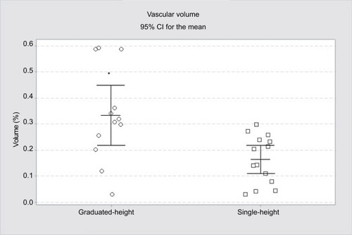
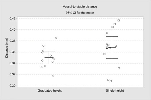
Average perfusion volume was significantly higher with graduated-height staples (0.33% ± 0.18%) compared to single-height staples (0.16% ± 0.09%, P=0.011). Average vessel-to-staple line distance was not significant but trended lower with graduated-height staples (0.35±0.02 mm) compared to single-height staples (0.36±0.03 mm, P=0.18).
Discussion
Graduated-height staples had significantly higher perfusion volume than single-height staples, which likely has a downstream benefit on wound healing and clinical outcomes.
Conclusion
This study shows a higher perfusion volume around the staple lines using graduated-height staples as compared to single-height staples and this may contribute to better wound healing in patients.
Plain language summary
Staplers are commonly used for closing tissue in surgeries, such as when removing a piece of lung due to cancer or doing gastric bypass for weight loss. A challenge with this process is how much pressure the staples put on the tissue: too little pressure can lead to the wound opening again, whereas too much pressure can lead to the tissue not healing or further injury due to lack of blood flow. This study examined blood flow, or perfusion, to tissues after stapling using two different devices. One uses staples that are the same height, resulting in greater pressure in the tissue, while the other uses three different height staples that result in less pressure in the tissue but maintains strong closure. Examining blood flow has been a challenge at this small scale; however, the technique used in this paper allows the blood flow to be mapped with greater precision. Blood flow was higher when the three different height staples were used compared to the same-height staples. This supports that using three different height staples has lower pressure in the tissue and should lead to more successful tissue healing compared to same height staples.
Introduction
Surgical staplers have been used successfully for decades to appose, transect, and rejoin tissue. Stapler technology has continued to advance and reduce complication rates; however, they are still susceptible to clinical inconsistencies that may delay the recovery of patients and potentially be catastrophic.Citation1–Citation4 Minimizing stapler-introduced trauma to tissue (eg, hematoma, tearing) is a goal to positively impact their ability to heal efficiently. With advancing stapler technologies, the goal is to maximize wound healing to benefit patient outcomes; however, not all factors have been mapped due to technological limitations.
Two distinct stapler technologies which are commercially available may have differential results on wound healing. One uses graduated-height staples in each of the three rows to close the wound and are paired with a stepped cartridge face that applies a graduated lateral compression to tissue designed to apply the highest level of compression in the center of the cartridge nearest the knife path. Subsequently, the cut edge, with a reduction in compression moving away from the transection line ().Citation5,Citation6 This compression style applies the most compression at the cut edge for hemostasis, and the least compression away from the cut edge to aid the vascular supply and avoid cellular damage. Contrarily, the other stapler uses staples of identical height across the three rows along with a flat cartridge face, applying the same compression across the staple line.Citation5 While the graduated-height staples have been compared to single-height staples in other outcomes, such as staple formation,Citation7 wound healing measures such as perfusion have not been compared.
Figure 1 Tissue mock-up with staples and actual perfusion sampling.
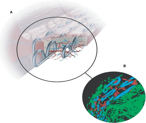
Conventional methods of assessing wound healing typically focus on gross measures (site observation, discoloration, temperature, and pulsation).Citation8–Citation10 Meanwhile, important quantitative characteristics known to be important for wound healing, such as perfusion, have been difficult to assess, particularly in surgical applications.Citation11,Citation12 To assess disruption to the blood flow in tissue following surgery, particularly with wound closure which can potentially impede healing, surgeons have turned to advanced techniques such as fluorescence angiography.Citation13 More progressive techniques such as Doppler signal and Indocyanine green techniques are useful for qualitative macro-perfusion.Citation11,Citation14 However, these techniques are restricted by tissue type and are unreliable for micro-perfusion detection. To overcome this, the present study utilized a quantitative method sensitive enough to measure micro-perfusion of stapled tissue: a contrast agent injected in the vasculature allowed for perfusion of stapled tissue to be measured radiographically.Citation15
A murine model targeting subtotal sleeve gastrectomy model was utilized to assess staple line perfusion between the two staplers at the microscopic level using a micro-computed tomography (micro-CT) high-resolution imaging technique. Micro-CT as a novel imaging method for perfusion quantification was designed for this study to compare staple lines between endoscopic linear surgical staplers that utilize either graduated-height or single-height staples for wound closure.
Materials and methods
Animals
Animals in this study were housed in an AAALAC-accredited facility and handled according to the Guide for Care and Use of Laboratory Animals and the Animal Welfare Act. All procedures were approved by the Medtronic Institutional Animal Care and Use Committee (IACUC, protocol #R2187-301) prior to the study beginning. A total of 29 female Sprague Dawley rats (Jackson Labs, Bar Harbor, ME, USA) at age 2 months and weighing between 270 and 290 grams were utilized for this study.
Subtotal sleeve gastrectomy
Animals were euthanized prior to the procedure to ensure humane treatment of the animals and optimize perfusion of the tissue. Immediately upon euthanasia, to minimize potential coagulation, the rats were heparinized intravenously injected through the left ventricle of the heart and pushed through the circulatory system via a syringe pump. Next, each rat had a staple line fired across the stomach by either:
Graduated-height staples (Covidien Endo GIA™ Ultra Universal staplers with Covidien Endo GIA™ Reloads with Tri-Staple™ Technology Tan reloads, N=14) of 2 mm, 2.5 mm, and 3 mm heights.
Single-height staples (Ethicon Endo Surgery Echelon Flex™ Endopath® White reloads, N=15) of 2.5 mm heights.
The staplers were clamped and fired on the stomach tissue according to the manufacturers’ instructions for use. Following the staple firing, a contrast barium agent was injected via the perfusion needle and the stomachs were dissected and immediately fixed in formalin within a tissue cassette.
All stomachs were determined to be in the indicated compressed tissue thickness range for either graduated-height or single-height as determined by measurements from a proprietary spring-loaded caliper. Stomach tissue thickness had little variability across animals, with a median of 0.045 inches and a range of 0.040–0.050 inches for both graduated-height and single-height groups, respectively (Table S1).
Samples were sent to a third party for blinded staple-line vascular perfusion radiographic analysis.
Staple-line vascular perfusion analysis
3-D volumes of barium-infused, stapled rat stomach vasculature (for example, see ) were acquired using micro-CT at 20 µm voxel resolution with a GE Explore Locus scanner (360 views, two frame averaging, 490 µA, 80 kVp, 1800 ms exposure time). Post-acquisition, staples and vasculature were segmented in a fully automated fashion from the volumes using spectral based filters that preferentially enhance voxels that are brighter than background within a predefined neighborhood/window (size tuned to the average staple or vessel diameter respectively). Utilization of this approach prevents segmentation errors that result from application of traditional fixed thresholding techniques (ie, over-segmentation of background when low intensity/incompletely perfused vessels are targeted or under-segmentation of under-perfused vasculature when only bright/well-perfused vasculature is targeted). To remove contribution of partial volume artifacts around the staples to the vascular segmentation volume, the output binary staple volume was “dilated” by five voxels and removed/subtracted from the segmented vascular volume. Subsequently, vessel and staple binary volumes were skeletonized using 3D thinning algorithms that reduced these structures to single voxel thick lines (MatLab R2011b, MathWorks, Natick, MA, USA). This was performed to prevent possible over-estimation of perfusion when thicker or clumped vessels are prevalent and potentially dominate volumetric measurements. For delineation of a region of interest (ROI) for analysis, all three staple rows within the segmented staple volume were morphologically “closed” filling in spaces between rows to create a single solid object that “wraps” around all staples. Within this resultant ROI, ROI volume, vessel volume, staple volume, total skeletal vessel length, total skeletal staple length, mean skeletonized vessel voxel count from each skeletonized staple voxel for spheres of increasing diameter (80–1000 µm), and mean vessel-to-staple distance were analyzed and exported to Excel. Finally, segmented volumes were pseudo-colored and superimposed onto the original scanned volumes to provide visual confirmation that the segmentation performed by the automated, batched algorithm was accurate. These parameters were chosen since higher resolution scans would restrict field-of-view preventing visualization of the entire staple row. This technique was designed to mimic the examination of human vasculature, but will not be suitable for human microvasculature with diameters smaller than arterioles as these vessels may not allow perfusion of the contrast agent.
Statistics
Analyses were performed using Minitab 17 or higher (Minitab Inc., State College, PA, USA) and data were summarized by descriptive statistics (for continuous variables) or frequencies and percentages (for categorical variables). Statistical tests were 2-sided; statistical significance was accepted at P<0.05. For comparison of groups (graduated-height, single-height) a 2-sided t-test was employed to compare means of both vascular volume and mean vessel-to-staple distance. Summary plots are graphed including a 95% CI.
Results
Sample processing
Out of 29 total procedures, 2 out of 14 graduated-height samples and 1 out of 15 single-height samples were excluded from the study due to errors in the perfusion process, such as a subtotal amount of barium perfusate reaching the left ventricle of the heart.
Vascular volume within staple line
To normalize the variance in staple line length between rats, a 20 mm by 10 mm deep cropped volume () was quantified for each staple group. Graduated-height staples lines (n=12) had 0.33% ± 0.18% vascular volume while single-height staple lines (n=14) had 0.16% ± 0.09% vascular volume. This difference was statistically significant (=0.011).
Figure 2 Perfusion volume comparison of graduated-height and single-height reloads. *P=0.011.
Staple-to-vessel distance
The staple-to-vessel distance was measured to gage vasculature patency to the staple line ROI and defined by the average distance of patent vessels >20 µm in diameter and their distance to a staple. Graduated-height staple lines (n=12) had an average distance of 0.35±0.02 mm to patent blood vessels while single-height staples lines (n=14) had an average distance of 0.36±0.03 mm to patent blood vessels. A significant difference was not found between the two groups (=0.18).
Figure 3 Blood vessel distance to staple line comparison of graduated-height and single-height reloads. P=0.18.
Discussion
Stapling has gained popularity over alternate methods, such as suturing, due to its consistency, reduced complications, and ease of use.Citation16–Citation20 However, stapler evidence on perfusion’s role in outcomes is unclear due to methodological limitations but is important to investigate due to its established role in wound healing including supplying oxygen to the site of injury.Citation3,Citation10,Citation21 Using a novel micro-CT approach, this study attempts to quantify staplers that utilize different techniques for wound closure. We observed that perfusion volume and patent vasculature within the staple line is greater by using graduated-height staples that utilized stepped cartridge surfaces compared to single-height staples that utilize flat cartridge surfaces (), which likely contributes to improved wound healing for surgical patients.
Maximizing wound healing is essential for any surgical procedure and should be prioritized for the patient’s benefit. Perfusion to injured tissue has long been established as essential for wound healing.Citation1 Studies in rat skin closures or anastomosis procedures in canines showed improved blood flow to the sites of injuries with staples compared to traditional suture techniques.Citation22 Thus, optimizing perfusion is important for improving patient outcomes undergoing surgical procedures that utilize staple closures.
To date, gross qualitative measurements of perfusion have been used intraoperatively to make decisions about tissue integrity and ability to properly heal.Citation13 The novel method employed by this study allows for a quantitative measurement of the micro-vasculature imperative for the physiological progression of wound healing. Two different measures of perfusion were quantified: vascular volume and the distance between staple line and patent blood vessels. Volume is important because it directly assesses the amount of blood available at the region. We observed that graduated-height staples had a greater volume of perfusion compared with single-height staples. The distance between staple line and patent blood vessels quantifies how close the perfused blood is to the sites of injury, where lower distances should enable improved wound healing. Staple-to-patent vessel distance did not statistically differ between the two groups; however, there was a trend toward lower distances when using graduated-height staples. Both findings are supported by the graduated-height reload design, which has graduated lateral compression that promotes more patent vasculature closer to the staple line. Future studies using both human tissue and long-term wound healing assessments are needed to validate and expand on these findings.
Overall, our results indicate that the blood vascular volume, one measure of perfusion in the staple line, was statistically higher with graduated-height staples compared to single-height staples. This data supports stapler design features such as cartridge geometry and graduated-staple height benefit tissue perfusion, and likely subsequent healing events. The graduated-height staple reload is designed with a progressively increasing size of staple moving away from the cut line.Citation23 Previous data shows that graduated-height staples can perform similarly to single-height staples for air tightness.Citation24 However, graduated-height staples result in decreased stress in tissues compared to single-height staples,Citation25 which should enable greater blood ingress to the staple line and avoid ischemic tissue complications. This study shows a higher perfusion volume around the staple lines using graduated-height staples as compared to single-height staples and this may contribute to better wound healing in patients.
Acknowledgments
Medtronic provided statistical support (William Mulligan), graphic design support (Chris Switalski), and administrative support (Stephanie Herrmann-Stevenson and Brittany Eno).
This study was sponsored and funded by Medtronic (Minneapolis, MN, USA), which owns Covidien (Mansfield, MA, USA).
Supplementary material
Table S1 Stomach tissue thickness
Disclosure
Matthew Eschbach reports being employed by Medtronic during the conduct of the study. In addition, Mr Eschbach has medical device patents issued to Medtronic. Gregory M Sindberg, Marisha L Godek, Matthew Nagelschmidt, Nicholas Paquette, Michael Wegener, James Alberino and Jane Mayotte all report being employed by Medtronic during the conduct of the study. Amit Vasanji received financial support from ImageIQ (now ERT) to fund imaging and analysis activity for Medtronic-sponsored study. Andrew M Miesse reports being employed by Medtronic during the conduct of the study. In addition, Mr Miesse has medical device patents issued to Medtronic. The authors report no other conflicts of interest in this work.
References
- BakerRSFooteJKemmeterPBradyRVroegopTServeldMThe science of stapling and leaksObes Surg200414101290129815603641
- KivisaariJVihersaariTRenvallSNiinikoskiJEnergy metabolism of experimental wounds at various oxygen environmentsAnn Surg197518168238281138632
- SheridanWGLowndesRHYoungHLTissue oxygen tension as a predictor of colonic anastomotic healingDis Colon Rectum198730118678713677962
- SpiekerHDietrichABleeding complications in bariatric surgery: prophylaxis and therapyChirurg201586983384026099290
- CovidienEndo GIA™ Reload with Tri-staple™ TechnologyMansfieldMA, USA
- CovidienFEA of Linear StaplesMansfieldMA, USA2012
- OkamiJTokunagaTKanouTRandomized study comparing equal height staples with graduated height staples in bronchial closureAnn Thorac Surg201710431012101928551048
- PeacockEEWound Repair3rd edPhiladelphiaSaunders1984
- GuoSDipietroLAFactors affecting wound healingJ Dent Res201089321922920139336
- ReinkeJMSorgHWound repair and regenerationEur Surg Res2012491354322797712
- DargavilleTRFarrugiaBLBroadbentJAPaceSUptonZVoelckerNHSensors and imaging for wound healing: a reviewBiosens Bioelectron201341304223058663
- KondoTIshidaYMolecular pathology of wound healingForensic Sci Int20102031–3939820739128
- JamesDRRisFYeungTMFluorescence angiography in laparoscopic low rectal and anorectal anastomoses with pinpoint perfusion imaging – a critical appraisal with specific focus on leak risk reductionColorectal Dis201517Suppl 3162126394738
- NowakKSandra-PetrescuFPostSHorisbergerKIschemic and injured bowel evaluation by fluorescence imagingColorectal Dis201517Suppl 3121526394737
- ZhaoLLiuAGuoYUltra-low-dose CT coronary angiography using 128-slice dual source CT with low concentration contrast agent: initial experienceJpn J Radiol2017351272473229052025
- BragaMVignaliAGianottiLLaparoscopic versus open colorectal surgery: a randomized trial on short-term outcomeAnn Surg2002236675976612454514
- BraghettoMICardemilHGMandiolaBCMasiaLGGattiniSFImpact of minimally invasive surgery in the treatment of esophageal cancerArq Bras Cir Dig201427423724225626930
- DemmyTLCurtisJJMinimally invasive lobectomy directed toward frail and high-risk patients: a case-control studyAnn Thorac Surg199968119420010421140
- JensenEHPortschyPRChowaniecJTengMMeta-analysis of bioabsorbable staple line reinforcement and risk of fistula following pancreatic resectionJ Gastrointest Surg201317226727222948840
- MiyakeJComparison of wound healing of intestinal anastomosis by stapling devices and by Gambee’s suture under normal and abnormal conditions in dogNihon Geka Gakkai Zasshi19878843783893295511
- SchremlSSzeimiesRMPrantlLKarrerSLandthalerMBabilasPOxygen in acute and chronic wound healingBr J Dermatol2010163225726820394633
- NagamachiYBlood flow and tissue reaction at skin wound closuresWorld J Surg19881256356403072774
- HasegawaSNakayamaSHidaKKawadaKSakaiYEffect of tri-staple technology and slow firing on secure stapling using an endoscopic linear staplerDig Surg201532535336026228297
- ContiniEGodekMLWhiffenJMBronsonDGEx vivo pneumostasis evaluation of a variable-height staple designInnovations20138428428824145973
- NováčekVTranTNKlingeUFinite element modelling of stapled colorectal end-to-end anastomosis: advantages of variable height stapler designJ Biomech201245152693269722871347