
Abstract
Background
Sacroiliac joint (SI) pain is an often-overlooked cause of lower-back pain, due in part to a lack of specific findings on radiographs and a symptom profile similar to other back-related disorders. A minimally invasive surgical (MIS) approach to SI joint fusion using a series of triangular, titanium plasma spray-coated implants has shown favorable outcomes in patients with SI joint pain refractory to conservative care. The aim of this study was to provide a multicenter experience of MIS SI joint fusion using a patient-level analysis.
Patients and methods
We report a patient-level analysis from 144 patients with a mean of 16 months postoperative follow-up. Demographic information, perioperative measures, complications, and clinical outcomes using a visual analog scale for pain were collected prospectively. Random-effects regression models were used to account for intersite variability.
Results
The mean age was 58 years, 71% of patients were female, and 62% had a history of lumbar spinal fusion. Mean (95% confidence interval [CI]) operative time was 73 minutes (25.4–118), blood loss was minimal, and hospital stay was 0.8 days (0.1–1.5). At follow-up, mean (95% CI) visual analog scale pain scores improved by 6.1 points (5.7–6.6). Substantial clinical benefit, defined as a decrease in pain by >2.5 points or a score of 3.5 or less, was achieved in 91.9% of patients (95% CI 83.9%–96.1%), and 96% (95% CI 86.3%–98.8%) of patients indicated they would have the same surgery again.
Conclusion
When conservative measures fail to relieve symptoms resulting from degeneration or disruption of the SI joint, MIS SI joint fusion using a series of triangular, porous, titanium plasma spray-coated implants is a safe and effective treatment option.
Introduction
Chronic low back pain is well known as a public health epidemic. In highly developed countries, it is one of the top three causes of degradation in quality-adjusted life-years, along with ischemic heart disease and chronic obstructive pulmonary disease.Citation1 While lumbar spine pathology is an important cause of chronic low back pain, substantial evidence suggests that not all lower-back pain is in fact generated by lumbar spinal structures. The sacroiliac (SI) joint has been found to be a pain generator in up to 30% of patients diagnosed with lower-back pain.Citation2–Citation5 Disorders of the SI joint may be the result of trauma, pregnancy, inflammatory arthritis, osteoarthritis, or degeneration of the joint either de novo or after lumbar spinal fusion.Citation6,Citation7 Diagnosing the SI joint as the primary pain generator can be complex, as patients often present with a combination of lower-back, groin, gluteal, and/or leg pain.Citation2,Citation8 Furthermore, imaging studies are typically not sensitive to abnormalities in the absence of trauma, ankylosing spondylitis, tumors, or infection.Citation7
SI joint pain can be debilitating and treatment with conservative care is often unsuccessful. The economic burden of conservative care in this population is significant for Medicare, as well as commercial payer entities, at an estimated 3-year cost of US$1.6 billion per 100,000 commercial covered lives, and 5 year estimated cost of $270 million for Medicare beneficiaries.Citation9,Citation10 Furthermore, the impact of pain on persons living with the disease is similar to that associated with other prominent orthopedic conditions routinely treated surgically.Citation11
Open arthrodesis of the SI joint was commonly performed throughout the 1900s.Citation12,Citation13 However, this technique is less common now, as it requires a relatively large incision, significant bone harvesting, and lengthy hospital stay; moreover, patients must avoid weight-bearing for a prolonged period (up to several months) postoperatively.Citation14 A recent study comparing open and minimally invasive surgical (MIS) techniques for SI joint fusion demonstrated more favorable outcomes in the MIS cohort with respect to patient-reported outcomes, operative time, hospital stay, and rate of reoperation.Citation15
Herein, we report a patient-level meta-analysis of safety and effectiveness outcomes from a multicenter retrospective study of patients treated with MIS SI joint fusion using a series of triangular titanium, porous titanium plasma spray-coated implants (iFuse Implant System®; SI-BONE, Inc., San Jose, CA, USA).
Materials and methods
Consecutive patients who underwent MIS SI joint-fusion surgery at six sites were identified. Patients were included if preoperative and minimum 12-month follow-up data were available. Data extracted from medical charts included demographic information, medical history (including history of prior lumbar spinal fusion), length of hospital stay, surgical operating time, estimated blood loss, complications of surgery, SI joint pain measured on a 0–10 visual analog scale (VAS), and satisfaction with surgery. Institutional review board approval was obtained at all sites before beginning this study.
Diagnosis
Pain experienced in the lumbar region can arise from various anatomical structures and pathophysiological functions.Citation16 Portions of the sacral plexus from S1 and S2 innervate the SI joint on the dorsal side, and segments from L3 and S2 innervate the ventral side, resulting in possible dermatomal pain patterns anywhere from L2 to S4.Citation17 Therefore, differential diagnosis in this complex population is essential. A detailed clinical history coupled with a positive result on three or more physical provocation maneuvers, such as Gaenslen’s, flexion– abduction–external rotation, compression, distraction, and thigh thrust, were used as criteria for further testing of the SI joint.Citation18 Diagnostic imaging studies, such as X-ray, computed tomography (CT) and magnetic resonance imaging (MRI) were performed to assess pathology in the lumbopelvic hip complex. When clinical, physical, and imaging findings were concordant, image-guided diagnostic injections of the SI joint were performed as a final step in diagnosing the SI joint as the primary pain generator.Citation18,Citation19 A positive result was defined as a 75% reduction in pain immediately following injection of local anesthetic. All patients in this study failed a 6-month course of nonsurgical treatment consisting of a combination of medication optimization, activity modification, physical therapy, and SI joint injections.
Surgical technique
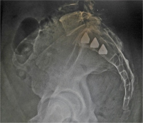
Minimally invasive SI joint surgery was performed on all patients using a series of triangular, titanium implants (iFuse Implant System) (). The implants are coated with a porous titanium plasma spray, an osteoconductive substrate that has been routinely used in total joint prostheses for decades to accommodate biologic fixation.Citation20 A radiolucent table was used to facilitate the use of intraoperative fluoroscopy. After general endotracheal anesthesia was administered, the patient was turned prone and prepped in the normal sterile fashion. A lateral incision (3 cm) was made into the gluteal region, positioned over the first sacral body as viewed on a lateral fluoroscopic image. The fascia was then bluntly dissected to reach the outer table of the ilium. A Steinmann pin was passed through the ilium across the SI joint to the center of the sacrum (lateral to the neural foramen). After a soft-tissue protector was passed over the pin, a hand drill was used to create a pathway through the ilium, across the SI joint, and into the sacrum. Finally, a triangular broach was used to further decorticate the bone and prepare a triangular channel to receive the first implant. Using a pin-guidance system, a total of three implants were placed in the majority of patients. The most cephalad implant was seated within the sacral ala above the first neural foramen. The second implant was located above or adjacent to the S1 foramen, and the third between the S1 and S2 foramen ( and ). The incision was irrigated, and the tissue layers were closed. Postoperatively, patients were instructed to ambulate with partial weight-bearing using the assistance of a walker. A variable program of gradual return to full weight-bearing was employed based on local practices and patient needs.
Figure 1 iFuse implants (iFuse Implant System®; SI-BONE, Inc., San Jose, CA, USA).
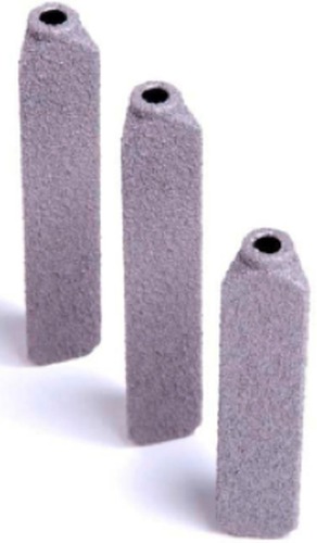
Figure 2 Anteroposterior view of three implants in position.
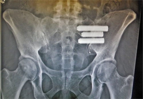
Figure 3 Lateral view of three implants in position.
Statistical analysis
Baseline demographic variables were summarized, where appropriate, with means, standard deviation, confidence intervals (CIs), and frequency tables. Changes in VAS pain scores were evaluated across sites using mixed models that accounted for each subject’s age, sex, history of prior lumbar fusion, and baseline pain score, and included study site as a random effect. Random-effects models assume that even after controlling for known covariates, outcomes are clustered within sites; models assume that the underlying effect at each site is a random variable rather than a fixed value. Similarly, random-effects logistic regression was used to summarize proportions across sites, controlling for age, sex, and history of prior lumbar fusion. Subgroup analysis was performed similarly, with predefined subgroups: prior lumbar fusion (yes versus no), age greater than or less than 65 years, and sex. When there was significant variation in a baseline characteristic by site, univariate random-effects models taking into account site only were used to report mean values (or proportions) and confidence limits. All analyses were performed using R software.Citation21
Clinical improvement was defined using well-accepted values for minimum clinically important difference (MCID) and substantial clinical benefit (SCB) available in the literature. MCID is defined as a change of >2.0 points, and SCB is defined as a 2.5-point decrease or raw score of <3.5.Citation22,Citation23
Results
The medical charts of 144 subjects were available for review. The mean follow-up time was 16 months (range 12–26) (). The majority (71%) of patients at all sites were female, and 62% of patients had a history of prior lumbar spinal fusion. Mean patient age varied across sites; the random-effects estimate was 57.7 years (95% CI 53.0–62.4, range 30–89), and 35% were over the age of 65 years. The mean baseline pain score was 8.6 (95% CI 8.1–9.1) (). At 1 year, patient-reported pain improved clinically and statistically. The mean VAS (pain) score dropped to 2.7 (95% CI 1.8–3.5), representing an improvement of 6.1 points (95% CI 5.7–6.6). Substantial clinical benefit was achieved in 92% of patients (95% CI 84%–96%) and MCID was achieved in 90% (95% CI 82%–95%) of patients. When controlling for age, sex, prior lumbar fusion, and study site, the proportion of patients who reported being satisfied or somewhat satisfied at 12 months was 95.7% (95% CI 86.3%–98.8%). Regression-coefficient modeling performed to assess the effect of age (>/<65 years), sex, and history of lumbar spinal fusion, was statistically significant (t>2) for sex only. Mean (95% CI) reduction in pain was −5.9 (−6.6 to −5.2) for men and −6.2 (−6.8 to −5.6) for women; there were no significant differences for age or history of prior lumbar fusion.
Table 1 Demographic information
Table 2 Results
While the majority of patients experienced significant improvement, a small percentage (15 patients, 10%), reported a change of 1 point or less on VAS; nine improved by 1 point, and six had no change. Two of these patients were revisions; prior SI joint fusion using percutaneous screws had failed. Both showed improvement on VAS scores from 5 to 4, were satisfied with surgery, and indicated they would have the same surgery again for the same result.
Operating time, available for 42 patients at three sites, averaged 73 minutes (95% CI 25.4–118) (). Estimated blood loss, available for all patients, was minimal: a mean of 31 cc (95% CI 25–37). The mean length of hospital stay, available for 109 patients, was 0.8 days (95% CI 0.1–1.5).
Table 3 Operative characteristics
Complications
No intraoperative complications occurred. A total of 28 postoperative sequelae were reported, the most common were falls (3.5%), trochanteric bursitis (2.8%), facet pain (2.1%), and piriformis syndrome (2.1%) (). Two patients returned with contralateral SI joint pain. Both patients reported complete relief of symptoms after undergoing subsequent MIS SI joint fusion on the contralateral side. One patient presented with symptoms of nerve-root impingement, confirmed on CT scan. The patient was returned to the operating room, the original implant was removed and replaced with a shorter implant, and the patient recovered without issue. The revision rate at 1 year was 0.7%.
Table 4 Adverse events
Discussion
Intermediate-term (>1 year) follow-up of a large number of patients who underwent MIS SI joint fusion using a series of triangular titanium implants showed high rates of pain relief and satisfaction and low rates of perioperative or intermediate-term complications. These findings are consistent with prior reportsCitation15,Citation24,Citation25 and a recently published prospective study.Citation26
As with any surgical procedure, an accurate diagnosis is imperative if one is to achieve positive clinical outcomes. Initial clinical presentation in the SI joint patient population can be misleading as several pathophysiologic conditions can present similarly. Structures in the lumbopelvic hip complex are interdependent, and kinematic changes in one area can affect surrounding structures.Citation17 A history of sleep disturbance, pain on prolonged sitting, leg instability, and pain in the lower back, buttock, hip, and groin, as well as the SI joint, are common. Furthermore, pain and degeneration of the SI joint after lumbar spinal fusion is common, with up to 43% of these patients experiencing SI joint pain and 75% showing radiographic changes.Citation27,Citation28 An accurate diagnosis requires a combination of history, physical examination maneuvers that stress the SI joint, and image-guided intra-articular diagnostic injections.
Multiple nonsurgical and surgical treatments for SI joint disorders are available. When nonsurgical management fails to provide adequate relief of symptoms, surgical stabilization is an option. A publication summarizing various arthrodesis techniques (both open and MIS) reports variable improvements in pain and function, with more invasive approaches reporting moderately high complications and nonunions.Citation15 Overall, MIS techniques have a record of significant improvements in pain and function, but results vary with implant and patient selection.
Similar to other reports, the majority of patients in the present cohort had a history of previous lumbar spinal fusion. It is unclear whether the degradation of the SI joint in these patients was a result of adjacent segment disease or de novo degeneration. However, in contrast to other technique reports,Citation29 clinical outcomes using the triangular implants used herein were not diminished in this patient population.
Favorable outcomes in patients with prior lumbar spinal fusion underscore the necessity to suspect the SI joint as a pain generator in patients with lower-back pain. The low success rate of spinal fusion combined with the high incidence of SI joint disorders discovered in patients presenting with lower-back pain leads one to suspect whether the SI joint is being overlooked as a pain generator in these patients.Citation3 Lower-back pain can obscure SI joint disorders, and current imaging technology may not be sensitive in detecting inciting pathology.
The type and number of postsurgical adverse events in our study was commensurate with other published studies using this device system.Citation15,Citation24–Citation26 The most commonly reported complications in the cohort reported herein were trochanteric bursitis and piriformis syndrome. These events are neither uncommon nor unexpected, and can be a result of altered gait pattern due to lower-back or hip pain, postoperative hip-abductor weakness, increased activity levels, and other trauma in the region. Miller et al conducted an analysis of complaints (adverse events) reported to the device manufacturer (SI-BONE, Inc.) as part of an ongoing postmarket surveillance program.Citation30 They reported a complication rate of 3.8% in 5,319 patients, and events included pain due to nerve impingement, hematoma at the operative site, iliac fracture, wound infection, device migration, and implant malposition. Revision surgery was reported in 1.8% of patients.
The current study has limitations. This retrospective chart review lacked patient-reported outcomes, such as the Oswestry Disability Index and Short Form (SF-36) Health Survey, available in controlled trials. Radiological outcomes were not assessed; bony bridging cannot be reliably assessed on plain-film radiographs.Citation6 Furthermore, in the absence of symptoms requiring further imaging, the cost and radiation exposure of CT scanning precludes such imaging studies from being performed routinely.
The study also has strengths. It was a large multicenter study with intermediate-term (greater than 1 year) outcomes. The patient-level meta-analysis provided a method of examining outcomes that accounted for differences in patient characteristics across sites, subsequently providing more accurate results.
Conclusion
For patients with SI joint pain recalcitrant to conservative treatment, minimally invasive surgical fusion of the SI joint using a series of triangular porous titanium plasma spray-coated implants is a safe surgical option that provides significant symptom relief with a high degree of patient satisfaction.
Disclosure
This study was sponsored by SI-BONE, Inc. No funds were received by the authors in support of this work. DS, JC, MG, TH, TG, and ANS receive teaching honoraria from SI-BONE, Inc. RC and DC are SI-BONE, Inc. employees.
References
- VosTFlaxmanADNaghaviMYears lived with disability (YLDs) for 1160 sequelae of 289 diseases and injuries 1990–2010: a systematic analysis for the Global Burden of Disease Study 2010Lancet201238098592163219623245607
- SchwarzerACAprillCNBogdukNThe sacroiliac joint in chronic low back painSpine (Phila Pa 1976)199520131377709277
- SembranoJNPollyDWHow often is low back pain not coming from the back?Spine (Phila Pa 1976)2009341E27E3219127145
- MaigneJYAivaliklisAPfeferFResults of sacroiliac joint double block and value of sacroiliac pain provocation tests in 54 patients with low back painSpine (Phila Pa 1976)19962116188918928875721
- BernardTNKirkaldy-WillisWHRecognizing specific characteristics of nonspecific low back painClin Orthop Relat Res19872172662802951048
- DarGPelegSMasharawiYSacroiliac joint bridging: demographical and anatomical aspectsSpine (Phila Pa 1976)20053015E429E43216094261
- BroadhurstNABondMJPain provocation tests for the assessment of sacroiliac joint dysfunctionJ Spinal Disord19981143413459726305
- FoleyBSBuschbacherRMSacroiliac joint pain: anatomy, biomechanics, diagnosis, and treatmentAm J Phys Med Rehabil20068512997100617117004
- AckermanSCummingsJPollyDKnightTSchneiderKHoltTComparison of the costs of nonoperative care to minimally invasive surgery for sacroiliac joint disruption and degenerative sacroiliitis in a United States Medicare population: potential economic implications of a new minimally-invasive technologyClinicoecon Outcomes Res20132013557558724348055
- AckermanSJPollyDWJrKnightTHoltTCummingsJJrNonoperative care to manage sacroiliac joint disruption and degenerative sacroiliitis: high costs and medical resource utilization in the United States Medicare populationJ Neurosurg Spine201420435436324527824
- CherDPollyDBervenSSacroiliac joint pain: burden of diseaseMed Devices (Auckl)20147738124748825
- MooreMRSurgical treatment of chronic painful sacroiliac joint dysfunctionVleemingAMooneyVSnijdersCJDormanTAStoeckartRMovement, Stability, and Low Back Pain: The Essential Role of the PelvisNew YorkChurchill Livingstone1997563572
- WaisbrodHKrainickJUGerbershagenHUSacroiliac joint arthrodesis for chronic lower back painArch Orthop Trauma Surg198710642382402956935
- LorioMPPollyDWJrNinkovicILedonioCGHallasKAnderssonGUtilization of minimally invasive surgical approach for sacroiliac joint fusion in surgeon population of ISASS and SMISS membershipOpen Orthop J201481624551025
- SmithAGCapobiancoRCherDOpen versus minimally invasive sacroiliac joint fusion: a multi-center comparison of perioperative measures and clinical outcomesAnn Surg Innov Res2013711424172188
- SizerPSJrPhelpsVMatthijsOPain generators of the lumbar spinePain Pract20011325527317134409
- SizerPSPhelpsVThompsenKDisorders of the sacroiliac jointPain Pract200221173417134467
- SzadekKMvan der WurffPvan TulderMWZuurmondWWPerezRSDiagnostic validity of criteria for sacroiliac joint pain: a systematic reviewJ Pain200910435436819101212
- LaslettMEvidence-based diagnosis and treatment of the painful sacroiliac jointJ Man Manip Ther200816314215219119403
- MalloryTHHeadWCLombardiAVJrEmersonRHJrEberleRWMitchellMBClinical and radiographic outcome of a cementless, titanium, plasma spray-coated total hip arthroplasty femoral component. Justification for continuance of useJ Arthroplasty19961166536608884439
- R Project [website on the Internet] Available from: http://www.r-project.orgAccessed July 18, 2014
- CopayAGGlassmanSDSubachBRBervenSSchulerTCCarreonLYMinimum clinically important difference in lumbar spine surgery patients: a choice of methods using the Oswestry Disability Index, Medical Outcomes Study questionnaire Short Form 36, and pain scalesSpine J20088696897418201937
- GlassmanSDCopayAGBervenSHPollyDWSubachBRCarreonLYDefining substantial clinical benefit following lumbar spine arthrodesisJ Bone Joint Surg20089091839184718762642
- RudolfLSacroiliac joint arthrodesis-MIS technique with titanium implants: report of the first 50 patients and outcomesOpen Orthop J20126149550223284593
- SachsDCapobiancoRMinimally invasive sacroiliac joint fusion: one-year outcomes in 40 patientsAdv Orthop2013201353612823997957
- DuhonBCherDWineKLockstadtHKovalskyDSooCLSafety and 6-month effectiveness of minimally invasive sacroiliac joint fusion: a prospective studyMed Devices (Auckl)2013621922924363562
- SlinkardNAgelJSwiontkowskiMFDocumentation of outcomes for sacroiliac joint fusion: does prior spinal fusion influence the outcome?Eur Spine J201322102318232423975440
- LiliangPCLuKLiangCLTsaiYDWangKWChenHJSacroiliac joint pain after lumbar and lumbosacral fusion: findings using dual sacroiliac joint blocksPain Med201112456557021463470
- MasonLWChopraIMohantyKThe percutaneous stabilisation of the sacroiliac joint with hollow modular anchorage screws: a prospective outcome studyEur Spine J201322102325233123686478
- MillerLRecklingWCBlockJEAnalysis of postmarket complaints database for the iFuse SI Joint Fusion System: a minimally invasive treatment for degenerative sacroiliitis and sacroiliac joint disruptionMed Devices (Auckl)20136778423761982