
Abstract
Chronic sacroiliac (SI) joint-related low back pain (LBP) is a common, yet under-diagnosed and undertreated condition due to difficulties in accurate diagnosis and highly variable treatment practices. In patients with debilitating SI-related LBP for at least 6 months duration who have failed conservative management, arthrodesis is a viable option. The SImmetry® SI Joint Fusion System is a novel therapy for SI joint fusion, not just fixation, which utilizes a minimally invasive surgical approach, instrumented fixation for immediate stability, and joint preparation with bone grafting for a secure construct in the long term. The purpose of this report is to describe the minimally invasive SI Joint Fusion System, including patient selection criteria, implant characteristics, surgical technique, postoperative recovery, and biomechanical testing results. Advantages and limitations of this system will be discussed.
Introduction
Low back pain (LBP) is one of the most common physical ailments worldwide with a point prevalence of 18% and a 1-year prevalence of 38%.Citation1 While most cases of acute LBP eventually resolve, one in five cases will persist for more than 1 month. Patients with chronic LBP often present a diagnostic dilemma to the clinician. More than 85% of LBP cases are non-specific since they cannot be readily attributed to a specific disease or spinal abnormality.Citation2 Furthermore, there is little consensus on appropriate clinical evaluation methods for LBP,Citation3 which is evident by studies showing large variations in the utilization of diagnostic tests.Citation4,Citation5
The contribution of the sacroiliac (SI) joint in chronic LBP has been recognized for decades. The reported prevalence of LBP originating from the SI joint ranges from 13% to 30%,Citation6–Citation10 with the largest series reporting a 23% prevalence in 1,300 well-selected patients.Citation6 Despite the high prevalence of chronic SI joint-related LBP, accurate diagnosis of this condition remains challenging and has historically resulted in suboptimal and misdirected treatment offerings. Nonsurgical therapies for chronic LBP of SI origin, including analgesics, physical therapy, spinal manipulation, intra-articular steroid injections, and radiofrequency denervation, provide modest and temporary pain relief.Citation11 In chronically debilitating cases refractory to at least 6 months of nonsurgical care, SI joint fusion becomes a viable treatment option in well-selected patients.Citation12
Traditional SI joint fusion is a complex and invasive procedure involving open exposure of the joint, decortication of the articular surfaces, bone graft harvesting, and instrumented fixation using plates, screws, and/or rods. In addition to less than ideal patient outcomes reported in many series,Citation13 open SI joint fusion is associated with significant procedural complications such as blood loss (average: 290 mL),Citation14 neurovascular injury, disruption of musculoligamentous structures, autograft harvest-related morbidity, extended hospitalization (average: 5 days),Citation14 and protracted time to return to work (average: 5 months).Citation13–Citation17 In order to lower surgical morbidity associated with SI joint fusion, minimally invasive surgical approaches for SI joint fixation and fusion have recently been developed. The purpose of this report is to describe a novel minimally invasive system (SImmetry® SI Joint Fusion System; Zyga Technology Inc., Minnetonka, MN, USA) intended for SI joint arthrodesis, including patient selection criteria, implant characteristics, surgical technique, postoperative recovery, and biomechanical testing results.
Patient selection criteria
Accurate differential diagnosis and careful patient selection are mandatory for therapeutic success with SI joint treatments. Correct identification of SI joint-generated LBP involves multiple ordered diagnostic steps, since SI joint pain often mimics discogenic or lumbar radicular symptoms.Citation18 Typical patient symptoms indicative of chronic SI joint pathology include pain with sitting or lying that is intensified by climbing hills or stairs, dull ache below L5 unilaterally, and/or buttock pain possibly radiating to the groin or thigh. Careful review of a patient’s medical history will often reveal a history of trauma, significant leg-length disparities, or prior lumbar fusion. Physical examination includes a general back examination and magnetic resonance imaging to rule out other pathologies. If suspicion of SI joint-originating LBP remains, the patient is evaluated by conducting a series of SI joint-specific provocative tests, including pelvic compression, thigh thrust, flexion abduction external rotation (FABER), distraction, and Gaenslen’s test. Fortin’s Finger Test is also regularly employed. The SI joint is suspected to be the primary pain generator if at least three of five tests are positive.Citation19 Finally, SI joint dysfunction is confirmed with two separate fluoroscopically guided, intra-articular diagnostic SI joint blocks that provide >75% immediate symptom relief.Citation20
Implant characteristics
The SImmetry® SI Joint Fusion System includes titanium implants that are available in lengths ranging from 30 mm to 70 mm and are characterized by a spiral thread design and self-tapping corticocancellous threads. The threaded design allows controlled insertion and removal of the implants, and the deep threads provide a macroscale scaffold for long-term fixation. A microscale texture is produced by blasting with biocompatible calcium phosphate, which later dissolves, leaving a titanium surface free from embedded media. This resorbable blast media (RBM) surface treatment provides a 2–4 μm surface roughness, which has been demonstrated to be optimal for osteointegration in contrast to either smoother or rougher finishes.Citation21,Citation22 The RBM finish is immune to delamination during insertion, ongrowth, or removal, unlike hydroxyapatite-coated or titanium plasma-sprayed (TPS) surfaces.Citation23
Surgical technique
The surgical procedure involves four main steps: minimally invasive lateral access, joint preparation, bone graft insertion, and implant delivery (). Surgeons must be intimately familiar with the relevant lumbosacral spine and iliac anatomy, including dysplastic variations, in order to ensure accurate instrument trajectory and to avoid iatrogenic neurovascular injury. Under general anesthesia, the patient is positioned prone on a radiolucent operating table and the pelvis is prepped and draped for a lateral incision on the buttocks. A C-arm image intensifier is prepared to obtain anteroposterior, lateral, pelvic inlet, pelvic inlet-oblique, pelvic outlet and pelvic outlet-oblique projections.
Figure 1 Major procedural steps including: (A) access, (B) joint preparation, (C) bone grafting, and (D) fixation.
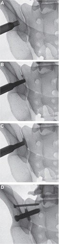
Under anteroposterior, outlet, and lateral fluoroscopic views, the skin is marked to identify the ideal access and trajectory. The lateral view should utilize a gantry which superimposes the sacral slopes on a single image. A 1.5–2 cm longitudinal incision is made, with the skin and subcutaneous tissue divided down to the gluteal fascia. Under lateral fluoroscopic guidance, a 6 mm dilator with internal obturator is advanced to the planned entry point on the outer ilium. The obturator is then exchanged for a guide pin, which is drilled just enough to maintain its position within the outer ilium cortex. After accurate pin trajectory is confirmed with inlet, inlet-oblique, and outlet-oblique fluoroscopic views, the guide pin is advanced into the SI joint, just touching the sacral cortex. The pin should have a trajectory as perpendicular to the SI joint as possible. The lateral ilium cortex is then sequentially dilated to 9 mm to allow advancement of a working cannula to approximately 1 cm beyond the lateral cortex. Proper intraosseous trajectory and depth are confirmed with pelvic inlet and outlet projections. Next, a 9 mm cannulated drill creates an osseous tunnel though the ilium over the guide pin, and care is taken with radiographic guidance to avoid drilling across the sacral cortex of the SI joint. Ilium drillings are collected and incorporated into bone graft material for later grafting, which is advantageous since it avoids direct iliac crest bone graft harvesting, which can cause complications such as iliac crest fracture, chronic pain, and nerve damage.Citation24
SI joint preparation begins by clearing cartilage to allow a decorticator to be fully seated on the sacral cortex of the SI joint. Joint surface preparation is accomplished by use of a unique flexible decorticator that extends out and follows the undulating surface of the joint while denuding cartilage and partially decorticating the joint. The joint is prepared well beyond the margins of the 12.5 mm threaded implant, with an affected region of approximately 5 cm2 of the ilium and 5 cm2 of the sacrum, exclusive of the implant. This represents approximately 50% of the articular joint area.Citation25 Irrigation and suction are used intermittently throughout the process to extract joint tissue. Approximately 5 cc bone graft, including autologous bone from the ilium drillings (typically 2–3 cc), is then packed into the denuded cavity to promote bony fusion. Dilators are reinserted to allow accurate advancement of the drill pin to a safe depth and trajectory perpendicular to the joint. A 9 mm pilot hole is drilled through the sacral cortex, over which the initial 12.5 mm cannulated implant is advanced over the pin until fully seated. Fixation is obtained with at least one large diameter 12.5 mm implant and one or more additional smaller 6.5 mm implants to ensure rotational rigidity.
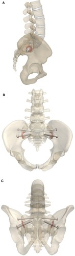
The 6.5 mm anti-rotation implant(s) are inserted just superior to the 12.5 mm implant or into S2 without the use of joint preparation and bone graft, in an otherwise similar fashion. Upon completion of placement of the implants, final lateral, inlet, inlet-oblique, outlet, and outlet-oblique images are obtained (). The deep tissues and skin incision may be infiltrated with bupivacaine and epinephrine for postoperative pain control.
Figure 2 Schematic representation of the implanted SImmetry SI Joint Fusion System in (A) lateral, (B) inlet, and (C) outlet views.
Several patient-related factors may complicate imaging interpretation and/or implant placement, including obesity, low bone density, sacral dysplasia, or small frame. The importance of preoperative planning cannot be overstated in such patients. In cases where dysmorphic patient anatomy will not allow for safe traverse medial to the neural foramen of the S1 or S2 bony corridors, the implants should be inserted only within the ala. A minimum of 1.5 cm implant purchase into the sacrum is recommended for adequate implant stability.
Postoperative recovery
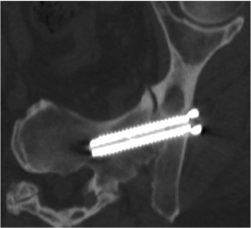
Patients may be discharged on the same day, although a 1–2 day hospitalization may be required for pain control; pain medications are prescribed as needed. Patients may progress to full weight bearing as tolerated, typically prior to hospital discharge. Fusion status is assessed with computed tomography 6–12 months after the procedure ().
Figure 3 Six-month postoperative orthogonal view demonstrating bridging bone with no evidence of lucency.
Biomechanical testing
The SImmetry® SI Joint Fusion System implants were developed specifically to limit SI joint motion by achieving immediate fixation with threaded titanium implants and promoting long-term fusion by arthrodesis. The use of a two-implant construct has traditionally been utilized for fixation of stable and unstable pelvic fractures,Citation26 with the second iliosacral screw providing significant fixation benefits.Citation27–Citation29 van Zwienen et alCitation28 evaluated iliosacral screw constructs in a cadaveric fracture model. These authors reported a 28% and 44% reduction in fracture site motion and a 170% and 300% increase in rotational stiffness with the addition of a second screw for S2 and S1 placements, respectively. Yinger et alCitation27 evaluated the effectiveness of multiple fixation constructs, including SI screws, sacral bars, and tension band plates in a hard plastic model with an Orthopaedic Trauma Association (OTA) 61-C1.2 fracture. The addition of a second iliosacral screw reduced fracture site translation by 50% and rotation by 40%. In fact, the two transverse screw construct provided rigidity that was only slightly exceeded by the strongest construct consisting of a screw and multiple anterior plates. This design is advantageous, since shear loads across the SI joint can exceed 600 N,Citation30 and multiple implants can more effectively share loads and resist rotation. Additionally, the implant procedure requires only a posterior approach.
An additional advantage of the 12.5 mm implant relates to implant strength and durability when compared to a 6.5 mm trauma screw. Using the American Society for Testing and Materials (ASTM) standard tests, this implant had 2% greater pullout force, 22% greater failure torque, and 17% greater bending yield. Notably, this 12.5 mm implant underwent 5 million cycles in bending fatigue at 7.2 Nm with no failure, whereas the trauma screw failed at 30,000 cycles. The 12.5 mm diameter implant is six-fold stronger under shear and bending forces with double the frontal area when compared to a 6.5 mm implant. It can therefore be assumed that the use of both implants likely provides more than adequate fixation and strength to tolerate typical long-term physiological loads borne by the SI joint.
Discussion
Chronic LBP originating from the SI joint represents a diagnostic and therapeutic challenge due to complexities in differential diagnosis and the suboptimal therapeutic options available. Failure to recognize and optimally treat SI joint pathology results in undertreatment of a significant portion of chronic LBP sufferers. With the recent development of highly sensitive and specific diagnostic algorithms, awareness of the role of the SI joint in chronic LBP is increasing.
Despite recent efforts to characterize the role of the SI joint in chronic LBP, no definitive conservative, interventional, or surgical management options exist for managing chronic SI joint pain.Citation11 Instrumented SI joint arthrodesis via large anterior or posterior surgical approaches is limited by significant risk of iatrogenic neurovascular and muscular injury, requirement for iliac crest bone harvesting, and unsatisfactory long-term patient outcomes.Citation13 Consequently, minimally invasive techniques for fixation of the SI joint have been introduced over the last few years with encouraging early results. Several case series have reported promising mid-term outcomes with SI joint fixation using titanium plasma spray-coated implants.Citation31–Citation35 However, the long-term outcomes of this method are unknown, and since implants are placed without use of supplemental bone graft, the risk of late implant loosening remains a concern.Citation36
True minimally invasive SI joint arthrodesis, accomplished with small incisions, joint decortications, and instrumented fixation supplemented with bone graft for long-term stability, is a relatively novel concept. Mason et alCitation37 treated 73 patients with intractable SI joint pain resistant to conservative care using a percutaneous hollow modular anchorage screw supplemented with demineralized bone matrix. Throughout a mean of 3 years follow-up, back pain severity decreased by 44% on average, although fusion status was not reported. Although these results are promising, additional human studies are needed to accurately assess the therapeutic potential for this technique.
In summary, a true minimally invasive SI joint arthrodesis technique is possible in well-selected patients, combining the advantages of a minimally invasive surgical approach, proper joint preparation with decortication and bone grafting, and optimized titanium implants designed to provide a stable long-term construct.
Disclosure
LEM and JEB received financial support from Zyga Technology, Inc. (Minnetonka, MN, USA). The authors declare no other conflicts of interest in this work.
References
- HoyDBainCWilliamsGA systematic review of the global prevalence of low back painArthritis Rheum20126462028203722231424
- van TulderMWAssendelftWJKoesBWBouterLMSpinal radiographic findings and nonspecific low back pain. A systematic review of observational studiesSpine (Phila Pa 1976)19972244274349055372
- CherkinDCDeyoRAWheelerKCiolMAPhysician variation in diagnostic testing for low back pain. Who you see is what you getArthritis Rheum199437115228129759
- CherkinDCDeyoRALoeserJDBushTWaddellGAn international comparison of back surgery ratesSpine (Phila Pa 1976)19941911120112068073310
- VolinnEMayerJDiehrPVan KoeveringDConnellFALoeserJDSmall area analysis of surgery for low-back painSpine (Phila Pa 1976)19921755755811535726
- BernardTNJrKirkaldy-WillisWHRecognizing specific characteristics of nonspecific low back painClin Orthop Relat Res19872172662802951048
- SchwarzerACAprillCNBogdukNThe sacroiliac joint in chronic low back painSpine (Phila Pa 1976)199520131377709277
- MaigneJYAivaliklisAPfeferFResults of sacroiliac joint double block and value of sacroiliac pain provocation tests in 54 patients with low back painSpine (Phila Pa 1976)19962116188918928875721
- DePalmaMJKetchumJMSaulloTWhat is the source of chronic low back pain and does age play a role?Pain Med201112222423321266006
- KatzVSchoffermanJReynoldsJThe sacroiliac joint: a potential cause of pain after lumbar fusion to the sacrumJ Spinal Disord Tech2003161969912571491
- HansenHManchikantiLSimopoulosTTA systematic evaluation of the therapeutic effectiveness of sacroiliac joint interventionsPain Physician2012153E247E27822622913
- AshmanBNorvellDCHermsmeyerJTChronic sacroiliac joint pain: fusion versus denervation as treatment optionsEvid Based Spine Care J201013354422956926
- SchützUGrobDPoor outcome following bilateral sacroiliac joint fusion for degenerative sacroiliac joint syndromeActa Orthop Belg200672329630816889141
- BuchowskiJMKebaishKMSinkovVCohenDBSieberANKostuikJPFunctional and radiographic outcome of sacroiliac arthrodesis for the disorders of the sacroiliac jointSpine J200555520528 discussion 52916153580
- SimpsonLAWaddellJPLeightonRKKellamJFTileMAnterior approach and stabilization of the disrupted sacroiliac jointJ Trauma19872712133213393694724
- DabeziesEJMilletCWMurphyCPAckerJHRobicheauxRED’AmbrosiaRDStabilization of sacroiliac joint disruption with threaded compression rodsClin Orthop Relat Res19892461651712766606
- WaisbrodHKrainickJUGerbershagenHUSacroiliac joint arthrodesis for chronic lower back painArch Orthop Trauma Surg198710642382402956935
- SembranoJNPollyDWJrHow often is low back pain not coming from the back?Spine (Phila Pa 1976)2009341E27E3219127145
- SzadekKMvan der WurffPvan TulderMWZuurmondWWPerezRSDiagnostic validity of criteria for sacroiliac joint pain: a systematic reviewJ Pain200910435436819101212
- SimopoulosTTManchikantiLSinghVA systematic evaluation of prevalence and diagnostic accuracy of sacroiliac joint interventionsPain Physician2012153E305E34422622915
- WennerbergAAlbrektssonTEffects of titanium surface topography on bone integration: a systematic reviewClin Oral Implants Res200920Suppl 417218419663964
- SchwartzZRazPZhaoGEffect of micrometer-scale roughness of the surface of Ti6Al4V pedicle screws in vitro and in vivoJ Bone Joint Surg Am200890112485249818978418
- FranchiMOrsiniEMartiniDDestination of titanium particles detached from titanium plasma sprayed implantsMicron200738661862517084088
- ArringtonEDSmithWJChambersHGBucknellALDavinoNAComplications of iliac crest bone graft harvestingClin Orthop Relat Res19963293003098769465
- MahatoNKVariable positions of the sacral auricular surface: classification and importanceNeurosurg Focus2010283E1220192657
- TileMPelvic ring fractures: should they be fixed?J Bone Joint Surg Br19887011123276697
- YingerKScaliseJOlsonSABayBKFinkemeierCGBiomechanical comparison of posterior pelvic ring fixationJ Orthop Trauma200317748148712902785
- van ZwienenCMvan den BoschEWSnijdersCJKleinrensinkGJvan VugtABBiomechanical comparison of sacroiliac screw techniques for unstable pelvic ring fracturesJ Orthop Trauma200418958959515448446
- KorovessisPGMagnissalisEADeligianniDBiomechanical evaluation of conventional internal contemporary spinal fixation techniques used for stabilization of complete sacroiliac joint separation: a 3-dimensional unilaterally isolated experimental stiffness studySpine (Phila Pa 1976)20063125E941E95117139210
- PelJJSpoorCWPool-GoudzwaardALHoek van DijkeGASnijdersCJBiomechanical analysis of reducing sacroiliac joint shear load by optimization of pelvic muscle and ligament forcesAnn Biomed Eng200836341542418204902
- CummingsJJrCapobiancoRAMinimally invasive sacroiliac joint fusion: one-year outcomes in 18 patientsAnn Surg Innov Res2013711224040944
- RudolfLMIS fusion of the SI joint: does prior lumbar spinal fusion affect patient outcomes?Open Orthop J2013716316823730380
- RudolfLSacroiliac joint arthrodesis-MIS technique with titanium implants: report of the first 50 patients and outcomesOpen Orthop J2012649550223284593
- SachsDCapobiancoROne year successful outcomes for novel sacroiliac joint arthrodesis systemAnn Surg Innov Res2012611323270468
- SachsDCapobiancoRMinimally invasive sacroiliac joint fusion: one-year outcomes in 40 patientsAdv Orthop2013201353612823997957
- ShaffreyCISmithJSStabilization of the sacroiliac jointNeurosurg Focus201335Suppl 2 Editorial
- MasonLWChopraIMohantyKThe percutaneous stabilisation of the sacroiliac joint with hollow modular anchorage screws: a prospective outcome studyEur Spine J201322102325233123686478