
Abstract
Objective
To investigate the underlying regional homogeneity (ReHo) of brain-activity abnormalities in patients with comitant strabismus (CS) and their relationship with behavioral performance.
Methods
Twenty patients with CS (ten men and ten women) and 20 (ten men and ten women) age-, sex-, and education-matched healthy controls (HCs) underwent resting-state functional magnetic resonance imaging scans. The ReHo method was used to assess local features of spontaneous brain activities. Patients with CS were distinguished from HCs by receiver operating characteristic curve. Correlation analysis was performed to explore the relationship between the observed mean ReHo values of the different brain areas and behavioral performance.
Results
Compared to HCs, the patients with CS showed significantly increased ReHo values in the right inferior temporal cortex/fusiform gyrus/cerebellum anterior lobe, right lingual gyrus, and bilateral cingulate gyrus. We did not find any relationship between the observed mean ReHo values of the different brain areas and behavioral performance.
Conclusion
CS causes dysfunction in many brain regions, which may explain the fusion compensation in CS.
Introduction
Strabismus is an ocular alignment disorder characterized by a horizontal, vertical, and/or torsional deviation of one eye relative to the other, which may result in compromised binocular vision and amblyopia.Citation1,Citation2 Strabismus is classified roughly into comitant and incomitant types. According to a study conducted in Central China, strabismus was prevalent in 108 (5.0%) out of the 2,363 eligible students.Citation3 Clinically, strabismus also exhibits stereopsis impairment. At present, surgery is the major method of strabismus correction.Citation4
Optimal eye movements and alignment require a functional oculomotor system. Extraocular muscle (EOM) dysfunction is an important factor in strabismus. Initially, it was reported that EOM dystrophy can lead to squint,Citation5 and subsequently, Duane’s retraction syndrome was found to be closely related to muscular malnutrition.Citation6 Moreover, abnormal positions of the EOMs can also lead to strabismus.Citation7 Additionally, dysfunction of the EOM pulleys had been shown to be involved in incomitant strabismus.Citation8,Citation9 Strabismus can affect the binocular visual function, resulting in impaired visual fusion in strabismus patients.Citation10
Functional magnetic resonance imaging (fMRI) has been widely used to study strabismus. A previous report described reduced gray matter volume in the occipital eye field (OEF) and parietal eye field (PEF) and an increase in the gray matter volume in the frontal eye field (FEF) of adults with strabismus.Citation11 The FEF is an area of the frontal cortex that can trigger eye movements.Citation12 Another group demonstrated reduced functional connections between the V1 and V2 areas in macaque monkeys with strabismic amblyopia.Citation13 It has been shown that the mean diffusivity of the occipital tracts was increased in patients with strabismic amblyopia.Citation14
The regional homogeneity (ReHo) method, a resting-state fMRI (rs-fMRI) measurement method, is widely used to determine the local synchronization of spontaneous fMRI signals, and it provides important information on the activities of the brain.Citation15,Citation16 The ReHo method has been successfully deployed to investigate pathological mechanisms of neural diseases such as optic neuritis,Citation17 sleep disorders,Citation18 and Parkinson’s disease.Citation19 However, it has not yet been used to explore the pathophysiological changes in comitant strabismus (CS).
To our knowledge, our study is the first to evaluate regional spontaneous brain activity in subjects with CS and demonstrate its relationship with behavioral performances.
Subjects and methods
Subjects
Ten men and ten women with CS were recruited from the Ophthalmology Department of the First Affiliated Hospital of Nanchang University Hospital. The diagnostic criteria for congenital strabismus included the following: 1) strabismus from birth; 2) stereopsis defects (no visual fusion); 3) binocular uncorrected or corrected visual acuity (VA) being equal; and 4) with alternated cover. The angles of the strabismus group were equal, and the large squint angle range was 50–60 delta. Subjects were excluded if they had any one of the following conditions: 1) acquired strabismus, incomitant strabismus, concealed oblique; 2) conditions of eye diseases, trauma, or optic neuropathy (eg, infection, inflammation, ischemic disease, compression damage, eye surgery, intraocular placeholder lesions); 3) psychiatric disorders, diabetes, cardiovascular disease, cerebral infarction disease; and 4) drug or alcohol addiction.
Twenty healthy controls (HCs; ten men, ten women) with similar age range, sex ratio, and education status were also recruited for this study. All HCs met the following requirements: 1) normal brain parenchyma on cranial MRI; 2) no ocular disease with uncorrected or corrected VA >1.0; 3) no psychiatric disease; and 4) accessible to the MRI scanning (eg, no cardiac pacemaker or implanted metal devices). This study was authorized and approved by the First Affiliated Hospital of Nanchang ethics committee. All research methods followed the Declaration of Helsinki and conformed to the principles of medical ethics. All subjects participated voluntarily and were informed of the purposes, methods, and potential risks before signing an informed consent form.
MRI parameters
MRI scanning was performed with a 3-T MR scanner (Trio, Siemens, Germany). T1-weighted images were acquired with a three-dimensional spoiled gradient-recalled sequence in an axial orientation. We obtained 176 high-resolution images taking 5 minutes (repetition time =1,900 ms, echo time =2.26 ms, thickness =1.0 mm, gap =0.5 mm, acquisition matrix =256×256, field of view =250×250 mm, flip angle =9°) and 240 functional images covering the whole brain taking 10 minutes (repetition time =2,000 ms, echo time =30 ms, thickness =4.0 mm, gap =1.2 mm, acquisition matrix =64×64, flip angle =90°, field of view =220×220 mm, 30 axial slices with gradient-recalled echo-planar imaging pulse sequence).
fMRI data analysis
The 240 functional images were analyzed as described previously.Citation17 Briefly, the data were filtered by MRIcro (Nottingham University, Nottingham, UK) and preprocessed using SPM8 (The MathWorks, Inc., Natick, MA, US) and DPARSFA (Institute of Psychology, CAS., Beijing, People’s Republic of China) software. Then, the data were processed with slice timing, head motion correction, spatial normalization, and smoothening with a Gaussian kernel of 6×6×6 mm3 full-width at half-maximum (FWHM). After spatially normalized to the Montreal Neurological Institute (MNI) space using the standard echoplanar image template and resampled at a resolution of 3×3×3 mm3, the fMRI images were detrended and bandpass-filtered (0.01–0.08 Hz) to reduce the effects of low-frequency drift and physiological high-frequency respiratory and cardiac noise.
Based on Kendall’s coefficient of concordance (KCC), ReHo computation was performed with the REST software (Center for Cognition and Brain Disorders, HZNU., Hangzhou, Zhejiang, People’s Republic of China), as previously described.Citation17
Statistical analysis
To investigate the group differences in the ReHo values between patients with CS and HCs, fMRI data were fitted with a general linear model (GLM) with the SPM8 toolkit. We used the subject’s group membership as regressors. P<0.05 was considered statistically significant and corrected with random field (Gaussian random field) theory with minimum z >2.3. Two-sample Student’s t-test was used for continuous data for behavioral performances. All statistical analyses were done with the IBM SPSS Statistics 20.0 software (IBM Corporation, Armonk, NY, USA).
Brain–behavior correlation analysis
Brain regions with different between-group ReHo values were classified as regions of interest (ROIs) with the REST software. For each ROI, the mean ReHo value was calculated by averaging over all voxels. Finally, correlation analysis was performed to investigate the relationship between the mean ReHo value in each of those different areas and the related behavioral performances. P<0.05 was considered statistically significant.
Clinical data analysis
All the clinical data of the patients with CS were collected, including the course of the disease and the best-corrected VA.
Results
Demographics and visual measurements
As shown in , there were no obvious differences in weight (P=0.453), age (P=0.705), best-corrected VA–right (P=0.316), and best-corrected VA–left (P=0.705) between the patients with CS and the HCs.
Table 1 Demographic information and clinical measures for Stra and HCs
ReHo differences
Compared to HCs, patients with CS had significantly increased ReHo values in the right inferior temporal cortex (RITC)/right fusiform gyrus (RFG)/right cerebellum anterior lobe (RCAL), right lingual gyrus (RLG), and bilateral cingulate gyrus (BCG) ( [red areas] and ). Specifically, the mean ReHo values of altered ReHo regions between the CS group and HCs are shown in and .
Figure 1 Significant differences in ReHo values between the Stra group and HCs.
Abbreviations: BCG, bilateral cingulate gyrus; HCs, healthy controls; RCAL, right cerebellum anterior lobe; ReHo, regional homogeneity; RFG, right fusiform gyrus; RITC, right inferior temporal cortex; RLG, right lingual gyrus; Stra, strabismus.
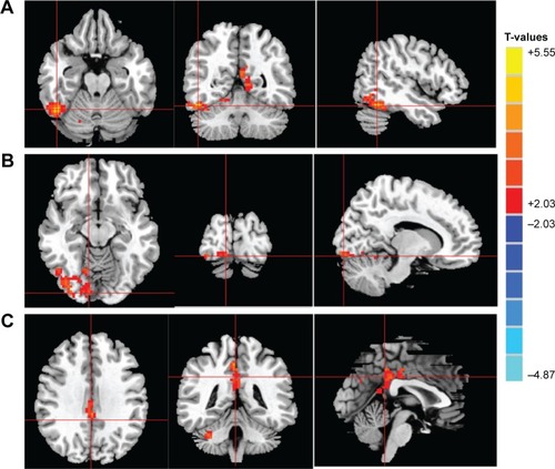
Figure 2 Mean of altered ReHo values between the Stra group and HCs.
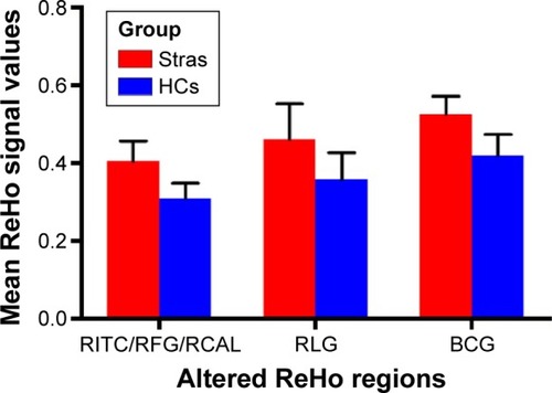
Table 2 Brain regions with significantly different ReHo values between the Stra and HCs
Receiver operating characteristic curve
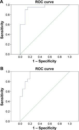
The differences of ReHo values in altered ReHo regions between the CS and HC groups may serve as diagnostic markers for strabismus. To test this possibility, we extracted the mean ReHo values of different brain regions and performed the receiver operating characteristic (ROC) curve analysis. In our study, the values of the area under the curve (AUC) for the RITC/RFG/RCAL () and the cluster of BCG () were 0.938 and 0.940, respectively.
Figure 3 ROC curve analysis of the mean ReHo signal values for altered brain regions.
Abbreviations: BCG, bilateral cingulate gyrus; CI, confidence interval; RCAL, right cerebellum anterior lobe; ReHo, regional homogeneity; RFG, right fusiform gyrus; RITC, right inferior temporal cortex; ROC, receiver operating characteristic.
Discussion
To our knowledge, this is the first study to evaluate the effect of CS on resting-state brain activity using the ReHo technique. Compared to HCs, patients with CS showed significantly increased ReHo values in the RITC/RFG/RCAL, RLG, and BCG.
The human inferior temporal cortex consists of the inferior temporal gyrus, the middle temporal gyrus, and the fusiform gyrus. The inferior temporal cortex is not only responsible for visual shape selectivity but also involved in the classification of visual information.Citation20,Citation21 Previous studies have demonstrated that the inferior temporal gyrus responds selectively to 3D structures defined by binocular disparity.Citation22,Citation23 Yan et al observed that patients with comitant extropia showed decreased white matter volumes in the right inferior temporal gyrus.Citation24 In agreement with this finding, we found significantly increased ReHo values in the RITC in patients with CS. Another complication often occurring in CS patients is the stereovision impairment.Citation25 The ReHo values in the RITC were higher in CS patients, which may explain the compensation of stereovision in these patients.
The cerebellum is involved in balance and motor control, as well as the execution of accurate eye movements.Citation26 Consistently, one group reported that the cerebellum is involved in the execution of eye and hand movements.Citation27 A previous study has shown that cerebellar vermis activation is related to visually guided saccades.Citation28 Joshi and DasCitation29 found that the posterior interposed nucleus (PIN) in the cerebellum plays an important role in conjugate eye movements in strabismic monkeys. In support of these findings, we also observed higher ReHo values in the cerebellum anterior lobe of patients with CS, indicating a stronger cerebellar activity than HCs. This suggests that functional reorganization may be occurring in the cerebellum anterior lobe to compensate for the conjugate eye movement abnormalities.
The lingual gyrus and fusiform gyrus are related to vision processing.Citation30 A previous study reported that the lingual gyrus was a stable brain region in the visual system network and that the area involved in color and visual motion (area V4) is located in the lingual and fusiform gyri of the prestriate cortex.Citation31,Citation32 “Visual snow” is also related to the dysfunction of lingual gyrus.Citation33 The fusiform gyrus, a fusiform face area (FFA), is a hub for face processing.Citation34 Yang et alCitation35 reported that the lingual visual cortex was highly activated in patients with infantile esotropia, suggesting a potential role in optical fusion. In our study, we also found that the ReHo values in the RLG and fusiform gyrus were significantly higher, which may be related to the compensation of stereovision in CS.
The cingulate gyrus is an integral part of the limbic system, which is involved with emotion formation, depression, and pain.Citation36–Citation38 The limbic system is closely related to memory and emotion.Citation39 In the present study, we also found that patients with CS had higher ReHo values in areas in the BCG, which may suggest that strabismus has a significant changes in the limbic system. Besides, the higher ReHo values in these areas may be related to compensation by the limbic system.
Conclusion
We found that patients with CS had abnormal spontaneous activity in specific regions of the brain, which may be related to the compensation of stereovision in CS. These findings provide important information for the understanding of neural changes in CS. However, there are some limitations to our study, such as the relatively small sample size. Moreover, for some subjects, the scan time was too long, so body movement could have affected the ReHo findings. Moreover, the clinical characteristics were not strict; we included both exo- and esotropic patients.
Future research should distinguish between different types of CS to more accurately assess brain function activity changes.
Acknowledgments
This study was supported by the National Natural Science Foundation of China (81160118 and 81400372); the Jiangxi Province Voyage Project (2014022); the Jiangxi Province Degree and Postgraduate Education Reform Project (2015); the Science and Technology Platform Construction Project of Jiangxi Province (2013116); the Youth Science Foundation of Jiangxi Province (20151BAB215016); the Technology and Science Foundation of Jiangxi Province (20151BBG70223); the Hunan Province Education Department Outstanding Youth Science Foundation (GJJ14170); the Health Development Planning Commission Science Foundation of Jiangxi Province (20155154); and the Scholar Project of Ganjiang River (2015).
Disclosure
The authors report no conflicts of interest in this work.
References
- KocFErtenYYurdakulNSDoes restoration of binocular vision make any difference in the quality of life in adult strabismusBr J Ophthalmol201397111425143024037610
- AdamsDLEconomidesJRHortonJCContrasting effects of strabismic amblyopia on metabolic activity in superficial and deep layers of striate cortexJ Neurophysiol201511393337334425810480
- FuJLiSMLiuLRPrevalence of amblyopia and strabismus in a population of 7th-grade junior high school students in Central China: the Anyang Childhood Eye Study (ACES)Ophthalmic Epidemiol201421319720324742059
- SimonszHJKollingGHUnnebrinkKFinal report of the early vs. late infantile strabismus surgery study (ELISSS), a controlled, prospective, multicenter studyStrabismus200513416919916361188
- LewandowskiKBStrabismus as a possible sign of subclinical muscular dystrophy predisposing to rhabdomyolysis and myoglobinuria: a study of an affected familyCan Anaesth Soc J19822943723767104807
- HondaYYoshiokaMDuane’s retraction syndrome associated with muscular dystrophyJ Pediatr Ophthalmol Strabismus1978153157159739346
- DickmannAPetroniSSalerniAEffect of vertical transposition of the medial rectus muscleon primary position alignment in infantile esotropia with A- or V-pattern strabismusJ AAPOS2011151141621397800
- ClarkRAThe role of extraocular muscle pulleys in incomitant non-paralytic strabismusMiddle East Afr J Ophthalmol201522327928526180464
- OhSYClarkRAVelezFRosenbaumALDemerJLIncomitant strabismus associated with instability of rectus pulleysInvest Ophthalmol Vis Sci20024372169217812091413
- ReadJCStereo vision and strabismusEye (Lond)201529221422425475234
- ChanSTTangKWLamKCChanLKMendolaJDKwongKKNeuroanatomy of adult strabismus: a voxel-based morphometric analysis of magnetic resonance structural scansNeuroimage200422298699415193630
- VernetMQuentinRChanesLMitsumasuAValero-CabréAA frontal eye field, where art thou? Anatomy, function, and non-invasive manipulation of frontal regions involved in eye movements and associated cognitive operationsFront Integr Neurosci201486625202241
- BiHZhangBTaoXHarwerthRSSmithEL3rdChinoYMNeuronal responses in visual area V2 (V2) of macaque monkeys with strabismic amblyopiaCereb Cortex20112192033204521263036
- DuanYNorciaAMYeatmanJDMezerAThe structural properties of major white matter tracts in strabismic amblyopiaInvest Ophthalmol Vis Sci20155695152516026241402
- ZangYJiangTLuYHeYTianLRegional homogeneity approach to fMRI data analysisNeuroimage200422139440015110032
- TononiGMcIntoshARRussellDPEdelmanGMFunctional clustering: identifying strongly interactive brain regions in neuroimaging dataNeuroimage1998721331499558645
- ShaoYCaiFQZhongYLAltered intrinsic regional spontaneous brain activity in patients with optic neuritis: a resting-state functional magnetic resonance imaging studyNeuropsychiatr Dis Treat2015113065307326715848
- DaiXJGongHHWangYXGender differences in brain regional homogeneity of healthy subjects after normal sleep and aftersleep deprivation: a resting-state fMRI studySleep Med201213672072722503940
- LiYLiangPJiaXLiKAbnormal regional homogeneity in Parkinson’s disease: a resting state fMRI studyClin Radiol2016711e28e3426628410
- FreedmanDJRiesenhuberMPoggioTMillerEKExperience-dependent sharpening of visual shape selectivity in inferior temporal cortexCereb Cortex200616111631164416400159
- SigalaNVisual categorization and the inferior temporal cortexBehav Brain Res200414911714739004
- VerhoefBEVogelsRJanssenPInferotemporal cortex subserves three-dimensional structure categorizationNeuron201273117118222243755
- JanssenPVogelsROrbanGAMacaque inferior temporal neurons are selective for disparity-defined three-dimensional shapesProc Natl Acad Sci U S A199996148217822210393975
- YanXLinXWangQDorsal visual pathway changes in patients with comitant extropiaPLoS One201056e1093120532166
- Serrano-PedrazaIManjunathVOsunkunleOVisual suppression in intermittent exotropia during binocular alignmentInvest Ophthalmol Vis Sci20115252352236421220559
- HerzfeldDJKojimaYSoetedjoRShadmehrREncoding of action by the Purkinje cells of the cerebellumNature2015526757343944226469054
- NitschkeMFArpTStavrouGErdmannCHeideWThe cerebellum in the cerebro-cerebellar network for the control of eye and hand movements – an fMRI studyProg Brain Res200514815116415661188
- HayakawaYNakajimaTTakagiMFukuharaNAbeHHuman cerebellar activation in relation to saccadic eye movements: a functional magnetic resonance imaging studyOphthalmologica2002216639940512566881
- JoshiACDasVEMuscimol inactivation of caudal fastigial nucleus and posterior interposed nucleus in monkeys with strabismusJ Neurophysiol201311081882189123883862
- BogousslavskyJMiklossyJDeruazJPAssalGRegliFLingual and fusiform gyri in visual processing: a clinico-pathologic study of superior altitudinal hemianopiaJ Neurol Neurosurg Psychiatry19875056076143585386
- YangYLDengHXXingGYXiaXLLiHFBrain functional network connectivity based on a visual task: visual information processing-related brain regions are significantly activated in the task stateNeural Regen Res201510229830725883631
- ZekiSWatsonJDLueckCJFristonKJKennardCFrackowiakRSA direct demonstration of functional specialization in human visual cortexJ Neurosci19911136416492002358
- SchankinCJManiyarFHSprengerTChouDEEllerMGoadsbyPJThe relation between migraine, typical migraine aura and “visual snow”Headache201454695796624816400
- GhumanASBrunetNMLiYDynamic encoding of face information in the human fusiform gyrusNat Commun20145567225482825
- YangXZhangJLangLGongQLiuLAssessment of cortical dysfunction in infantile esotropia using fMRIEur J Ophthalmol201424340941624170518
- HadlandKARushworthMFGaffanDPassinghamREThe effect of cingulate lesions on social behaviour and emotionNeuropsychologia200341891993112667528
- LozanoAMMaybergHSGiacobbePHamaniCCraddockRCKennedySHSubcallosal cingulate gyrus deep brain stimulation for treatment-resistant depressionBiol Psychiatry200864646146718639234
- RobinAMPabaneyAHMitsiasPDSchwalbJMFurther evidence for a pain pathway involving the cingulate gyrus: a case of chronic cluster headache cured by glioblastomaStereotact Funct Neurosurg201593319419825833317
- LövbladKOSchallerKVargasMIThe fornix and limbic systemSemin Ultrasound CT MR201435545947325217299