
Abstract
Cognitive dysfunction is prevalent in psychiatric disorders. Deficits are observed in multiple domains, including working memory, executive function, attention, and information processing. Disability caused by cognitive dysfunction is frequently as debilitating as the prominent emotional disturbances. Interactions between the hippocampus and the prefrontal cortex are increasingly appreciated as an important link between cognition and emotion. Recent developments in optogenetics, imaging, and connectomics can enable the investigation of this circuit in a manner that is relevant to disease pathophysiology. The goal of this review is to shed light on the contributions of this circuit to cognitive dysfunction in neuropsychiatric disorders, focusing on Alzheimer’s disease and depression.
Introduction
A major goal of preclinical investigations has been to parse the relative importance of specific brain regions and subregions in the context of neuropsychiatric disorders. While this approach is useful in understanding the contribution of specific cellular phenotypes, identifying key signal-transduction pathways and individual molecules, it does not integrate disease-induced pathophysiological changes that influence multiple brain regions and their functional interactions. The ability to define mental health diseases as circuit disorders and identify the circuits that are relevant to particular diseases can lead us to a perspective that is closer to the clinical disease state and enable us to consider interventions that can repair these circuits and restore the brain-activity network. A potential advantage with the circuit approach is that the same circuit is likely to be impaired across disorders when there is overlap in the expression of behavioral deficits.
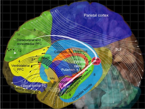
The hippocampal formation (HPF) and prefrontal cortex (PFC) interact in a bidirectional manner to regulate several cognitive functions and process emotional information. They are critically important structures in the brain’s memory system, facilitating fast encoding of new information, consolidation, retrieval, and organization of the memory network.Citation1 There is strong neural synchrony between the two regions during behavior, and their functional interaction is regulated by oscillations.Citation2,Citation3 The neural ensemble formed by the cells in the hippocampus and PFC mutually influences each other in a circuit-like fashion in modulating emotional and cognitive processes. Connections between the hippocampus and PFC have been demonstrated in rodents,Citation4–Citation7 primates,Citation8 and humans.Citation9 The rodent and human hippocampal prefrontal connections are illustrated schematically in and respectively. The HPF and PFC are connected by both monosynaptic and polysynaptic connections.Citation10 Retrograde-labeling studies revealed that projections from the medial PFC (mPFC) robustly label neurons in the ventral hippocampus and subiculum.Citation10,Citation11 Anterograde labeling revealed connections between the CA1 region of the hippocampus and the subiculum with the mPFC.Citation4 Indirect multisynaptic connections between the HPF to mPFC include projections through the nucleus accumbens, ventral tegmental area, amygdala, entorhinal cortex, and midline thalamus.Citation12–Citation14 These complex multisynaptic pathways are involved in higher cognitive functions and dysregulated in various neuropsychiatric disorders.Citation15 We review the influence of aberrations in the hippocampus–PFC circuit on cognitive dysfunction in depression and Alzheimer’s disease (AD).
Figure 1 The rodent hippocampal–prefrontal cortex (PFC) circuit.
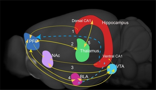
Figure 2 Human prefrontal hippocampal connections.
Hippocampal–PFC circuit in depression
Depression afflicts 298 million people globally.Citation16 Untreated depression is the leading cause of suicide in the 15- to 24-year age-group.Citation17 A better understanding of disease pathology and more efficacious treatments are needed, as currently available prescription antidepressants are effective in only about 60% of affected individuals.Citation18 Although chronic stress and trauma, particularly in early life, has been connected with vulnerability to depression,Citation19,Citation20 the precise neurobiological underpinnings are yet to be understood. A complex interaction between genetic and environmental factors is also involved in disease pathophysiology.
Multiple brain regions have been studied to understand the etiology of depression, including the hippocampus, amygdala, striatum, insula, medial thalamus, and several frontal cortex (FC) regions.Citation21 Significant attention has been focused on the hippocampus, due to its central role in the stress-induced dysregulation of the hypothalamic–pituitary–adrenal axis.Citation22 Additionally, imaging studies in patients with depression have shown a reduction in hippocampal volume.Citation22–Citation24 It is also the most frequently noted brain structure alteration in depression.Citation25 Smaller but consistent reductions have also been reported in the PFC.Citation26 Elevated corticosterone levels and dysregulation of the hypothalamic–pituitary–adrenal axis are ascribed as contributing factors in the hippocampal shrinkage that occurs in major patients with depressive disorder (MDD), but the precise molecular and cellular mechanisms involved are not known. Support for examining the hippocampus also comes from preclinical antidepressant studies demonstrating that hippocampal neurogenesis, which occurs in the subgranular zone of the dentate gyrus (DG), is important for the behavioral effects of antidepressants.Citation27,Citation28 Interestingly, the DG is also vulnerable to the effects of stress, evidenced by a decline in subgranular zone neurogenesis.Citation29
The fact that the hippocampus is also central in memory functions makes it a crucial brain region that mediates both dysregulated mood and cognitive dysfunction in depression. A rapidly accumulating body of evidence indicates that a segmentation of the hippocampus along its axis, dorsoventral in rodents and posterioanterior in primates, is responsible for its dual functionality in regulating both emotion and cognition, the dorsal hippocampus being involved in cognitive function and the ventral in mood and anxiety.Citation30
Hippocampal subfields in depression
There is significant interest in understanding the role of specific hippocampal subfields in depression, as these have the potential to inform the precision of brain stimulation-based therapeutic interventions,Citation31 and deciphering hippocampal neuroplasticity in disease states and response to antidepressant treatment.Citation32,Citation33 Immunohistochemical analysis of postmortem brains from depressed patients revealed lower granule-cell number and smaller volume of the granule-cell layer in the anterior DG of untreated patients.Citation32 No significant changes were noted in the posterior DG. Structural analysis reported the strongest volumetric decreases in the DG of unmedicated MDD patients, which were partially rescued by antidepressant treatment.Citation34 Imaging brain regions as a function of depressive episodes showed that illness progression led to reduction in DG volume and thinning of the left mPFC, with no effect on amygdala volume.Citation35 A consensus picture that emerges from clinical imaging studies is that the DG is the most vulnerable hippocampal subfield in depression. It is however not known whether this is purely disease-related or if there are also predisposing genetic factors that result in the DG being preferentially affected in depression. Animal-stress studies have demonstrated that the DG is particularly sensitive to elevations in glucocorticoid levels and reduced neurogenesis in the subgranular zone. Although a reduction in neurogenesis could contribute to a reduction in DG volume, it is unlikely to be the only factor.Citation36 The findings give rise to several questions that will require clinical and preclinical investigation. As antidepressants (primarily selective serotonin-reuptake inhibitors) are able partially to rescue volumetric deficits, it would be important to investigate the cellular and molecular mechanisms involved in volume recovery. The correlation between volume recovery and mood improvement also needs to be examined.
Cognitive deficits in depression
Cognitive dysfunction in depression has become a topic of recent interest as the field has increasingly recognized that these deficits impede functional recovery and are independent of emotional disturbances.Citation37,Citation38 Patients with MDD frequently exhibit difficulty with attention,Citation39 information processing,Citation40 working memory,Citation41 and executive function.Citation42 It should be stressed that these deficits are not uniformly present in all depressed patients, as some abnormalities have been reported in specific MDD subgroups. However, the persistence of these deficits despite improvement in moodCitation43,Citation44 calls for efforts to understand how cognitive dysfunction is intertwined with depression and to develop treatments that specifically target this aspect of the illness.
Rodent studies have provided support for the hypothesis that functional interactions between the hippocampus and PFC are reflected in synchronized neuronal activity.Citation3,Citation4,Citation45 Interestingly, there is an increase in θ-frequency synchronization between the ventral hippocampus and the mPFC in mice exposed to an anxiogenic environment, implicating this circuit in behavioral inhibition, which is characterized by reduced exploration during anxiety.Citation46 Impaired synaptic plasticity of this circuit was suggested in another neural oscillation study that employed the chronic unpredictable stress paradigm to generate a depression-like phenotype.Citation47 Hippocampus–PFC connectivity-dependent memory consolidation was impaired in depressed patients in a functional magnetic resonance imaging (fMRI) study that showed reduced hippocampal connectivity to the PFC.Citation48 Early-life stress exposure is a known risk factor for developing depression later in life.Citation19 The heightened vulnerability of the brain to emotional and psychological insults during childhood likely results in long-lasting changes in the brain that increase the risk of being afflicted by depression later in life. Understanding the link between childhood-stress exposure and brain function can thus provide critical insight into disease etiology. A significant interaction was found between childhood emotional neglect and reductions in hippocampal and PFC volumes in MDD patients,Citation49 which could signify an association between circuit and structural abnormalities. Can a faulty circuit lead to structural abnormalities? Perhaps they are independent defects that coexist under certain conditions, resulting in higher levels of functional impairment.
Autobiographical memory in depression
A bias toward recall of negative events is frequently seen in depressed patients. In particular, autobiographical memory (AM), the recollection of personal experiences, is impaired in depression. AM is important for effective interaction with the world, as it involves an individual’s perception of self and the ability to solve problems using information from past personal events.Citation50,Citation51 Suicidal patients have been noted to generalize their responses when queried about past events, and unable to recall event specifics.Citation52 This characteristic memory, termed “overgeneral” AM, is a persistent feature of major depression.Citation50 Due to the complex nature of AM, involving episodic memory, self-reflection, emotional valence, and visual recall, the activation of multiple brain regions would be expected.Citation53 High-resolution fMRI using multivoxel pattern analysis firmly implicated the ventromedial PFC and the hippocampus in AM representation.Citation54 A meta-analysis of AM functional neuroimaging studies noted that PFC activation was consistently reported, and over half the studies showed hippocampal activation.Citation53 Hippocampal activation frequently occurred in a core AM network that includes the parahippocampus, perirhinal, and entorhinal cortices. Event-related fMRI shed additional light on the various parameters of AM that influence hippocampal activation, and indicated that recollective qualities were an important component.Citation55 Neuroimaging fMRI analysis of AM retrieval in epilepsy patients with left hippocampal atrophy revealed significant deficits in task-related AM-network activity and strength of connections, suggesting that the hippocampus is a critical node in the AM network.Citation56
Hippocampal–PFC circuit in Alzheimer’s disease
The HPF is strongly implicated in AD pathology, with evidence from imaging, psychological testing, and postmortem analysis. The emergence of initial symptoms in AD corresponds to pathological changes in the hippocampus. A meta-analysis of structural imaging studies utilizing data from 700 patients concluded that at the time of AD diagnosis, hippocampal volume loss was at 23% in comparison to similar-age controls.Citation57 Longitudinal studies have shown that a specific reduction in the volume of the left amygdala–hippocampal complex is evident 5 years before AD diagnosis.Citation58 The AD Neuroimaging Initiative (ADNI), a major multicenter neuroimaging study, reported a reduction in hippocampal volume in patients with AD and mild cognitive impairment (MCI), a precursor to AD development.Citation59 It also documented substantial reduction in volume within a span of 6 months, and more accelerated volume loss in AD carriers of the ApoEε4 allele.Citation59 Furthermore, the rate of volume loss correlated with cognitive decline.Citation25,Citation29 The presence of the ApoEε4 allele is the strongest genetic risk factor for AD, producing higher AD pathology and structural and functional alterations in the brain.Citation60 Structural imaging revealed a specific reduction in left hippocampal volume in ApoEε4 carriers at early-stage AD.Citation61 An MRI marker capable of determining hippocampal texture was developed using scans from the large ADNI database and tested in three separate cohorts.Citation62 From the available prognostic results, it appears that hippocampal texture might be more sensitive than volume reduction in predicting CI and progression from MCI to AD.Citation62
Hippocampal subfields in AD
A recent approach to improve diagnostic predictive power is the imaging of hippocampal subfields. Extensive volumetric and shape analyses employing a variety of methods, such as manual segmentation of the entire hippocampus, voxel-based morphometry, and surface reconstruction, have yielded a consensus that subfield atrophy in AD is primarily in CA1.Citation63 This finding from live imaging investigations correlates well with previous histological examination of postmortem AD brain tissue showing highest neuronal loss in CA1 (68%), followed by the subiculum (47%) and hilus (25%) compared to the age-matched control group.Citation64 CA1 and subiculum atrophy were detectable in cognitively normal individuals 6 years prior to AD diagnosis,Citation65 indicating involvement of specific hippocampal subfields early in the disease process.
There are broad similarities in hippocampal volume loss in AD and depression, which could underlie the overlap in affective and cognitive deficits. Structural imaging of hippocampal subfields and immunohistochemical and histological analyses have shown that there is divergence in the specific subfields and cell types that are affected. Volumetric reduction in the DG is consistently reported in depression, although the precise mechanisms remain to be elucidated. In contrast, hippocampal atrophy in AD is caused by neuronal cell loss in the CA1 region and the entorhinal cortex, while the DG is mostly well preserved.Citation66 Whether these differences also influence how information flows through the hippocampal circuit in these diseases would be worthy of investigation.
Amyloid and neurofibrillary tangles, abnormally folded protein structures in the AD brain, initially accumulate in the entorhinal cortex and then spread to the hippocampus.Citation67 This phenomenon has the potential to disrupt local and global hippocampal-dependent connectivity.Citation68 Connectivity can also be impaired before the emergence of landmark pathology, such as in MCI patients who are at risk for AD, exhibiting significantly reduced connectivity between hippocampi.Citation69 The mPFC and lateral PFC have emerged as an AD-hub region responsible for linking functionally specialized regions.Citation70 As a site of high amyloid-β accumulation, its ability to process information would be increasingly affected with disease progression. Positron-emission tomography imaging of blood flow during a face-memory task indicated that memory deficits early in AD were due to a reduction in integrated network activity, with the PFC and hippocampus as principal components.Citation71 Functional connectivity of the hippocampus with several cortical regions was shown to be impaired in ApoEε4 carriers by a combination of neuropsychological testing and imaging analyses designed to decipher context-dependent alterations in hippocampal connectivity.Citation72 Early-stage AD ApoEε4 carriers also exhibited decreased functional connectivity between the hippocampus and the medial FC and parietal cortex.Citation61
Resting-state fMRI analysis in AD patients found that right hippocampal connectivity with the mPFC and the ventral anterior cingulate cortex was reduced, but connectivity of the left hippocampus with the dorsolateral PFC was enhanced.Citation73 The increased connectivity is likely to be a compensatory adjustment for recruiting additional resources to balance the decline in cognitive function in AD. A functional relationship between the posterior cingulate cortex and the hippocampus was seen in episodic memory fMRI in MCI patients. Interestingly, posterior cingulate cortex activation during episodic memory encoding was connected with right hippocampal activation, while episodic memory recognition was associated with left hippocampal activation, indicating the presence of hemisphere specialization.Citation74 In an AD study that found a strong correlation between hippocampal volume and memory impairment, left hippocampal volume predicted verbal recall while right hippocampal volume predicted spatial recall, providing additional support for hemispheric specialization of memory in AD.Citation75
Neuroimaging studies in AD and depression have progressed from focused analysis of specific anatomical regions involved in disease pathology to incorporating a network approach that can yield insight into global relationships between spatially distinct brain structures, spawning the field of macrolevel circuit analysis – connectomics.Citation76,Citation77 The understanding of brain wiring gained by clinical connectomic investigations can be complemented with preclinical optogenetic technology to examine specific circuits and cell types with high spatial and temporal resolution.Citation78 We expand on the applications of connectomic and optogenetic approaches to interrogate the hippocampal–PFC circuit in cognitive function.
Optogenetics sheds light on the hippocampal–PFC circuit
Optogenetics is rapidly becoming the method of choice to regulate neuronal circuits precisely in preclinical experiments. The method involves the use of light and light-sensitive opsin proteins to exert fine control over in vivo neuronal activity.Citation79,Citation80 Based on the goals of the investigation, specific opsin proteins can be chosen, either to depolarize or excite neurons (channelrhodopsin)Citation81 or hyperpolarize and inhibit (halorhodopsin and archaerhodopsin).Citation82 By utilizing animal models of neuropsychiatric disorders, optogenetics can enable the identification of disrupted brain circuits and complement conventional electrophysiological analysis. An elegant recent study employed optogenetic technology to facilitate the recall of old memories in a mouse model of AD.Citation83 It successfully achieved this by activating the cells involved in the formation of memory engrams, in the hippocampal circuit.Citation83 A similar approach was used by the same group to recall previous positive memories in a mouse model of depression. Optogenetic activation of the positive memory- engram cells in the hippocampal circuit was able to acutely reverse the depressed phenotype.Citation84 This experiment draws attention to the therapeutic potential of positive memory recall in depression, as activating memory of the positive experience was more effective in overcoming the adverse behavioral consequences of stress exposure than the experience itself.Citation85
The existence of an mPFC–thalamic nucleus reuniens–hippocampal CA1 loop for goal-directed spatial navigation was determined by optogenetically manipulating this circuit at multiple nodes to demonstrate the relative importance of sub-regions within the hippocampus.Citation86 The important role played by direct ventral hippocampus–mPFC afferents in encoding spatial cues during the performance of spatial working memory tasks was shown by employing a projection-specific optogenetic approach.Citation87 The ventral hippocampus–mPFC input was required only for encoding cues, not for retrieval or maintenance. γ-Frequency but not θ-frequency synchrony was essential in this circuit for successful encoding of cues, reinforcing previous work indicating that γ-synchrony could be essential for proper long-range connectivity.Citation88
Optogenetic stimulation of somatostatin and parvalbumin containing interneurons in the PFC helped clarify the distinct contribution of these interneuron subtypes in working memory tasks and reward processing. This provided new insight of prefrontal circuitry in cognitive function.Citation89 The combination of optogenetics and electrophysiology sheds light on the mechanism whereby the PFC regulates the processing of information for attention. This study strongly implicates fast-spiking mPFC parvalbumin neurons in guiding successful attention behavior.Citation90 The therapeutic potential of optogenetics for cognitive deficits was demonstrated by γ-frequency stimulation of PFC interneurons specifically at 40 or 60 Hz.Citation91 Interestingly, a single intervention was sufficient to produce cognitive enhancement that lasted over a week. Precise and selective activation of glutamatergic neurons in the mPFC improved associative recognition memory, an important aspect of cognitive function.Citation92 However, it is important to note that stimulation was effective only if it occurred during the delay phase, and simply elevating glutamate release was ineffective. This observation illustrates how the precision of optogenetic stimulation can inform drug development by providing key details for consideration. A major application of optogenetic research in neuropsychiatry will be to translate the important preclinical findings into useful noninvasive therapies in the clinic. When examining the success of circuit-level optogenetic analyses and behavioral function, it is quite clear that a high degree of spatial and temporal resolution is involved. Currently, therapeutic interventions with comparable precision are not available for clinical use. Several novel approaches have recently been proposed for potential clinical translation.Citation93 These include viral transduction, where virus particles are used to over-express proteins of interest in a spatially restricted manner. Paramagnetic proteins, primarily ferritin, fused to a channel receptor function as an endogenous iron nanoparticle and enable the use of a magnetic field to modulate channel activity temporally.Citation94,Citation95 Low-intensity focused ultrasound has been recently used to non-invasively alter neuronal activity with high spatial resolution.Citation96 Dysregulation of specific molecular targets has been identified in the entorhinal–hippocampal circuit.Citation93,Citation97,Citation98 It is however likely that the most promising therapies would require some testing in primates before commencing human clinical trials.
Connectomics
In contrast to the highly precise, neuronal subtype-level resolution of optogenetics, connectomics focuses on the major neural highways in the brain. The Human Connectome Project aims to map neural connections in the brain at a scale that has not been previously attempted. The goal is to obtain insight into the anatomical and functional complexity in the human brain at the level of long-range connections. A combination of diffusion MRI to trace white-matter tracts for structural connectivity and resting-state fMRI for functional activity is being employed.Citation77 The hope is that conducting these analyses in over a thousand individuals will provide accurate baselines and enable identification of disease-induced alterations in brain-network connectivity with high confidence.Citation77 Connectomic analyses have begun to provide new insight into the pathophysiology of neuropsychiatric disorders, and it is now possible to define certain illnesses as connectivity disorders. Reduced functional connectivity, determined by functional connectivity MRI, has been reported in advanced AD, with patients demonstrating a striking decrease in connectivity between the hippocampus and FC.Citation99 Altered functional connectivity is also evident early in AD,Citation73 when plaque accumulation would be minimal. Long-distance connectivity was shown to be selectively vulnerable in AD, and deterioration correlated with cognitive decline. Graph theory-based topological analysis revealed that attenuation in long-distance connectivity also reduced the efficiency of the global brain network, leading to more widespread clinical cognitive deficits.Citation100
In patients afflicted with CI and depression, there is substantial overlap of connectivity abnormalities. Network-topology analyses reveal a reduction in network strength, efficiency, and regional connectivity in FC structures, collectively indicating disruption of white-matter integrity.Citation54 In patients with a first episode of untreated depression, white-matter abnormalities can be a potential biomarker of pathophysiology. If compromised white-matter integrity and function are indeed precipitating factors in depression, it would be important to examine the role of antidepressants in rescuing or reversing these deficits. Ketamine has attracted much attention in recent years, due to its rapid-acting antidepressant properties.Citation101 Its psychomimetic effects and abuse potential can however interfere with its use as a mainstream antidepressant in the clinic. Understanding its precise antidepressant mechanism is thus a high priority to develop next-generation compounds that do not carry undesirable side effects. A recent study on the role of ketamine in MDD patients demonstrated its ability to normalize disconnectivity between the PFC and the rest of the brain, suggesting that actions on white matter could be involved in its antidepressant mechanism.Citation102 Although this suggests that impaired connectivity can be rescued, it is critical to understand ketamine’s impact on white matter. Efforts should be aimed at distinguishing between acute and chronic effects, as chronic ketamine usage has been shown to disrupt connectivity between the caudate and PFC.Citation103 Focusing attention on understanding the molecular substrates that influence white-matter integrity would be an important research avenue. It can yield useful information regarding circuit neurobiology, and also potentially identify molecules that can restore connectivity deficits in multiple psychiatric and neurodegenerative disorders.
Studying brain-network connectivity is likely to be challenging when developmental changes are ongoing, as the complex and dynamic maturation windows can influence the consistency of neuroimaging results. However, it is important to obtain structural and functional connectivity information of the brain during its most vulnerable state.Citation104 Depression frequently surfaces during adolescence,Citation105 a critical developmental period where an increase in white matter and reduction in cortical gray matter occurs.Citation106 In unmedicated, first-episode adolescent patients with depression, both structural and functional connectivity in the PFC–hippocampal circuit was abnormal, with decreased functional connectivity in multiple PFC regions.Citation107 Adolescent depression studies have also reported hyperconnectivity of the default network and better functional connectivity than healthy controls between the mPFC and posterior cingulate cortex during the processing of cognitive information.Citation108 Hyperconnectivity was also noted during goal-directed emotional processing.Citation108 The adolescent brain can be influenced by hormones, reward valence, and social interactions, which are also likely to impact network connectivity.Citation104 It will thus be interesting to see the results of follow-up studies designed to examine treatment effects on connectivity.
A common molecular target in AD and depression
Dysregulation of the hippocampus–PFC circuit and similarities in hippocampal volume reduction in AD and depression could point to common deficits in cellular signaling. Identifying disease-related molecules that regulate key nodal points in the intracellular signaling network can yield deep insight into disease pathophysiology and provide opportunities to test the link between molecular dysfunction and behavioral deficits. Transcription factors that function as crucial molecules downstream of cell signaling have an important role in the investigation of molecular mechanisms of neuropsychiatric disorders. They are functionally positioned at critical nodal points where disease and drug-induced signal transduction converge. They are also capable of regulating an entire program of gene expression when activated, and thereby influence several distinct cellular processes and mechanisms. The cAMP response element binding (CREB) protein transcription factor has been actively investigated in memory,Citation109,Citation110 depression,Citation111,Citation112 and antidepressant activity,Citation113,Citation114 providing important understanding into the molecular basis of disease-relevant behavioral responses. CREB is activated by phosphorylation and drives the expression of different target genes based on the brain region where it is activated.Citation115 It is interesting to note that CREB is also emerging as an important molecule in AD research.Citation116,Citation117 Postmortem studies on AD brain tissue have reported down-regulation of CREB in the hippocampus.Citation118 Recent postmortem analysis found levels of CREB and activated phospho-CREB to be reduced in the PFC, and rather intriguingly discovered that CREB/phospho-CREB levels were also similarly reduced in peripheral blood mononuclear cells.Citation119 Since reduction in CREB occurred prior to amyloid deposition and can also be detected in blood, it would be worthwhile to investigate CREB levels in clinical populations. CREB could emerge as an important molecular regulator of cognition in both depression and AD, and also serve as a useful biomarker.
Conclusion
The recent shift in focus from imaging gray-matter volumes to integrating brain-network data and analysis has created considerable excitement, and promises to reveal new insight into brain development, neuropsychiatric disorders, and brain function. The macrolevel understanding gained by connectomic studies can be investigated at the level of individual neurons and neural ensembles via optogenetic methodologies. These conceptual and technological advances have enabled neuroscientists to consider psychiatric disorders as circuit malfunctions. The value of investigating a particular circuit such as the hippocampus–PFC is enhanced by the fact that it involves regions that are strongly implicated in depression and AD and also share overlap in cognitive deficits. Determining precise molecular and genetic mechanisms will provide additional resolution and strengthen the rationale for utilizing circuit and connectivity information in disease diagnosis and treatment. Major dividends can ensue in terms of treatments that effectively target primary circuit deficits involved in multiple neuropsychiatric disorders.
Acknowledgments
This work was supported by US Public Health Service grant MH106640 and the use of facilities at the Sioux Falls VA Healthcare system.
Disclosure
The authors report no conflicts of interest in this work.
References
- PrestonAREichenbaumHInterplay of hippocampus and prefrontal cortex in memoryCurr Biol20132317R764R77324028960
- ColginLLOscillations and hippocampal-prefrontal synchronyCurr Opin Neurobiol201121346747421571522
- GordonJAOscillations and hippocampal-prefrontal synchronyCurr Opin Neurobiol201121348649121470846
- JayTMWitterMPDistribution of hippocampal CA1 and subicular efferents in the prefrontal cortex of the rat studied by means of antero-grade transport of Phaseolus vulgaris-leucoagglutininJ Comp Neurol199131345745861783682
- SwansonLWKöhlerCAnatomical evidence for direct projections from the entorhinal area to the entire cortical mantle in the ratJ Neurosci1986610301030233020190
- ThierryAMGioanniYDégénétaisEGlowinskiJHippocampo-prefrontal cortex pathway: anatomical and electrophysiological characteristicsHippocampus200010441141910985280
- VarelaCKumarSYangJYWilsonMAAnatomical substrates for direct interactions between hippocampus, medial prefrontal cortex, and the thalamic nucleus reuniensBrain Struct Funct2014219391192923571778
- ZhongYMYukieMRocklandKSDistinctive morphology of hippocampal CA1 terminations in orbital and medial frontal cortex in macaque monkeysExp Brain Res2006169454955316328292
- CroxsonPLJohansen-BergHBehrensTEQuantitative investigation of connections of the prefrontal cortex in the human and macaque using probabilistic diffusion tractographyJ Neurosci200525398854886616192375
- HooverWBVertesRPAnatomical analysis of afferent projections to the medial prefrontal cortex in the ratBrain Struct Funct2007212214917917717690
- JayTMGlowinskiJThierryAMSelectivity of the hippocampal projection to the prelimbic area of the prefrontal cortex in the ratBrain Res198950523373402598054
- RussoSJNestlerEJThe brain reward circuitry in mood disordersNat Rev Neurosci201314960962523942470
- MarenSSeeking a spotless mind: extinction, deconsolidation, and erasure of fear memoryNeuron201170583084521658578
- WolffMAlcarazFMarchandARCoutureauEFunctional heterogeneity of the limbic thalamus: from hippocampal to cortical functionsNeurosci Biobehav Rev20155412013025446945
- GodsilBPKissJPSpeddingMJayTMThe hippocampal-prefrontal pathway: the weak link in psychiatric disorders?Eur Neuropsychopharmacol201323101165118123332457
- FerrariAJCharlsonFJNormanREBurden of depressive disorders by country, sex, age, and year: findings from the global burden of disease study 2010PLoS Med20131011e100154724223526
- GalaifERSussmanSNewcombMDLockeTFSuicidality, depression, and alcohol use among adolescents: a review of empirical findingsInt J Adolesc Med Health2007191273517458321
- RushAJTrivediMHWisniewskiSRAcute and longer-term outcomes in depressed outpatients requiring one or several treatment steps: a STAR*D reportAm J Psychiatry2006163111905191717074942
- HeimCNewportDJMletzkoTMillerAHNemeroffCBThe link between childhood trauma and depression: insights from HPA axis studies in humansPsychoneuroendocrinology200833669371018602762
- HammenCStress and depressionAnnu Rev Clin Psychol2005129331917716090
- PandyaMAltinayMMaloneDAJrAnandAWhere in the brain is depression?Curr Psychiatry Rep201214663464223055003
- CampbellSMacqueenGThe role of the hippocampus in the pathophysiology of major depressionJ Psychiatry Neurosci200429641742615644983
- ShelineYISanghaviMMintunMAGadoMHDepression duration but not age predicts hippocampal volume loss in medically healthy women with recurrent major depressionJ Neurosci199919125034504310366636
- OpelNRedlichRZwanzgerPHippocampal atrophy in major depression: a function of childhood maltreatment rather than diagnosis?Neuropsychopharmacology201439122723273124924799
- VidebechPRavnkildeBHippocampal volume and depression: a meta-analysis of MRI studiesAm J Psychiatry2004161111957196615514393
- KoolschijnPCvan HarenNELensvelt-MuldersGJHulshoff PolHEKahnRSBrain volume abnormalities in major depressive disorder: a meta-analysis of magnetic resonance imaging studiesHum Brain Mapp200930113719373519441021
- SantarelliLSaxeMGrossCRequirement of hippocampal neurogenesis for the behavioral effects of antidepressantsScience2003301563480580912907793
- MalbergJEEischAJNestlerEJDumanRSChronic antidepressant treatment increases neurogenesis in adult rat hippocampusJ Neurosci200020249104911011124987
- GouldEMcEwenBSTanapatPGaleaLAFuchsENeurogenesis in the dentate gyrus of the adult tree shrew is regulated by psychosocial stress and NMDA receptor activationJ Neurosci1997177249224989065509
- FanselowMSDongHWAre the dorsal and ventral hippocampus functionally distinct structures?Neuron201065171920152109
- SamuelsBALeonardoEDHenRHippocampal subfields and major depressive disorderBiol Psychiatry201577321021125542516
- BoldriniMSantiagoANHenRHippocampal granule neuron number and dentate gyrus volume in antidepressant-treated and untreated major depressionNeuropsychopharmacology20133861068107723303074
- AbbottCCJonesTLemkeNTHippocampal structural and functional changes associated with electroconvulsive therapy responseTransl Psychiatry20144e48325405780
- HuangYCouplandNJLebelRMStructural changes in hippocampal subfields in major depressive disorder: a high-field magnetic resonance imaging studyBiol Psychiatry2013741626823419546
- TreadwayMTWaskomMLDillonDGIllness progression, recent stress, and morphometry of hippocampal subfields and medial prefrontal cortex in major depressionBiol Psychiatry201577328529425109665
- StockmeierCAMahajanGJKonickLCCellular changes in the postmortem hippocampus in major depressionBiol Psychiatry200456964065015522247
- JaegerJBernsSUzelacSDavis-ConwaySNeurocognitive deficits and disability in major depressive disorderPsychiatry Res20061451394817045658
- MarazzitiDConsoliGPicchettiMCarliniMFaravelliLCognitive impairment in major depressionEur J Pharmacol20106261838619835870
- KeilpJGGorlynMOquendoMABurkeAKMannJJAttention deficit in depressed suicide attemptersPsychiatry Res20081591–271718329724
- SimonsCJJacobsNDeromCCognition as predictor of current and follow-up depressive symptoms in the general populationActa Psychiatr Scand20091201455219133876
- TavaresJVClarkLCannonDMEricksonKDrevetsWCSahakianBJDistinct profiles of neurocognitive function in unmedicated unipolar depression and bipolar II depressionBiol Psychiatry200762891792417825802
- CastanedaAEMarttunenMSuvisaariJThe effect of psychiatric comorbidity on cognitive functioning in a population-based sample of depressed young adultsPsychol Med2010401293919413917
- Paelecke-HabermannYPohlJLeplowBAttention and executive functions in remitted major depression patientsJ Affect Disord2005891–312513516324752
- FavaMGravesLMBenazziFA cross-sectional study of the prevalence of cognitive and physical symptoms during long-term antidepressant treatmentJ Clin Psychiatry200667111754175917196056
- SiapasAGLubenovEVWilsonMAPrefrontal phase locking to hippocampal theta oscillationsNeuron200546114115115820700
- AdhikariATopiwalaMAGordonJASynchronized activity between the ventral hippocampus and the medial prefrontal cortex during anxietyNeuron201065225726920152131
- ZhengCZhangTSynaptic plasticity-related neural oscillations on hippocampus-prefrontal cortex pathway in depressionNeuroscience201529217018025684752
- GenzelLDreslerMCornuMMedial prefrontal-hippocampal connectivity and motor memory consolidation in depression and schizophreniaBiol Psychiatry201577217718625037555
- FrodlTReinholdEKoutsoulerisNReiserMMeisenzahlEMInteraction of childhood stress with hippocampus and prefrontal cortex volume reduction in major depressionJ Psychiatr Res2010441379980720122698
- WilliamsJMBarnhoferTCraneCAutobiographical memory specificity and emotional disorderPsychol Bull2007133112214817201573
- ConwayMAPleydell-PearceCWThe construction of autobiographical memories in the self-memory systemPsychol Rev2000107226128810789197
- WilliamsJMBroadbentKAutobiographical memory in suicide attemptersJ Abnorm Psychol19869521441493711438
- SvobodaEMcKinnonMCLevineBThe functional neuroanatomy of autobiographical memory: a meta-analysisNeuropsychologia200644122189220816806314
- BaiFShuNYuanYTopologically convergent and divergent structural connectivity patterns between patients with remitted geriatric depression and amnestic mild cognitive impairmentJ Neurosci201232124307431822442092
- AddisDRMoscovitchMCrawleyAPMcAndrewsMPRecollective qualities modulate hippocampal activation during autobiographical memory retrievalHippocampus200414675276215318333
- AddisDRMoscovitchMMcAndrewsMPConsequences of hippocampal damage across the autobiographical memory network in left temporal lobe epilepsyBrain2007130Pt 92327234217681983
- ShiFLiuBZhouYYuCJiangTHippocampal volume and asymmetry in mild cognitive impairment and Alzheimer’s disease: meta-analyses of MRI studiesHippocampus200919111055106419309039
- BernardCHelmerCDilharreguyBTime course of brain volume changes in the preclinical phase of Alzheimer’s diseaseAlzheimers Dement2014102143151.e124418054
- SchuffNWoernerNBoretaLMRI of hippocampal volume loss in early Alzheimer’s disease in relation to ApoE genotype and biomarkersBrain2009132Pt 41067107719251758
- LiuCCLiuCCKanekiyoTXuHBuGApolipoprotein E and Alzheimer disease: risk, mechanisms and therapyNat Rev Neurol20139210611823296339
- WangXWangJHeYApolipoprotein E ε4 modulates cognitive profiles, hippocampal volume, and resting-state functional connectivity in Alzheimer’s diseaseJ Alzheimers Dis201545378179525624419
- SørensenLIgelCHansenNLEarly detection of Alzheimer’s disease using MRI hippocampal textureHum Brain Mapp20163731148116126686837
- de FloresRLa JoieRChetelatGStructural imaging of hippocampal subfields in healthy aging and Alzheimer’s diseaseNeuroscience2015309295026306871
- WestMJColemanPDFloodDGTroncosoJCDifferences in the pattern of hippocampal neuronal loss in normal ageing and Alzheimer’s diseaseLancet199434489257697727916070
- ApostolovaLGMosconiLThompsonPMSubregional hippocampal atrophy predicts Alzheimer’s dementia in the cognitively normalNeurobiol Aging20103171077108818814937
- MuellerSGSchuffNYaffeKMadisonCMillerBWeinerMWHippocampal atrophy patterns in mild cognitive impairment and Alzheimer’s diseaseHum Brain Mapp20103191339134720839293
- BraakHBraakENeuropathological stageing [sic] of Alzheimer-related changesActa Neuropathol19918242392591759558
- SupekarKMenonVRubinDMusenMGreiciusMDNetwork analysis of intrinsic functional brain connectivity in Alzheimer’s diseasePLoS Comput Biol200846e100010018584043
- SorgCRiedlVMuhlauMSelective changes of resting-state networks in individuals at risk for Alzheimer’s diseaseProc Natl Acad Sci U S A200710447187601876518003904
- BucknerRLSepulcreJTalukdarTCortical hubs revealed by intrinsic functional connectivity: mapping, assessment of stability, and relation to Alzheimer’s diseaseJ Neurosci20092961860187319211893
- GradyCLFureyMLPietriniPHorwitzBRapoportSIAltered brain functional connectivity and impaired short-term memory in Alzheimer’s diseaseBrain2001124Pt 473975611287374
- HarrisonTMBurggrenACSmallGWBookheimerSYAltered memory-related functional connectivity of the anterior and posterior hippocampus in older adults at increased genetic risk for Alzheimer’s diseaseHum Brain Mapp201637136638026503161
- WangLZangYHeYChanges in hippocampal connectivity in the early stages of Alzheimer’s disease: evidence from resting state fMRINeuroimage200631249650416473024
- PapmaJMSmitsMde GrootMThe effect of hippocampal function, volume and connectivity on posterior cingulate cortex functioning during episodic memory fMRI in mild cognitive impairmentEur Radiol Epub2017313
- de Toledo-MorrellLDickersonBSullivanMPSpanovicCWilsonRBennettDAHemispheric differences in hippocampal volume predict verbal and spatial memory performance in patients with Alzheimer’s diseaseHippocampus200010213614210791835
- FristonKJFrithCDLiddlePFFrackowiakRSFunctional connectivity: the principal-component analysis of large (PET) data setsJ Cereb Blood Flow Metab19931315148417010
- FornitoAZaleskyABreakspearMThe connectomics of brain disordersNat Rev Neurosci201516315917225697159
- LeeJHDurandRGradinaruVGlobal and local fMRI signals driven by neurons defined optogenetically by type and wiringNature2010465729978879220473285
- ZemelmanBVLeeGANgMMiesenböckGSelective photostimulation of genetically chARGed neuronsNeuron2002331152211779476
- BoydenESZhangFBambergENagelGDeisserothKMillisecond-timescale, genetically targeted optical control of neural activityNature Neurosci2005891263126816116447
- ZhangFVierockJYizharOThe microbial opsin family of optogenetic toolsCell201114771446145722196724
- HanXBoydenESMultiple-color optical activation, silencing, and desynchronization of neural activity, with single-spike temporal resolutionPloS One200723e29917375185
- RoyDSAronsAMitchellTIPignatelliMRyanTJTonegawaSMemory retrieval by activating engram cells in mouse models of early Alzheimer’s diseaseNature2016531759550851226982728
- RamirezSLiuXMacDonaldCJActivating positive memory engrams suppresses depression-like behaviourNature2015522755633533926085274
- DranovskyALeonardoEDNeuroscience: the power of positivityNature2015522755629429526085266
- ItoHTZhangSJWitterMPMoserEIMoserMBA prefrontal-thalamo-hippocampal circuit for goal-directed spatial navigationNature20155227554505526017312
- SpellmanTRigottiMAhmariSEFusiSGogosJAGordonJAHippocampal-prefrontal input supports spatial encoding in working memoryNature2015522755630931426053122
- YamamotoJSuhJTakeuchiDTonegawaSSuccessful execution of working memory linked to synchronized high-frequency gamma oscillationsCell2014157484585724768692
- KimDJeongHLeeJDistinct roles of parvalbumin- and somatostatin-expressing interneurons in working memoryNeuron201692490291527746132
- KimHAhrlund-RichterSWangXDeisserothKCarlénMPrefrontal parvalbumin neurons in control of attentionCell20161641–220821826771492
- ChoKKHochRLeeATPatelTRubensteinJLSohalVSGamma rhythms link prefrontal interneuron dysfunction with cognitive inflexibility in Dlx5/6+/− miceNeuron20158561332134325754826
- BennABarkerGRStuartSAOptogenetic stimulation of prefrontal glutamatergic neurons enhances recognition memoryJ Neurosci201636184930493927147648
- RajasethupathyPFerencziEDeisserothKTargeting neural circuitsCell2016165352453427104976
- StanleySAKellyLLatchaKNBidirectional electromagnetic control of the hypothalamus regulates feeding and metabolismNature2016531759664765027007848
- StanleySASauerJKaneRSDordickJSFriedmanJMRemote regulation of glucose homeostasis in mice using genetically encoded nanoparticlesNat Med2015211929825501906
- YuanYYanJMaZLiXNoninvasive focused ultrasound stimulation can modulate phase-amplitude coupling between neuronal oscillations in the rat hippocampusFront Neurosci20161034827499733
- GazzaleyAHSiegelSJKordowerJHMufsonEJMorrisonJHCircuit-specific alterations of N-methyl-D-aspartate receptor subunit 1 in the dentate gyrus of aged monkeysProc Natl Acad Sci U S A1996937312131258610179
- SmithTDAdamsMMGallagherMMorrisonJHRappPRCircuit-specific alterations in hippocampal synaptophysin immunoreactivity predict spatial learning impairment in aged ratsJ Neurosci200020176587659310964964
- AllenGBarnardHMcCollRReduced hippocampal functional connectivity in Alzheimer diseaseArch Neurol200764101482148717923631
- LiuYYuCZhangXImpaired long distance functional connectivity and weighted network architecture in Alzheimer’s diseaseCereb Cortex20142461422143523314940
- MonteggiaLMZarateCJrAntidepressant actions of ketamine: from molecular mechanisms to clinical practiceCurr Opin Neurobiol20153013914325562451
- AbdallahCGAverillLACollinsKAKetamine treatment and global brain connectivity in major depressionNeuropsychopharmacology20174261210121927604566
- RobertsRECurranHVFristonKJMorganCJAbnormalities in white matter microstructure associated with chronic ketamine useNeuropsychopharmacology201439232933823929545
- MenonVDevelopmental pathways to functional brain networks: emerging principlesTrends Cogn Sci2013171262764024183779
- KesslerRCWaltersEEEpidemiology of DSM-III-R major depression and minor depression among adolescents and young adults in the National Comorbidity SurveyDepress Anxiety1998713149592628
- GieddJNBlumenthalJJeffriesNOBrain development during childhood and adolescence: a longitudinal MRI studyNature Neurosci199921086186310491603
- GengHWuFKongLDisrupted structural and functional connectivity in prefrontal-hippocampus circuitry in first-episode medication-naïve adolescent depressionPloS One2016112e014834526863301
- HoTCConnollyCGHenje BlomEEmotion-dependent functional connectivity of the default mode network in adolescent depressionBiol Psychiatry201578963564625483399
- KidaSSeritaTFunctional roles of CREB as a positive regulator in the formation and enhancement of memoryBrain Res Bull2014105172424811207
- SilvaAJKoganJHFranklandPWKidaSCREB and memoryAnnu Rev Neurosci1998211271489530494
- MuschampJWVan’t VeerAParsegianAActivation of CREB in the nucleus accumbens shell produces anhedonia and resistance to extinction of fear in ratsJ Neurosci20113183095310321414930
- BreuillaudLRossettiCMeylanEMDeletion of CREB-regulated transcription coactivator 1 induces pathological aggression, depression-related behaviors, and neuroplasticity genes dysregulation in miceBiol Psychiatry201272752853622592058
- NewtonSSThomeJWallaceTLInhibition of cAMP response element-binding protein or dynorphin in the nucleus accumbens produces an antidepressant-like effectJ Neurosci20022224108831089012486182
- NibuyaMNestlerEJDumanRSChronic antidepressant administration increases the expression of cAMP response element binding protein (CREB) in rat hippocampusJ Neurosci1996167236523728601816
- TanisKQDumanRSNewtonSSCREB binding and activity in brain: regional specificity and induction by electroconvulsive seizureBiol Psychiatry200863771072017936724
- MüllerMCárdenasCMeiLCheungKHFoskettJKConstitutive cAMP response element binding protein (CREB) activation by Alzheimer’s disease presenilin-driven inositol trisphosphate receptor (InsP3R) Ca2+ signalingProc Natl Acad Sci U S A201110832132931329821784978
- BartolottiNSeguraLLazarovODiminished CRE-induced plasticity is linked to memory deficits in familial Alzheimer’s disease miceJ Alzheimers Dis2015502477489
- PugazhenthiSWangMPhamSSzeCIEckmanCBDownregulation of CREB expression in Alzheimer’s brain and in Aβ-treated rat hippocampal neuronsMol Neurodegener201166021854604
- BartolottiNBennettDALazarovOReduced pCREB in Alzheimer’s disease prefrontal cortex is reflected in peripheral blood mononuclear cellsMol Psychiatry20162191158116627480489
- PhanKLOrlichenkoABoydEPreliminary evidence of white matter abnormality in the uncinate fasciculus in generalized social anxiety disorderBiol Psychiatry200966769169419362707
- YasminHNakataYAokiSDiffusion abnormalities of the uncinate fasciculus in Alzheimer’s disease: diffusion tensor tract-specific analysis using a new method to measure the core of the tractNeuroradiology200850429329918246334
- TaylorWDMacFallJRGerigGKrishnanRRStructural integrity of the uncinate fasciculus in geriatric depression: relationship with age of onsetNeuropsychiatr Dis Treat20073566967419300596
- UngerleiderLGGaffanDPelakVSProjections from inferior temporal cortex to prefrontal cortex via the uncinate fascicle in rhesus monkeysExp Brain Res19897634734842792241
- MakrisNPandyaDNThe extreme capsule in humans and rethinking of the language circuitryBrain Struct Funct2009213334335819104833
- PetridesMPandyaDNAssociation fiber pathways to the frontal cortex from the superior temporal region in the rhesus monkeyJ Comp Neurol1988273152662463275
- SchmahmannJDPandyaDNThe complex history of the fronto-occipital fasciculusJ Hist Neurosci200716436237717966054
- MakrisNKennedyDNMcInerneySSegmentation of sub-components within the superior longitudinal fascicle in humans: a quantitative, in vivo, DT–MRI studyCereb Cortex200515685486915590909
- PolettiCECreswellGFornix system efferent projections in the squirrel monkey: an experimental degeneration studyJ Comp Neurol19771751101128407267
- KleinJCRushworthMFSBehrensTETopography of connections between human prefrontal cortex and mediodorsal thalamus studied with diffusion tractographyNeuroimage201051255556420206702
- BarbasHBlattGJTopographically specific hippocampal projections target functionally distinct prefrontal areas in the rhesus monkeyHippocampus1995565115338646279
- Allen Institute for Brain ScienceAllen Mouse Brain Atlas Available from: http://mouse.brain-map.org/Accessed March 1, 2017
- Allen Institute for Brain ScienceAllen Human Brain Atlas2010 Available from: http://human.brain-map.org/Accessed March 1, 2017