
Abstract
Objective
Impairments in emotion regulation, and more specifically in cognitive reappraisal, are thought to play a key role in the pathogenesis of anxiety disorders. However, the available evidence on such deficits is inconsistent. To further illustrate the neurobiological underpinnings of anxiety disorder, the present meta-analysis summarizes functional magnetic resonance imaging (fMRI) findings for cognitive reappraisal tasks and investigates related brain areas.
Methods
We performed a comprehensive series of meta-analyses of cognitive reappraisal fMRI studies contrasting patients with anxiety disorder with healthy control (HC) subjects, employing an anisotropic effect-size signed differential mapping approach. We also conducted a subgroup analysis of medication status, anxiety disorder subtype, data-processing software, and MRI field strengths. Meta-regression was used to explore the effects of demographics and clinical characteristics. Eight studies, with 11 datasets including 219 patients with anxiety disorder and 227 HC, were identified.
Results
Compared with HC, patients with anxiety disorder showed relatively decreased activation of the bilateral dorsomedial prefrontal cortex (dmPFC), bilateral dorsal anterior cingulate cortex (dACC), bilateral supplementary motor area (SMA), left ventromedial prefrontal cortex (vmPFC), bilateral parietal cortex, and left fusiform gyrus during cognitive reappraisal. The subgroup analysis, jackknife sensitivity analysis, heterogeneity analysis, and Egger’s tests further confirmed these findings.
Conclusions
Impaired cognitive reappraisal in anxiety disorder may be the consequence of hypo-activation of the prefrontoparietal network, consistent with insufficient top-down control. Our findings provide robust evidence that functional impairment in prefrontoparietal neuronal circuits may have a significant role in the pathogenesis of anxiety disorder.
Introduction
Anxiety disorders are globally a very common and disabling group of mental disorders,Citation1,Citation2 the cause of particularly high economic burden and clinically significant personal distress.Citation3 Many studies have sought to understand the neural basis of how pathological anxiety is triggered and maintained. Using emotion regulation questionnaires, psychological studies have indicated that anxiety disorders may entail impaired emotion regulation.Citation4,Citation5 Models of anxiety have highlighted that impaired regulation of negative affectivity plays a significant role in the pathogenesis of anxiety disorders,Citation6,Citation7 explaining the onset and maintenance of anxiety.Citation7
Consequently, a clear understanding of the neural systems underlying emotion dysregulation in anxiety disorders is important for identifying biological targets to improve the specificity and efficacy of diagnostic and therapeutic interventions for anxiety disorder.
Emotion regulation has been conceptualized as the process by which individuals modify the expression, experience, and physiology of their emotions.Citation8 According to models of emotion regulation, the most studied strategy is cognitive reappraisal, a type of antecedent-focused emotion regulation strategy that is achieved by altering one’s interpretation or appraisal of affective events.Citation9,Citation10 Cognitive reappraisal has been proven to be a high-efficiency way of regulating affect and physiological arousal,Citation11 costing less in terms of cognitive resourceCitation12,Citation13 compared with response-focused strategies (eg, expressive suppression) and having longer-lasting effects than attention-focused strategies (eg, distraction).Citation14,Citation15 First-line intervention approaches to mental disorder (eg, cognitive behavioral therapy) are closely related to cognitive reappraisal.Citation16 Therefore, a better understanding of the brain mechanisms underlying cognitive reappraisal is crucial for translating basic and translational advances into clinical application.
In the past decades, the rapid growth of literature on fMRI neuroimaging studies has focused on neural correlates of cognitive reappraisal.Citation3,Citation10,Citation17 A well-known behavior and fMRI study demonstrated that cognitive reappraisal resulted in prefrontal cortex responses and decreased negative emotion ratings in healthy control (HC).Citation18 Recently, three meta-analyses showed that implementing cognitive reappraisal consistently activates a large regulatory network, including the high activated dorsolateral prefrontal cortex (dlPFC), dmPFC, dACC, vmPFC, SMA, and inferior/superior parietal cortex, as well as the hypo-activated amygdala and insula, during emotion down-regulation.Citation10,Citation15,Citation17,Citation19 This demonstrates that conscious cognitive reappraisal recruits a classic frontoparietal control network to modulate emotional responding in the amygdala.Citation10,Citation17,Citation19 Although most previous cognitive reappraisal studies of healthy individuals describe a consistent neural regulatory network, whether and how these substrates differ in individuals with anxiety disorders remains poorly understood.
Recent investigations have begun to fill in these gaps, finding that impaired cognitive reappraisal of emotion is observable across anxiety disorders.Citation3 Compared with healthy individuals, patients with social anxiety disorder (SAD) and generalized anxiety disorder (GAD) have been found to show less consistent activation of the dmPFC and dACC when down-regulating negative images using reappraisal.Citation20–Citation22 Elsewhere, a study of post-traumatic stress disorder (PTSD) found less dlPFC activation in patients compared with HC during cognitive reappraisal.Citation23 Such findings show impaired top-down control of the prefrontal cortex during emotion regulation in patients with anxiety disorder. However, not all are entirely consistent. For example, one study using a similar task paradigm for SAD conversely showed increased activation of the dlPFC.Citation20 Furthermore, another recent study found no significant differences in frontoparietal activity during reappraisal between groups of SAD patients and healthy participants.Citation5 As this demonstrates, while there is some understanding of the neural mechanisms underlying anxiety disorders it has become increasingly difficult to aggregate and synthesize neuroimaging findings to reach conclusive agreement. Instead, further brain regions have been found to be associated with anxiety disorders (eg, the SMA and parietal cortex).Citation3 One the one hand, anxiety disorder is often accompanied by multiple psychological and physical symptoms whose neural mechanisms cannot be explained by abnormalities in a single brain region.Citation24 On the other, small and heterogeneous samples and substantial methodological differences between studies are likely to generate false positive results,Citation25 while consistent results obtained by independent studies from different research groups are relatively robust. A comprehensive quantitative analysis (meta-analysis) of the results of multiple studies to obtain convergent experimental evidence could therefore be hugely beneficial.Citation26 Hence, this study attempts to locate the core cognitive neuropsychology mechanisms that are specific to anxiety disorder, based on a meta-analysis of the extant literature.
To our knowledge, only one study has previously performed a meta-analysis of cognitive reappraisal fMRI studies in populations of patients with mood and anxiety disorders.Citation27 However, in that study, only four studies of patients with anxiety disorder were investigated. Other subtypes of anxiety disorder, such as GAD or panic disorder (PD), were not included. Thus, the findings cannot be generalized to other anxiety disorders. Moreover, major depressive disorders and anxiety disorders were combined in the sample of Picó-Pérez et al.Citation27 Two recent reviews have shown major depressive disorders and anxiety disorders to have different disorder-specific deficits in the neural mechanisms of emotion regulationCitation28 involving cognitive reappraisal.Citation3 For example, a malfunction of the dlPFC is more predictive of and specific to depression disorder (relative to anxiety disorders) with an inability to down-regulate emotion reactions,Citation28 while reduced activity of the dACC is specific to anxiety disorders during down-regulation of negative emotion.Citation3 Further, other potentially influential factors, such as anxiety disorder subtype, data-processing software, and MRI field strengths, were not considered in the prior meta-analysis. Therefore, the core neural mechanisms of cognitive reappraisal underlying anxiety disorders remain unclear. Since further original cognitive reappraisal studies of anxiety disorders have been published in recent years, it is worthwhile performing an updated fMRI meta-analysis to investigate the abnormal brain activity underlying reappraisal and to explore other clinical profiles of alterations in brain function.
Thus, the goals of this study were threefold. First, we conducted a pooled meta-analysis to identify the most prominent and replicable brain regions of impaired cognitive reappraisal in patients with anxiety disorders. To do this we used anisotropic effect-size signed differential mapping (AES-SDM) software – a newly developed meta-analytic method with increased specificity, sensitivity, and reliability of analyses – taking reported peak coordinates, statistical parametric maps, or both from original articles to reconstruct effect-size maps of the differences between patients and controls.Citation29 With this method, unlike other coordinate-based meta-analytic approaches such as ALE,Citation30 both positive and negative coordinates are recreated on the same map, which is a significant feature that prevents a particular voxel from erroneously appearing to be positive and negative at the same time.Citation31 Second, subgroup meta-analyses were conducted to explore the heterogeneity and robustness of the main results. Specifically, we performed subgroup meta-analyses to compare treatment-naive/medicine-free anxiety disorder patients with HC. A stratified meta-analysis of the subtype of anxiety disorders was subsequently conducted. We also conducted subgroup meta-analyses on data-processing software (Analysis of Functional NeuroImages [AFNI]) and MRI field strengths (3.0 T). Third, meta-regression analyses assessed the effects of gender and other relevant clinical profiles, such as age and comorbidity.
Methods
Search strategies
A comprehensive systematic literature search was conducted using PubMed, the Cochrane Library, Web of Science, and Embase databases to identify functional magnetic resonance imaging literature on anxiety disorders, published before September 2017 and including “in press” articles. The search keywords were “panic disorder,” “agoraphobia,” “specific phobia,” “social phobia,” “social anxiety disorder,” “SAD,” “posttraumatic stress disorder,” “post-traumatic stress disorder,” “PTSD,” “obsessive compulsive disorder,” “obsessive-compulsive disorder,” “OCD,” “acute stress disorder,” “generalized anxiety disorder,” “generalized anxiety,” “GAD,” and “anxiety,” plus “functional magnetic resonance imaging,” “fMRI,” and “functional MRI,” as well as “reappraisal” and “cognitive reappraisal.” The reference lists of the identified trials and review articles were also manually checked to identify any other relevant papers.
Study eligibility criteria
We considered studies that compared the blood oxygen level dependent responses of patients with anxiety disorder with those of HC, when performing a reappraisal task. In this kind of task, participants undergo fMRI during a cognitive reappraisal task that requires them to reappraise (ie, reduce) or maintain emotional responses to affective pictures (eg, a negative picture). The affective pictures come from the International Affective Picture System (IAPS)Citation32 or other databases. The contrasts of interest are the comparison of two conditions (reappraise and maintain) between clinical and healthy participants. Articles were included in the meta-analysis if they also met the following criteria: (1) patients in the studies had an anxiety disorder that met the criteria of the fourth edition of the Diagnostic and Statistical Manual of Mental Disorders (DSM-IV);Citation33 (2) the articles were published in a peer-reviewed journal; (3) whole-brain analyses were applied to both patients and HC; (4) the results of whole-brain analyses were reported; (5) significant thresholds for data were used that were corrected for multiple comparisons. The corresponding authors of the studies qualifying for inclusion were contacted by email for additional information.
Articles were excluded if they met the following criteria: (1) anxiety disorder patients were not compared with HC; (2) they were conference abstracts or reviews; (3) they only reported region of interest results instead of whole-brain findings; (4) task activation analyses using the fMRI method were not used; (5) either the patient or HC group included ≤10 patients; and (6) where the same patient group was used in different publications, only the study with the largest sample was included.
Quality assessment and data extraction
We performed the literature searches, reviewed the retrieved articles, and extracted and cross-checked data. All the studies included were quality-assessed using a 12-point checklist () that focused on both the clinical and demographic characteristics of the study samples and the imaging-specific methodology used. This checklist was based on a prior meta-analytic studyCitation34,Citation35 that looked at structural MRI measures, but its critical variables are also significant for assessing task-based fMRI studies. Where inconsistent results were obtained, they were discussed and resolved by consensus review. For all studies entered into the meta-analysis, name of first author, publication year, basic demographic information (sample size, age, gender, and percentage of females), patient data (symptom severity, percentage of comorbidity, medication status), task stimulus material, data-processing method, MRI parameters, and three-dimensional coordinates, were extracted according to the AES-SDM method.Citation36
Voxel-wise meta-analysis
Regional differences in activation between patients and HC during fMRI tasks were identified in the selected studies using AES-SDM software in a standard process. Systematic whole-brain jackknife sensitivity analysis was performed to examine the robustness (specifically the sensitivity) of the results. Using this method, where a previously significant brain region remains significant in all or most of the combinations of studies, it can be concluded that the finding is highly replicable.Citation31 Recognizing the possible influence of medication status, subtype of anxiety disorder, and methodological differences between studies, we then performed several subgroup meta-analyses including medicine-naive patients, patients with SAD, patients with GAD and SAD, data processing software (AFNI), and MRI field strengths (3.0 T). Given the variability that exists in treatment status, we defined the sample of medicine-naive patients as participants who had been free of psychotropic medication for at least two months before the scan.Citation37
All the above analytical processes were performed on the selected studies using AES-SDM software,Citation38,Citation39 which has previously been used with psychiatric patientsCitation31,Citation40 and is described in detail elsewhere.Citation29,Citation31 Briefly, we used a relatively wide full-width at half-maximum (FWHM; 20 mm) to assign indicators of proximity to the reported three-dimensional coordinates, as this can optimally balance sensitivity and specificity in the AES-SDM.Citation41 We employed the standard threshold (p<0.005 with peak Z>1, extent threshold of clusters >10 voxels) as recommended by the AES-SDM software developers. This is proposed to optimally balance the sensitivity and specificity of results and to be an approximate equivalent to corrected p-value=0.05 (indeed 0.025) in AES-SDM for the effect-size used. This method showed adequate sensitivity and excellent control of false positive results (~2.5%) compared with other meta-analytical procedures.Citation29 Significant clusters obtained from AES-SDM analysis were overlaid on an MRIcron high-resolution brain template for display purposes.Citation31 Publication bias was assessed using Egger’s test, as implemented in the AES-SDM software (version 5.141).
Heterogeneity and publication bias analysis
We examined the statistical (between-studies) heterogeneity of clusters using a random effects model with Q statistics (chi-square distribution converted to z values and tested with both permutations, p<0.005, cluster extent=10 voxels). Moreover, the possibility of publication bias associated with brain regions activated differently during reappraisal between patients and HC was examined using Egger’s test.
Meta-regression analysis
Some basic demographic and clinical information, such as the percentage of female patients, the percentage of comorbidity, sample size, mean age, race (white), and symptom severity (Liebowitz Social Anxiety Scale-Self Rated and Spielberger State-Trait Anxiety Inventory) were entered into the regression analysis to investigate their association with regional differences in activation between patients and HC during cognitive reappraisal. The main output for each variable above was the map of the regression slope. As described in previous meta-analytic studies, we set a more conservative probability threshold (p<0.0005) in order to reduce detection of spurious relationships as much as possible.
Results
Included studies and sample characteristics
The search strategy described in the methods section yielded a total of 47 studies. Of these, eight studies,Citation5,Citation20–Citation23,Citation42–Citation44 with 11 relevant patients vs HC comparisons, ultimately met the inclusion criteria. In total they included 219 patients with anxiety disorder (70 GAD, 36 PD, 14 PTSD, 82 SAD, 17 comorbid SAD/GAD), and 227 HC. In eight studies, one study contained a down-regulate negative and positive affect,Citation22 other studies only included a down-regulate negative affect. One studyCitation5 that found no significant differences between SAD patients and HC during a cognitive reappraisal task was also included – in this case we used a textfile named “Gaebler.no_peaks” in order to report no activation peaks for a given contrast, as described in the meta-analysis tutorial.Citation29,Citation31,Citation36 Patients with anxiety disorder and HC in the studies included were generally comparable in terms of age and gender. Regarding medication status, four studies with five relevant patients vs HC comparisons included patients undergoing treatment or who had undergone a “washout” less than two months before the scan. presents a flow diagram showing how we identified relevant studies. summarizes the clinical and demographic data from all the enrolled studies.
Figure 1 Search strategy used for the inclusion of the studies considered in the meta-analysis.
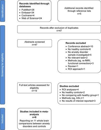
Table 1 Demographic and clinical characteristics of the task-based fMRI studies included in the current meta-analysis
Meta-analysis of functional brain abnormalities during reappraisal in anxiety disorder
The results from the AES-SDM analysis are summarized in and . After correcting for multiple comparisons, compared with HC, patients with anxiety disorder showed significantly decreased activity during cognitive reappraisal in (1) the bilateral dmPFC (bilateral superior frontal gyrus, Brodmann area [BA] 9/32) and extending into the bilateral dACC (bilateral dorsal anterior cingulate, dorsal BA 24/32) and bilateral SMA (bilateral supplementary motor area, medial BA 6); (2) the left inferior parietal; (3) the right superior parietal gyrus; (4) the left amygdala; (5) the left fusiform gyrus; and (6) the left vmPFC (middle frontal gyrus, medial BA 11). Definitions of the dmPFC and dACC can be found in previous reviews.Citation3,Citation45 However, we detected no significantly enhanced activity during reappraisal in patients with anxiety disorder compared with HC.
Figure 2 Regions of decreased (blue) activation in individuals with anxiety disorder compared with HC during cognitive reappraisal and subgroup analysis of medication naive group, 3 T MRI group, AFNI software group, SAD group and GAD and SAD group.
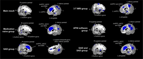
Table 2 Altered regional differences in activation during cognitive reappraisal in patients with anxiety disorder compared to healthy controls
Jackknife sensitivity analysis
A whole-brain jackknife sensitivity analysis was performed to examine the replicability of the findings of the pooled meta-analysis. As shown in , the results indicate that the decreased activation in the dmPFC, dACC, SMA and left inferior parietal was highly replicable, preserved as it was throughout all 11 combinations of studies. Results for the left amygdala, left fusiform gyrus, and left middle frontal gyrus remained significant in all but one combination, while those for the right superior parietal gyrus were significant in all but two combinations. Detailed results are listed in .
Subgroup meta-analyses
Given the possible influence of medication status, subtype of anxiety disorder, and methodological differences between studies, several subgroup meta-analyses were performed. As shown in and , these analyses suggest that the above results remained largely unchanged. In the subgroup of studies using 3.0 T MRI (eight datasets), all clusters in the combined analysis remained significant except for the right superior parietal gyrus. In the subgroup of studies using AFNI software (seven datasets), all clusters in the combined analysis remained significant except for the left amygdala and left middle frontal gyrus. In the subgroup of medication-naive patients (six datasets), the clusters of the dmPFC, dACC, SMA, left inferior parietal, right superior parietal gyrus, and left fusiform gyrus remained significant. In the subgroup of SAD (five datasets), the clusters of the dmPFC, dACC, SMA, left amygdala, and right superior parietal gyrus remained significant. In the subgroup of both GAD and SAD (eight datasets), the clusters of the dmPFC, dACC, SMA, left inferior parietal, left amygdala, left fusiform gyrus and right superior parietal gyrus remained significant.
Table 3 Subgroup analyses of the included studies
Heterogeneity analysis and publication bias
An analysis of heterogeneity revealed that a few regions with altered activation (left amygdala, left fusiform gyrus, and left middle frontal gyrus) showed significant statistical heterogeneity between studies (p<0.005). No strong evidence for publication bias was revealed by Egger’s test (p>0.05), except for the left amygdala (p=0.006).
As the left amygdala was absent in two of the five subgroup analyses, and showed a significant heterogeneity and publication bias, we excluded it from the core brain region contributing to impaired cognitive reappraisal in anxiety disorders.
Meta-regression analyses
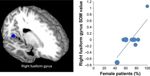
As shown in , the percentage of female patients had a positive relationship with activation in the right fusiform gyrus (Montreal Neurological Institute coordinate: x=20, y=−64, z=−12; AES-SDM value=2.369, p=0.000125587; 77 voxels). However, this result should be interpreted with caution as it is based on only three studies. Percentage of comorbidity, sample size, and mean age of patients with anxiety disorder were not associated with decreased brain regions. Race (white) and symptom severity (Liebowitz Social Anxiety Scale-Self Rated and Spielberger State-Trait Anxiety Inventory) could not be explored because only six and five datasets respectively were available from the studies included.Citation31
Figure 3 Results of the meta-regression analysis showing the percentage of female patients with a positive relationship with activation in the right fusiform gyrus. In the graphs, AES-SDM values needed to create this plot were extracted from the peak of maximum slope significance, and each study is represented as a dot, whose size reflects sample size. The regression line (meta-regression signed differential mapping slope) is shown.
Discussion
We believe that this is the first study to independently explore regional neurofunctional activation and simultaneously investigate subgroup meta-analyses and meta-regression analyses in patients with anxiety disorder (including most subtypes) during cognitive reappraisal. First, the pooled meta-analysis identified significantly decreased activity during cognitive reappraisal in the bilateral dmPFC, bilateral dACC, bilateral SMA, bilateral parietal cortex, left vmPFC, and left fusiform gyrus in patients with anxiety disorder compared with HC. Second, subgroup analyses indicated that the above findings remained largely unchanged. The robustness of the main findings was further demonstrated by the jackknife sensitivity analysis, heterogeneity analysis, and Egger tests. Finally, meta-regression analysis showed that activation in the right fusiform gyrus was positively correlated with percentage of female patients. These findings suggest that impaired cognitive reappraisal in anxiety disorder may be the consequence of hypo-activation in the prefrontal cortex, dACC, and parietal cortex during emotion regulation, consistent with insufficient top-down control.
The most common finding of the current study was decreased activation in the bilateral dmPFC of patients with anxiety disorder during cognitive reappraisal. A large number of cognitive reappraisal studies have shown that healthy individuals can activate the dmPFC to achieve successful emotion regulation,Citation10 whereas here dmPFC activation was lower in patients with anxiety disorder during reappraisal. According to the results of cognitive emotion regulation questionnaire and behavioral research, anxiety disorder had impaired cognitive emotion regulation strategies involving cognitive reappraisal.Citation44,Citation46 Neuroimaging studies have found that decreased dmPFC activity in anxiety disorder might be associated with reduced emotion regulation capability.Citation21 Furthermore, studies of healthy adults have found that activation in the dmPFC is associated with perceiving one’s affective statesCitation47,Citation48 and self-related processing.Citation49 A previous meta-analysis examining large samples of healthy subjects in cognitive reappraisal studies has suggested that the dmPFC may support semantic and self-reflective processes relevant to elaborating the meaning of affective stimuli or drawing inferences about one’s emotional state.Citation10 According to the findings discussed above, one possible explanation for the decreased dmPFC is that patients are unable to recruit the dmPFC to assist in monitoring and reflecting on the meaning of altering their affective states during reappraisal, and that this contributes to a reduced ability to down-regulate negative reactivity. Thus, insufficient recruitment of dmPFC activity could be an important neural correlate of emotion regulation deficits in anxiety disorder.
Comparing patients with anxiety disorder and HC during reappraisal revealed clusters of decreased activity in the dmPFC and extending into the dACC and SMA. The dACC lies on the medial surface of the frontal lobes and plays a significant role in regulating top-down cognitive control and assigning appropriate control to other areas of the brain.Citation50,Citation51 The dACC has also been recognized as a key region of the emotion-regulation network,Citation10,Citation17,Citation19 which is thought to mediate several functions including monitoring for conflict and detecting the likelihood of error commission, particularly involving attentional and executive control during the appraisal of negative emotion.Citation50,Citation52,Citation53 Poor emotion regulation may involve impaired attention control. Theories based on cognitive and neuroscience research have indicated that deficient attentional control is associated with anxiety.Citation54,Citation55 A previous review also found that anxiety disorder shows impaired recruitment of the dACC during emotion regulation.Citation3 Consequently, our findings suggest that anxiety disorder has a reduced ability to recruit the dACC implicated in top-down attention control to engage the emotion-regulation network. In addition, the SMA has also been consistently implicated as a core region of the emotion-regulation network in healthy people, and is associated with successful down-regulation of negative emotion.Citation10,Citation17 During reconceptualization (changing an appraisal) the pre-SMA may reflect the execution of this reconceptualization by reformulating mental representations through language and memory processing, with the anterior and posterior cluster of the SMA related to cognitive and executive aspects of motor behavior.Citation17 Thus, an impaired SMA may reflect reduced recruitment of the executive and cognitive aspects at different stages, which contributed to emotion dysregulation in anxiety disorders.
The parietal cortex is one of the brain regions most commonly identified in fMRI studies of emotion regulation in anxiety disorders.Citation20,Citation22 This brain region is a part of the frontoparietal control regionCitation56 related to sensory information, with top-down response-related information to facilitate flexible, goal-directed behavior.Citation57 Meta-analyses of neuroimaging studies have indicated that the parietal cortex is mainly involved in selective attention and might assist in holding reappraisals in mind.Citation14,Citation15 Compared with HC, patients with PTSD have a reduced ability to recruit the parietal cortex, implicated in top-down attentional control in the affective number Stroop task.Citation58 In one structural neuroimaging study, the SAD patients showed increased cortical thickness in the frontoparietal network, which fitted with findings at a functional level showing frontoparietal networks to be associated with executive-controlling and attentional functions.Citation59 Together, these results indicate that abnormalities in the parietal cortex may result in the dysfunction of top-down attentional control in anxiety disorder.
Another brain region showing abnormally reduced activity during appraisal was the left vmPFC. Because direct connections between the frontoparietal control region and amygdala associated with emotional response are relatively sparse compared with connections between vmPFC and the amygdala,Citation60 frontoparietal areas are likely to influence the amygdala indirectly by modulating activity in the vmPFC.Citation14,Citation52 Based on animal and human research, vmPFC plays an important role in fear extinction and appraisal of negative emotion.Citation10,Citation52 A prominent neurobiological model of the vmPFC highlights top-down inhibition of the amygdala through the vmPFC as a crucial neural mechanism that may be defective in certain mood and anxiety disorders.Citation61 Therefore, it might be speculated that patients with anxiety disorder cannot recruit the vmPFC through frontoparietal areas, in turn modulating the emotional response of the amygdala.
Besides cognitive and attentional regions, the left fusiform gyrus was found to have decreased functional activation in anxiety disorder during reappraisal. The fusiform gyrus is associated with the perception of affective stimuli.Citation62 Commonly used emotion stimuli for probing neurobiological responses to cognitive reappraisal mainly come from the IAPSCitation3 and involve face stimuli.Citation32 During emotional face processing, healthy samples show greater neural activity in the fusiform gyrus,Citation63 while highly anxious individuals show impaired ability to process emotional faces.Citation64 Furthermore, SAD patients show significantly weaker activation in the left fusiform gyrus for emotional face perception, as compared with HC.Citation65 Emotion perception is a significant part of emotion regulation; meta-analytic connectivity modeling analysis has shown the bilateral fusiform gyrus to be involved in cognitive emotion regulation.Citation17 Thus, our findings suggest that an impaired fusiform gyrus may be an important neural correlate in anxiety disorders of cognitive emotion dysregulation. Notably, the meta-regression analysis identified a positive association between percentage of female patients and activation of the right fusiform gyrus. Female patients with anxiety disorder seem to have distinct neurobiological underpinnings compared with male patients.Citation66 A previous neuroimaging study reported that the fusiform gyrus showed increased activity in females with stress compared with males with stress, when perceiving emotional faces.Citation67 This result might reflect the fact that female patients with anxiety disorder have less difficulty recognizing and distinguishing emotions compared with male patients with anxiety disorder.
However, heterogeneity analysis showed that there was significant, unexplained between-study variability concerning the left vmPFC and left fusiform gyrus. In our meta-analytic study, besides heterogeneity analysis, we also attempted to control the quality of the studies included by applying strict selection criteria and performing jackknife sensitivity analysis, subgroup analyses, and meta-regression analyses in order to reduce heterogeneity as much as possible. Although the left vmPFC and left fusiform gyrus showed significant heterogeneity, a quantitative assessment, as measured by Egger’s test, was not significant. The jackknife sensitivity analysis further confirmed the robustness of the findings. Therefore, in our view, these two brain regions remain significant in our meta-analysis.
Interestingly, while our meta-analytic study found an abnormal neural network contributing to impaired cognitive emotion regulation, an altered dlPFC, typically found in cognitive reappraisal,Citation10,Citation17 was not found. The dlPFC has been proposed to play an important role in cognitive emotion regulation.Citation15 This brain area was cognitive control center is related to action inhibition, working memory, reasoning, and social cognition.Citation17 There are some explanations for the non-significant changes in the dlPFC in the present meta-analysis. First, although the dlPFC is a significant part of the frontoparietal control network, a flexible and superordinate system supporting adaptive behavioral control,Citation57,Citation68 different brain regions of the frontoparietal network have shared and unique functions across a broad range of cognitive demands.Citation68 Normal activation in the dlPFC may not be affected because of help from other brain areas of the frontoparietal network. Behavior studies have shown that patients with anxiety disorder can successfully down-regulate their negative emotion,Citation43,Citation44 which may be in line with the above putative mechanism by which the activity of the dlPFC is not affected. Second, although in their review Zilverstand et alCitation3 reported abnormal activity in the dlPFC in anxiety disorders, they did not conduct a meta-analysis to provide a comprehensive description of the neurobiological underpinnings of cognitive emotion dysregulation. Furthermore, some of the articles in their review did not have sufficient statistical power. For example, the article reported decreased activity in the dlPFC in anxiety disorder compared with HC during reappraisal on the basis of a comparison of group differences, and no task effect or group × task interactions were found in the prefrontal cortex.Citation44
Compared with the study of Picó-Pérez et al,Citation27 decreased activation in ventrolateral prefrontal cortex (vlPFC) and increased activation in insula, cerebellum, precentral and inferior occipital gyri were not observed in our study. These findings could be explained by two aspects. First, the vlPFC may support selection and inhibition of appropriate appraisals.Citation10 As a significant part of the limbic network, the insula is critically involved in the perception and encoding of aversive stimuli.Citation69 Previous systematic reviews suggested that dysfunction of lateral prefrontal cortex was predictive and specific to depression disorderCitation28 and major depressive patients could not recruit lateral prefrontal cortical region (ie, vlPFC) to attenuate activity in the limbic network during cognitive reappraisal.Citation45 In addition, relative to anxiety disorders, enhanced responses of limbic system during downregulation of emotion were specific findings to major depressive disorder.Citation3 Second, enhanced responses of limbic system contributed to excessive emotional processing and emotional experience,Citation3,Citation45 the regions including cerebellum, precentral and inferior occipital gyri might be associated with the emotional experience.Citation27 Therefore, these regions that were not observed in our study might reflect specific deficits of major depressive disorder in neural mechanisms of emotion regulation. These findings could be useful in distinguishing anxiety disorder from major depression.
Limitations
There are several limitations to our study that should be highlighted. First, the number of fMRI studies included was small; the literature search yielded only eight studies with 11 relevant patients vs controls comparisons. This could affect the generalizability of our results, particularly in the subgroup meta-analyses and meta-regressions analyses. Second, our meta-analysis was based on coordinates from published studies rather than raw statistical maps, which might reduce its accuracy.Citation29 Third, the heterogeneity of the data acquisition and analysis techniques, including MRI field strengths, slice thickness, voxel size, and data-processing software, may reduce the accuracy of these results. Fourth, our meta-analysis included studies of medicine-naive patients who had undergone a medication washout period before scanning, so that longer-term influences of medication on brain function could not be completely excluded. Although we conducted a subgroup meta-analysis of the medicine-naive, these results should be interpreted with caution. Finally, some patients with anxiety disorder had co-morbid major depression. Anxiety and major depressive disorders may have different disorder-specific deficits in the neural mechanisms of cognitive reappraisal.Citation3,Citation28 Although the patients fulfilled our criteria for comorbid-major depression with anxiety disorder being the primary diagnosis, the influence of major depression cannot be completely ruled out.
Conclusion
In this meta-analysis, we identified the most robust functional neuroimaging findings on cognitive reappraisal in anxiety disorder. The results demonstrated that patients with anxiety disorder could not recruit the prefrontoparietal network, including the dmPFC, dACC, SMA, vmPFC, and parietal cortex, to down-regulate their emotion response. Our findings provide robust evidence that impairment of prefrontoparietal neuronal circuits may play an important role in the pathogenesis of anxiety disorder. This finding may provide novel targets for medical or cognitive-behavioral interventions and neuromodulation approaches (eg, transcranial magnetic stimulation). With longitudinal data, future investigations should further explore whether these functional abnormities are associated with structural changes or influenced by disease severity and medication status.
Author contributions
Hai-Yang Wang was the main author of this study and participated in the conception and design of the study, literature search, methods, analysis and manuscript write-up; Xiao-Xia Zhang, Cui-Ping Si, Yang Xu, Qian Liu, He-Tao Bian, and Bing-Wei Zhang contributed to reviewing studies, extracting data and feedback on analysis; Xue-Lin Li and Zhong-Rui Yan, corresponding authors, participated in the design of the study, analysis and interpretation of data. All authors contributed toward data analysis, drafting and critically revising the paper, gave final approval of the version to be published, and agree to be accountable for all aspects of the work.
Acknowledgments
The authors wish to thank all investigators who have provided additional information regarding their studies upon our request. This work was supported by National Natural Science Foundation of China (grant no. 81401486) and the Natural Science Foundation of Shandong Province of China (ZR2015HL039).
Supplementary materials
Table S1 Imaging methodology quality assessment checklist (when criteria were partially met, 0.5 points were assigned)
Table S2 Sensitivity analyses of task-based fMRI studies of regional differences in activation in patients with anxiety disorder compared with healthy controls
References
- NewASFanJMurroughJWA functional magnetic resonance imaging study of deliberate emotion regulation in resilience and post-traumatic stress disorderBiol Psychiatry200966765666419589502
- GaeblerMDanielsJKLamkeJPFydrichTWalterHBehavioural and neural correlates of self-focused emotion regulation in social anxiety disorderJ Psychiatry Neurosci201439424925824690369
- ZivMGoldinPRJazaieriHHahnKSGrossJJEmotion regulation in social anxiety disorder: behavioral and neural responses to three socio-emotional tasksBiol Mood Anxiety Disord2013312024517388
- BlairKSGeraciMSmithBWReduced dorsal anterior cingulate cortical activity during emotional regulation and top-down attentional control in generalized social phobia, generalized anxiety disorder, and comorbid generalized social phobia/generalized anxiety disorderBiol Psychiatry201272647648222592057
- GoldinPRManberTHakimiSCanliTGrossJJNeural bases of social anxiety disorder: emotional reactivity and cognitive regulation during social and physical threatArch Gen Psychiatry200966217018019188539
- BallTMRamsawhHJCampbellsillsLPaulusMPSteinMBPrefrontal dysfunction during emotion regulation in generalized anxiety and panic disordersPsychol Med20134371475148623111120
- ReineckeAFilippiniNBernaCEffective emotion regulation strategies improve fMRI and ECG markers of psychopathology in panic disorder: implications for psychological treatment actionTransl Psychiatry20155e67326529426
- FitzgeraldJMPhanKLKennedyAEShankmanSALangeneckerSAKlumppHPrefrontal and amygdala engagement during emotional reactivity and regulation in generalized anxiety disorderJ Affect Disord201721839840628501740
Disclosure
The authors report no conflicts of interest in this work.
References
- KesslerRCAguilargaxiolaSAlonsoJThe global burden of mental disorders: An update from the WHO World Mental Health (WMH) SurveysEpidemiol Psichiatr Soc2009181233319378696
- BaxterAJVosTScottKMFerrariAJWhitefordHAThe global burden of anxiety disorders in 2010Psychol Med201444112363237424451993
- ZilverstandAParvazMAGoldsteinRZNeuroimaging cognitive reappraisal in clinical populations to define neural targets for enhancing emotion regulation. A systematic reviewNeuroimage201715110511627288319
- ZhangBWXuJWangHYaoHZhangLLiuXWCognitive emotion regulation strategies in subjects with panic disorderChinese J Behav Med Brain Sci2014236484486 Chinese
- GaeblerMDanielsJKLamkeJPFydrichTWalterHBehavioural and neural correlates of self-focused emotion regulation in social anxiety disorderJ Psychiatry Neurosci201439424925824690369
- BarlowDHAllenLBChoateMLToward a unified treatment for emotional disorders–republished articleBehav Ther201647683885327993336
- CislerJMOlatunjiBOFeldnerMTForsythJPEmotion regulation and the anxiety disorders: an integrative reviewJ Psychopathol Behav Assess2010321688220622981
- GrossJJThe emerging field of emotion regulation: an integrative reviewRev Gen Psychol199823271299
- GrossJJThompsonRAEmotion regulation: conceptual foundationsGrossJJHandbook of Emotion RegulationNew YorkGuilford Press2007324
- BuhleJTSilversJAWagerTDCognitive reappraisal of emotion: a meta-analysis of human neuroimaging studiesCereb Cortex201424112981299023765157
- GrossJJAntecedent- and response-focused emotion regulation: divergent consequences for experience, expression, and physiologyJ Pers Soc Psychol19987412242379457784
- GrossJJEmotion regulation: affective, cognitive, and social consequencesPsychophysiology200239328129112212647
- YuanJLongQDingNLouYLiuYYangJSuppression dampens unpleasant emotion faster than reappraisal: Neural dynamics in a Chinese sampleSci China Life Sci201558548049125316046
- OchsnerKNGrossJJThe cognitive control of emotionTrends Cogn Sci20059524224915866151
- OchsnerKNSilversJABuhleJTFunctional imaging studies of emotion regulation: a synthetic review and evolving model of the cognitive control of emotionAnn N Y Acad Sci201212511E1E2423025352
- BeckATThe Current State of Cognitive Therapy: A 40-Year RetrospectiveArch Gen Psychiatry200562995395916143727
- KohnNEickhoffSBSchellerMLairdARFoxPTHabelUNeural network of cognitive emotion regulation – an ALE meta-analysis and MACM analysisNeuroimage201487Pt 234535524220041
- GoldinPRMcraeKRamelWGrossJJThe neural bases of emotion regulation: reappraisal and suppression of negative emotionBiol Psychiatry200863657758617888411
- FrankDWDewittMHudgenshaneyMEmotion regulation: quantitative meta-analysis of functional activation and deactivationNeurosci Biobehav Rev20144520221124984244
- GoldinPRManberTHakimiSCanliTGrossJJNeural bases of social anxiety disorder: emotional reactivity and cognitive regulation during social and physical threatArch Gen Psychiatry200966217018019188539
- ZivMGoldinPRJazaieriHHahnKSGrossJJEmotion regulation in social anxiety disorder: behavioral and neural responses to three socio-emotional tasksBiol Mood Anxiety Disord2013312024517388
- BlairKSGeraciMSmithBWReduced dorsal anterior cingulate cortical activity during emotional regulation and top-down attentional control in generalized social phobia, generalized anxiety disorder, and comorbid generalized social phobia/generalized anxiety disorderBiol Psychiatry201272647648222592057
- NewASFanJMurroughJWA functional magnetic resonance imaging study of deliberate emotion regulation in resilience and posttraumatic stress disorderBiol Psychiatry200966765666419589502
- Turk-BrowneNBFunctional interactions as big data in the human brainScience2013342615858058424179218
- WagerTDLindquistMKaplanLMeta-analysis of functional neuroimaging data: current and future directionsSoc Cogn Affect Neurosci20072215015818985131
- Van HornJDGraftonSTRockmoreDGazzanigaMSSharing neuroimaging studies of human cognitionNat Neurosci20047547348115114361
- Picó-PérezMRaduaJStewardTMenchónJMSoriano-MasCEmotion regulation in mood and anxiety disorders: A meta-analysis of fMRI cognitive reappraisal studiesProg Neuropsychopharmacol Biol Psychiatry2017799610428579400
- LiuYZLiuYZLinWJThe core neural mechanisms underlying depression disorder: a meta-analysis of fMRI studiesSci China2015451212141223
- RaduaJMataix-ColsDPhillipsMLA new meta-analytic method for neuroimaging studies that combines reported peak coordinates and statistical parametric mapsEur Psychiatry201227860561121658917
- TurkeltaubPEEdenGFJonesKMZeffiroTAMeta-analysis of the functional neuroanatomy of single-word reading: method and validationNeuroimage200216376578012169260
- RaduaJMataixcolsDVoxel-wise meta-analysis of grey matter changes in obsessive-compulsive disorderBrit J Psychiatry2009195539340219880927
- LangPJBradleyMMCuthbertBNInternational affective picture system (IAPS): Affective ratings of pictures and instruction manualTechnical Report A-82008
- American Psychiatric AssociationDSM-IV-TR: Diagnostic and statistical manual of mental disorders, text revisionWashington, DCAmerican Psychiatric Association2000
- DuMLiuJChenZBrain grey matter volume alterations in late-life depressionJ Psychiatry Neurosci201439639740624949867
- ChenGYiGZhuHIntrinsic disruption of white matter microarchitecture in first-episode, drug-naive major depressive disorder: A voxel-based meta-analysis of diffusion tensor imagingProg Neuropsychopharmacol Biol Psychiatry20177617918728336497
- RaduaJRubiaKCanales-RodríguezEJPomarol-ClotetEFusar-PoliPMataix-ColsDAnisotropic kernels for coordinate-based meta-analyses of neuroimaging studiesFront Psychiatry201451324575054
- IwabuchiSJKrishnadasRLiCAuerDRaduaJPalaniyappanLLocalized connectivity in depression: A meta-analysis of resting state functional imaging studiesNeurosci Biobehav Rev2015511778625597656
- RaduaJGrauMVan Den HeuvelOAMultimodal voxel-based meta-analysis of white matter abnormalities in obsessive–compulsive disorderNeuropsychopharmacology20143971547155724407265
- RubiaKAlegriaAACubilloAISmithABBrammerMJRaduaJEffects of stimulants on brain function in attention-deficit/hyperactivity disorder: a systematic review and meta-analysisBiol Psychiatry201476861662824314347
- NormanLJCarlisiCLukitoSStructural and functional brain abnormalities in attention-deficit/hyperactivity disorder and obsessive-compulsive disorder: a comparative meta-analysisJAMA Psychiatry201673881582527276220
- BoraEFornitoARaduaJNeuroanatomical abnormalities in schizophrenia: a multimodal voxelwise meta-analysis and meta-regression analysisSchizophr Res20111271–3465721300524
- ReineckeAFilippiniNBernaCEffective emotion regulation strategies improve fMRI and ECG markers of psychopathology in panic disorder: implications for psychological treatment actionTransl Psychiatry20155e67326529426
- FitzgeraldJMPhanKLKennedyAEShankmanSALangeneckerSAKlumppHPrefrontal and amygdala engagement during emotional reactivity and regulation in generalized anxiety disorderJ Affect Disord201721839840628501740
- BallTMRamsawhHJCampbellsillsLPaulusMPSteinMBPrefrontal dysfunction during emotion regulation in generalized anxiety and panic disordersPsychol Med20134371475148623111120
- RiveMMRooijenGVVeltmanDJPhillipsMLScheneAHRuhéHGNeural correlates of dysfunctional emotion regulation in major depressive disorder. A systematic review of neuroimaging studiesNeurosci Biobehav Rev20133722529255323928089
- ZhangBWXuJChangYWangHYaoHTangDImpaired cognitive reappraisal in panic disorder revealed by the late positive potentialNeuroreport20162729910326656936
- ParadisoSJohnsonDLAndreasenNCCerebral blood flow changes associated with attribution of emotional valence to pleasant, unpleasant, and neutral visual stimuli in a PET study of normal subjectsAm J Psychiatry1999156101618162910518175
- GallagherHLHappéFBrunswickNFletcherPCFrithUFrithCDReading the mind in cartoons and stories: an fMRI study of ‘theory of mind’ in verbal and nonverbal tasksNeuropsychologia2000381112110617288
- GusnardDAAkbudakEShulmanGLRaichleMEMedial prefrontal cortex and self-referential mental activity: relation to a default mode of brain functionProc Natl Acad Sci20019874259426411259662
- ShethSAMianMKPatelSRHuman dorsal anterior cingulate cortex neurons mediate ongoing behavioural adaptationNature2012488741021822122722841
- SunLPeräkyläJPolvivaaraMHuman anterior thalamic nuclei are involved in emotion–attention interactionNeuropsychologia201578825889426440152
- EtkinAEgnerTKalischREmotional processing in anterior cingulate and medial prefrontal cortexTrends Cogn Sci2011152859321167765
- ClemensBVoßBPawliczekCEffect of MAOA genotype on resting-state networks in healthy participantsCereb Cortex20152571771178124451655
- EysenckMWDerakshanNSantosRCalvoMGAnxiety and cognitive performance: attentional control theoryEmotion20077233635317516812
- BishopSJTrait anxiety and impoverished prefrontal control of attentionNat Neurosci2009121929819079249
- WangHYXuGQNiMFNeural mechanisms of implicit cognitive reappraisal: preceding descriptions alter emotional response to unpleasant imagesNeuroscience2017347657528192177
- DoddsCMSharonMZRobbinsTWDissociating inhibition, attention, and response control in the frontoparietal network using functional magnetic resonance imagingCerebral Cortex20112151155116520923963
- BlairKSVythilingamMCroweSLCognitive control of attention is differentially affected in trauma-exposed individuals with and without post-traumatic stress disorderPsychol Med2013431859522571775
- BrühlABHänggiJBaurVIncreased cortical thickness in a frontoparietal network in social anxiety disorderHum Brain Mapp20143572966297724039023
- GhashghaeiHTHilgetagCCBarbasHSequence of information processing for emotions based on the anatomic dialogue between prefrontal cortex and amygdalaNeuroimage200734390592317126037
- MyersschulzBKoenigsMFunctional anatomy of ventromedial prefrontal cortex: Implications for mood and anxiety disordersMol Psychiatry201217213214121788943
- GedayJGjeddeABoldsenASKupersREmotional valence modulates activity in the posterior fusiform gyrus and inferior medial prefrontal cortex in social perceptionNeuroimage200318367568412667845
- RossionBCaldaraRSeghierMSchullerAMLazeyrasFMayerEA network of occipito-temporal face-sensitive areas besides the right middle fusiform gyrus is necessary for normal face processingBrain2003126Pt 112381239512876150
- RossionBDricotLDevolderAHemispheric asymmetries for whole-based and part-based face processing in the human fusiform gyrusJ Cogn Neurosci200012579380211054921
- GentiliCGobbiniMIRicciardiEDifferential modulation of neural activity throughout the distributed neural system for face perception in patients with Social Phobia and healthy subjectsBrain Res Bull200877528629218771714
- KelimerLMMiladMRSex differences, gonadal hormones and the fear extinction network: implications for anxiety disordersBiol Mood Anxiety Disord201221322738383
- MatherMLighthallNRNgaLGorlickMASex differences in how stress affects brain activity during face viewingNeuroreport2010211493393720808182
- HardingIHYücelMHarrisonBJPantelisCBreakspearMEffective connectivity within the frontoparietal control network differentiates cognitive control and working memoryNeuroimage201510614415325463464
- OchsnerKNBungeSAGrossJJGabrieliJDRethinking feelings: an FMRI study of the cognitive regulation of emotionJ Cogn Neurosci20021481215122912495527