
Abstract
Background
Cognitive impairment in patients with cerebral small vessel disease (CSVD) is common, but the pathogenic mechanism is not well understood. The situation of non-breathing-related sleep fragmentation in CSVD patients and its influence on cognitive impairment is not clear. The aim of this study was to investigate the influence of non-breathing-related sleep fragmentation on cognitive function in patients with CSVD.
Methods
A group of 89 CSVD patients without breathing-related sleep disorders in the Department of Neurology, Third Affiliated Hospital of Sun Yat-sen University was enrolled. The patients underwent magnetic resonance scan, polysomnography, cognitive function evaluation using Montreal Cognitive Assessment scale (MoCA), and Mini-Mental State Examination. The patients were assigned to study group (arousal index [ArI] ≥26.8/hour) or control group (ArI <26.8/hour) based on the average level of ArI (mean =26.8, SD =7.5) at night, and the cognitive function of the patients in the two groups was analyzed.
Results
The total MoCA score, the subscale scores of visuospatial ability and delayed recall in the study group were significantly lower than that in the control group (P<0.05). The cognitive impairment measured by MoCA was positively related to ArI level and %N-3 sleep according to the results of logistic regression (P<0.05).
Conclusion
Non-breathing-related sleep fragmentation is associated with cognitive impairment in CSVD patients, especially executive function and delayed recall ability.
Introduction
Continuous, undisturbed sleep at night is significantly beneficial for one’s memory, immune regulation, and removal of neurotoxic byproducts that accumulate in the central nervous system during the awake period.Citation1 Sleep fragmentation, characterized by recurrent awakenings after sleep onset, often affects the elderlyCitation2 and could cause functional or even organic damage of the brain.Citation3 Sleep fragmentation can be breathing-related or non-breathing-related. Non-breathing-related sleep fragmentation is sleep fragmentation caused by reasons other than breathing problems, for example age, cerebrovascular diseases, Alzheimer’s, dementia, Parkinson’s disease, etc.Citation4
Cerebral small vessel disease (CSVD) is a syndrome of clinical and imaging findings that are thought to result from pathologies in perforating cerebral arterioles, capillaries, and venules, it is the most common cause of vascular cognitive impairment and dementia.Citation5 The pathogenic mechanism of cognitive impairment is not yet fully understood at the moment.Citation6 Research shows that non-breathing-related sleep fragmentation is related to pathological changes in iconology in CSVD,Citation7 but there is still no report on the relationship between non-breathing-related sleep fragmentation and cognitive impairment in this kind of patient. This research studied patients’ cognitive impairment affected by the severity of non-breathing-related sleep fragmentation, to help further investigate the pathological mechanism of CSVD-related cognitive impairment and to provide more evidence of CSVD prevention and treatments.
Methods
Design
This was a cross-sectional study. Patients were enrolled in The Third Affiliated Hospital of Sun Yat-sen University from August 2017 to July 2018. The study was approved by the ethics committee of the hospital, and was conducted according to the Declaration of Helsinki. All participants provided written informed consent.
Participants
One hundred and four consecutive patients with symptomatic CSVD were screened for recruitment following the inclusion/exclusion criteria detailed in the next paragraph.
Inclusion criteria were: 1) patients aged 35–70 years; 2) with one or more magnetic resonance (MR) markers of CSVD: lacunar infarcts (LI), white matter hyperintensities (WMH), cerebral microbleeds (CMB), and visible perivascular spaces (PVS);Citation8 3) without larger subcortical or cerebral Water-shed infarctions (>1.5 cm) on magnetic resonance imaging (MRI) as these are often embolic; 4) without large artery disease (carotid, vertebral, or intracranial stenosis >50%); 5) apnea–hypopnea index (AHI) <5/hour based on polysomnography (PSG).
Exclusion criteria were: 1) severe mental disorders such as schizophrenia, bipolar disorder or major depression, severe somatic or neurological disorders; dementia due to congenital mental disorders and other diseases; 2) presence of DSM-V Breathing-Related Sleep Disorders; 3) taking drugs that affect cognition or sleep; 4) unable to complete the scales due to objective conditions, such as deafness, hemiplegia, aphasia, visual impairment, etc; and 5) being pregnant.
Brain MRI scan
Patients underwent brain MRI on a 3-Tesla scanner (GE Discovery MR750) with a standard 8-channel head coil. Sequences included 3D TOF-MRA, T1Flair, T2WI, T2Flair, and SWI. Brain MRI was rated for the presence of four SVD makers: LI, WMH, CMB, and PVS independently. The presence of each SVD feature was summed in an ordinal “SVD burden score” (range 0–4).Citation8
PSG evaluation
All patients were evaluated for two consecutive nights in a sleep laboratory. The wireless telemetry PSG system (SOMNOscreen plus PSG+; SOMNOmedics GmbH, Randersacker, Germany, analysis software “DOMINO”) was used to monitor sleep. Sleep recordings were scored by an experienced PSG technologist blinded to the group assignments. The data were analyzed according to the American Academy of Sleep Medicine Manual for the Scoring of Sleep and Associated Events V.2.0.Citation9 PSG measures reported include: sleep onset latency (length of time in minutes that it takes to sleep); total sleep time (total duration of sleep in hours during the PSG recording); sleep efficiency ([SE] total sleep time divided by time in bed); wake after sleep onset ([WASO] the number of minutes the participant was awake after onset of persistent sleep); percentage of each sleep stage (N-1, N-2, N-3 [slow wave sleep] and rapid eye movement [REM] sleep); apnea–hypopnea index ([AHI] % time oxygen saturation <90%); and arousal index ([ArI] total number of arousals divided by the duration of sleep in hours. An arousal was defined as any sudden increase in electroencephalography frequency [to alpha or theta] that lasted >3 seconds).Citation10
Cognitive function measurement
Montreal Cognitive Assessment scale (MoCA)
The Chinese version of MoCA was used to evaluate cognitive function regarding aspects of visuospatial ability, executive function, naming, attention, language, abstraction, delayed recall, and orientation, with a total score between 0 and 30. If the educational level is <12 years, one point is added.Citation11,Citation12 Referring to the results of domestic research in People’s Republic of China, the cutoff point to identify cognitive impairment in CSVD is <23 points.Citation13
Mini-Mental State Examination (MMSE)
The Chinese version of MMSE is a 30-point questionnaire that is used to measure cognitive impairment regarding aspects of registration (repeating named prompts), attention and calculation, recall, language, ability to follow simple commands, and orientation. Any score ≥24 points (out of 30) indicates a normal cognition. Below this, scores can indicate severe (≤9 points), moderate (10–18 points), or mild (19–23 points) cognitive impairment.Citation14
Statistical methods
Statistical analyses for the present study were carried out with the SPSS software package (version 20.0). Descriptive statistics were presented as mean ± SD for continuous variables and as numbers or proportions for categorical variables. Student’s t-tests or Mann–Whitney was used when appropriate to compare the differences in sleep parameters and cognitive functions between the two groups. Binary logistic regression analyses were performed to assess the associations between total MoCA scores and sleep parameters (PSG). Regarding dependent variables, the total MoCA scores were dichotomized and coded as “dependent-1” (<23) and “independent-0” (≥23). The independent variables included SE, REM sleep ratio, N-3 sleep ratio, and WASO, which were included as continuous variables and ArI, coded as “dependent-0” (< mean) and “independent-1” (≥ mean) as categorical variables in the models. Two-sided P-values were considered to be statistically significant at ≤0.05.
Results
Clinical characteristics of patients
One hundred and four consecutive patients with symptomatic CSVD were screened for recruitment. Fifteen patients were excluded because they: 1) had breathing-related sleep disorders (n=10); 2) had delayed sleep phase disorder (n=1); and 3) had severe comorbid medical conditions (n=4). A group of 89 patients was thus recruited of which 56 patients met chronic insomnia criteria. The flow diagram of the study is presented in . The mean of ArI for all the patients was 26.8/hour (mean ArI =26.8, SD =7.5). Patients were divided into study group (ArI ≥26.8/hour) and control group (ArI <26.8/hour). There were no significant differences in demographic data, comorbidities, and SVD burden score between the two groups (P>0.05), as shown in .
Figure 1 Flow diagram of the study.
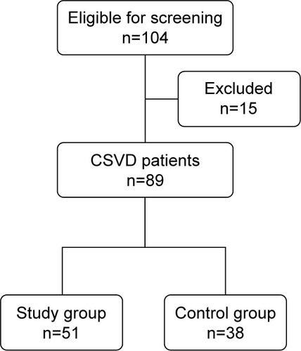
Table 1 Demographic and clinical data
Comparison of PSG parameters between the two groups
SE, REM ratio, and N-3 ratio of the patients in study group were lower than those in control group; WASO, N-1, and N-2 ratio of the patients in the study group were higher than those in the control group (P<0.05), with large effect sizes, as shown in .
Table 2 Comparison of PSG parameters between the two groups
Comparison of MMSE scores between the two groups
There was no significant difference in total score and subscale scores between study group and control group (P>0.05), as shown in .
Table 3 Comparison of Mini-Mental State Examination scores between the two groups
Comparison of MoCA scores between the two groups
The total score of MoCA, subscale scores of visuospatial ability and delayed recall in the study group were lower than those in the control group, with moderate to large effect sizes, as shown in .
Table 4 Comparison of Montreal Cognitive Assessment scale scores between the two groups
The associations between MoCA total scores and sleep parameters (PSG) based on binary logistic regression analyses
According to the results of logistic regression, those patients with higher ArI level were 3.542 (95% CI: 1.848–5.236, P<0.001) times more likely to have cognitive impairment than those with relatively lower ArL level. Those patients with shorter duration of N-3 sleep were 1.713 (95% CI: 1.251–2.517, P<0.05) times more likely to have cognitive impairment than those with relatively longer duration of N-3 sleep.
Discussion
Obstructive sleep apnea–hypopnea syndrome (OSAHS) is a common cause for sleep fragmentation and cognitive disorders.Citation15 This study showed, at first screening, only ten met OSAHS diagnostic criteria out of 104 CSVD patients, but 54% met chronic insomnia criteria. This ratio is much higher than that in older adults,Citation16 which means non-breathing-related sleep symptoms are more common in CSVD patients. The anatomy of the sleep–wake regulation system involves multiple areas of the brain, for example reticular ascending activation system, nucleus raphe, locus coeruleus, hypothalamus, thalamic reticular nucleus, basal forebrain, cerebral cortex, etc.Citation17 Small, deep cerebral vessels of CSVD patients show sclerosis, twisting, and self-regulating damage under the influence of vascular risk factors, which cause diffuse subcortical ischemia and periventricular fibrosis in the deep part of the brain and affects the sleep-related neural circuit, causing sleep disorder symptoms.Citation18 Although this study did not have a healthy control group, a past study showed an average ArI of older adults of 21.9/hour,Citation19 the average ArI of our CSVD subjects was 26.8/hour, showing a significant difference, which means non-breathing-related sleep fragmentation in CSVD is severe.
The relationship between non-breathing-related sleep disorders and cognitive function is controversial; there have been both positiveCitation20 and negative results.Citation21 The reason for this, apart from study design, might be the heterogeneity of sleep disorders. If we look further into the specific sleep parameters, some consistencies will emerge. For example, sleep duration (short or long sleep duration), WASO, and SE were found to be positively related to cognitive impairment.Citation22 Prolonged WASO and low SE could be the result of sleep fragmentation. This study discovered that sleep fragmentation was not only related to cognitive damage, but also directly related to WASO, SE, and changes in sleep structure, which indicates that sleep fragmentation could be a relatively stable sleep parameter for predicting cognitive impairment in CSVD patients.
In this study, the difference in cognitive functioning between the two groups was mainly reflected in two aspects: delayed recall and visuospatial ability. The impairment of both cognitive abilities could be related to sleep fragmentation-induced sleep structure disturbance. Non-rapid eye movement, especially N-3 sleep is crucial to declarative memory, while REM is important for enhancing short-term memory.Citation23,Citation24 The test of delayed recall in MoCA, which is one kind of declarative memory, is also relevant to short-term memory enhancing. The study group showed reduced N-3 sleep and REM ratio and impaired delayed recall, which indicates sleep fragmentation causing declarative memory impairment through sleep structure disturbance. Past experiments reported that sleep continuity disruption directly damaged the abilities of inhibitory control and working memory of healthy, senile people.Citation25 Inhibitory control is the ability to inhibit reacting to irrelevant stimulation when pursuing a specific cognitive goal. Working memory is temporary storage of information and correlates with many more general abilities and is linked to basic sensory processes.Citation26 This study found that the visuospatial ability impairment of the study group was significantly more serious than that of the control group, which may be the direct consequence of sleep fragmentation, because visuospatial ability is one of the main components of executive function.
Conclusion
This study indicates that non-breathing-related sleep fragmentation is common in patients with CSVD and could impair the ability of delayed recall and executive function. We suggest that reducing sleep fragmentation should be considered an important target in the prevention and treatment of cognitive impairment in CSVD patients. There are several limitations to the present study. First, because of the relatively small sample size, we did not conduct a multivariate analysis of sleep parameters and various cognitive fields, which may have led to confounding factors. We will increase the sample size for further analysis in future studies. Second, periodic leg movement is considered to be related to arousal during sleep, but it was not evaluated in this study because there were no participants who reported symptoms of restless leg syndrome. Finally, to improve our future studies, we will: 1) set up a normal control group and 2) prospectively observe changes in sleep fragmentation through treatment in CSVD patients and their effect on cognitive performance.
Disclosure
The authors report no conflicts of interest in this work.
References
- CedernaesJOsorioRSVargaAWKamKSchiöthHBBenedictCCandidate mechanisms underlying the association between sleep-wake disruptions and Alzheimer’s diseaseSleep Med Rev20173110211110.1016/j.smrv.2016.02.00226996255
- HuangYLLiuRYWangQSVan SomerenEJXuHZhouJNAge-associated difference in circadian sleep-wake and rest-activity rhythmsPhysiol Behav2002764–559760312126998
- TononiGCirelliCSleep and the price of plasticity: from synaptic and cellular homeostasis to memory consolidation and integrationNeuron2014811123410.1016/j.neuron.2013.12.02524411729
- SternALNaidooNWake-active neurons across aging and neurode-generation: a potential role for sleep disturbances in promoting diseaseSpringer Plus201542510.1186/s40064-014-0777-6
- PantoniLCerebral small vessel disease: from pathogenesis and clinical characteristics to therapeutic challengesLancet Neurol20109768970110.1016/S1474-4422(10)70104-620610345
- ShiYWardlawJMUpdate on cerebral small vessel disease: a dynamic whole-brain diseaseStroke Vasc Neurol201613839210.1136/svn-2016-00003528959468
- Del BruttoOHMeraRMZambranoMLamaJDel BruttoVJCastilloPRPoor sleep quality and silent markers of cerebral small vessel disease: a population-based study in community-dwelling older adults (The Atahualpa project)Sleep Med201516342843110.1016/j.sleep.2014.10.02325681211
- StaalsJMakinSDDoubalFNDennisMSWardlawJMStroke subtype, vascular risk factors, and total MRI brain small-vessel disease burdenNeurology201483141228123410.1212/WNL.000000000000083725165388
- RosenbergRSVan HoutSThe American Academy of Sleep Medicine inter-scorer reliability program: sleep stage scoringJ Clin Sleep Med201391819723319910
- American Sleep Disorders AssociationEEG arousals: scoring rules and examples: a preliminary report from the Sleep Disorders Atlas Task Force of the American Sleep Disorders AssociationSleep199215217318411032543
- NasreddineZSPhillipsNABédirianVThe montreal cognitive assessment, MoCA: a brief screening tool for mild cognitive impairmentJ Am Geriatr Soc200553469569910.1111/j.1532-5415.2005.53221.x15817019
- WenHBZhangZXNiuFSLiLThe application of Montreal cognitive assessment in urban Chinese residents of BeijingZhonghua Nei Ke Za Zhi2008471363918346324
- KongFBYangFChenWZhaoRLValue of the Montreal cognitive assessment for the detection of vascular cognitive impairment in cerebral small vessel diseaseChin J Clinicians201152369756980 Chinese
- WangZYZhangMYZhaiGYChenJXZhaoJApplication of Chinese version of Concise Mental State Examination (MMSE)Shanghai Arch Psychiatry198973108111 Available from: http://www.sinomed.ac.cn/zh/detail.do?ui=1994166490Accessed April 8, 2019
- PolsekDGildehNCashDObstructive sleep apnoea and Alzheimer’s disease: in search of shared pathomechanismsNeurosci Biobehav Rev20188614214910.1016/j.neubiorev.2017.12.00429223769
- PatelDSteinbergJPatelPInsomnia in the elderly: a reviewJ Clin Sleep Med20181461017102410.5664/jcsm.717229852897
- SchwartzMDKilduffTSThe neurobiology of sleep and wakefulnessPsychiatr Clin North Am201538461564410.1016/j.psc.2015.07.00226600100
- ChengCYTsaiCFWangSJHsuCYFuhJLSleep disturbance correlates with white matter hyperintensity in patients with subcortical ischemic vascular dementiaJ Geriatr Psychiatry Neurol201326315816410.1177/089198871349350323788613
- BonnetMHArandDLEEG arousal norms by ageJ Clin Sleep Med20073327127417561594
- ChenPLLeeWJSunWZOyangYJFuhJLRisk of dementia in patients with insomnia and long-term use of hypnotics: a population-based retrospective cohort studyPLoS One2012711e4911310.1371/journal.pone.004911323145088
- FoleyDMonjanAMasakiKDaytime sleepiness is associated with 3-year incident dementia and cognitive decline in older Japanese-American menJ Am Geriatr Soc200149121628163211843995
- YaffeKFalveyCMHoangTConnections between sleep and cognition in older adultsLancet Neurol201413101017102810.1016/S1474-4422(14)70172-325231524
- AckermannSRaschBDifferential effects of non-REM and REM sleep on memory consolidation?Curr Neurol Neurosci Rep201414243010.1007/s11910-013-0430-824395522
- AbelTHavekesRSaletinJMWalkerMPSleep, plasticity and memory from molecules to whole-brain networksCurr Biol20132317R774R78810.1016/j.cub.2013.07.02524028961
- WilckensKAWooSGKirkAREricksonKIWheelerMERole of sleep continuity and total sleep time in executive function across the adult lifespanAnnu Rev Psychol20136413516810.1146/annurev-psych-113011-14375023020641
- DiamondAExecutive functionsPsychol Aging201429365866510.1037/a003723425244484