Abstract
Introduction
Visual-motor skills are the basis for a great number of daily activities. To define a correct rehabilitation program for neurological patients who have impairment in these skills, there is a need for simple and cost-effective tools to determine which of the visual-motor system levels of organization are compromised by neurological lesions. In their 1995 book, The Visual Brain in Action (Oxford: Oxford University Press), AD Milner and MA Goodale proposed the existence of two pathways for the processing of visual information, the “ventral stream” and “dorsal stream,” that interact in movement planning and programming. Beginning with this model, our study aimed to validate a method to quantify the role of the ventral and dorsal streams in perceptual and visual-motor skills.
Subjects and methods
Nineteen right-handed healthy subjects (mean age 22.8 years ± 3.18) with normal or corrected-to-normal vision were recruited. We proposed that a delayed pointing task, a distance reproduction task, and a delayed anti-pointing task could be used to assess the ventral stream, while the dorsal stream could be evaluated with a grasping task and an immediate pointing task. Performance was recorded and processed with the video-analysis software Dartfish ProSuite.
Results
Results showed the expected pattern of predominance of attention for the superior left visual field, predominance of the flexor tone in proximal peri-personal space arm movements, tendency toward overestimation of short distances, and underestimation of long distances.
Conclusion
We believe that our method is advantageous as it is simple and easily transported, but needs further testing in neurologically compromised patients.
Introduction
Visual-motor skills are essential for the dexterity of the upper extremitiesCitation1 and for a wide range of daily activities involving movements of object achievement (reaching) and gripping (grasping), where vision guides the motor action.Citation2
Hand–eye coordination can be described as an ordered sequence consisting of: visualization of the target, focus of attention on it, identification and location of the target, planning and programming of the movement, activation of the upper limb muscles to start and perform the movement, and control of the action performed.Citation3 These processes require the integration of different systems, including the somato-sensory and perceptual system, central processing systems (especially those involved in monitoring, controlling, attention, and motivation), and the motor system. During these phases, visual and proprioceptive information is integrated to drive and support the movement of the upper limb, providing feed-back and feed-forward control.Citation4,Citation5
Given the importance of visual-motor skills in activities of daily living, the significance of their impairment can easily be understood. Such impairment is a rather common result of central nervous system damage. The proper planning of the rehabilitative treatment of these patients requires the development and application of assessment systems that are able to determine which of the organization levels of the visual-motor system are compromised and to quantify the results of intervention.Citation6
Information received in the visual areas of the occipital lobe is projected to several areas in the visual associative parietal and temporal lobes. Ungerleider and Mishkin identified, based on previous theories and experimental data obtained from cortical lesions of the cortex of monkeys, the presence of two pathways from the primary visual cortex to higher centers.Citation7 The first was called “dorsal stream” for its location in the dorsal convexity of brain cortex; it connects the primary visual area (V1) to the posterior parietal lobe. It was considered responsible for the localization of objects (the “where” stream). The second was called the “ventral stream”; it is centered on the V4 area and connects the V1 area to the inferior temporal lobe (ITL). It was considered responsible for the recognition of objects (the “what” stream).Citation8
Goodale and Milner later proposed a new interpretation of the fundamental organization of the visual system. Studying their patient with bilateral lesions in the occipito-lateral lobes and a small lesion in the left posterior parietal cortex, but with completely spared visual areas,Citation9 they showed a profound deficit in shape discrimination, but retained reaching ability.Citation10 Studies in healthy subjects also showed their ability, under certain experimental conditions, to act without being conscious of their action.Citation11 On the basis of such experimental evidence, Milner and Goodale have suggested that the fundamental difference between the two pathways is not in terms of percept type (space vs object) but in terms of the use of the information in the higher centers (action vs perception/recognition).Citation12
The ventral stream carries information necessary for the perception and recognition of the stimulus, while the dorsal stream carries that information required for controlling movements directed to the stimulus. A rather similar proposal had been advanced by Jeannerod, who suggested distinguishing a “semantic” from a “pragmatic” way of processing information for, respectively, the conscious understanding of the outside world and motor programming.Citation13
The dorsal stream carries the visual information concerning the position of objects moment by moment to coordinate limb movements in the visual space. Thus, it is more involved in the control and integration of stimuli in peripheral and lower visual fields, where hands, feet, and the ground are usually found.Citation14 The overexpression of lower visual fields in the dorsal stream is evident, for example, in the V6A area: it has been shown that pointing movements are faster and more accurate when they are carried out in the lower visual fields, compared with when the same movements are carried out in the upper visual field.Citation15
A functional deficit of the dorsal stream is responsible for optic ataxia, characterized by errors in the direction of reaching movements of the hand toward an object of interest, especially when this is placed in the periphery of the visual field.Citation16,Citation17 A lesion of the ventral stream can produce visual agnosia, in which the recognition of objects is not possible through visual information, and unilateral spatial neglect (neglect syndrome), in which the patient tends to ignore stimuli in the visual field opposite to the lesion.
The ventral and dorsal streams are not independent but functionally dissociated. The two streams are activated differently if the action is directed to a visible target or to one that has not been visible in the last 2 or more seconds.Citation18 Milner et al have shown that a patient with lesions of the dorsal stream and optic ataxia, improved their performance in pointing tasks when the stimulus disappeared more than 2 seconds before the start of the movement.Citation19
The objective of the study reported here was the validation of a method able to quantify the perceptual (ie, ventral stream) and visual-motor (ie, dorsal stream) skills in a sample of healthy subjects, to obtain a range of normality values. We expected that the obtained quantifications would confirm the functional patterns already described in scientific literature.
Subjects and methods
The study was performed in May 2012 in the Rehabilitation Unit of the Santa Maria alle Fonti Medical Center, part of the Don Carlo Gnocchi ONLUS Foundation.
Study sample
We recruited 19 right-handed healthy adults with normal or corrected vision (females:males, 14:5; age 22.8 ± 3.18 years; education 15.78 ± 1.35 years). All subjects were either students or residents at the University of Pavia. The exclusion criteria of this study were a history of psychiatric illness, anamnesis or actual alcohol abuse, recent trauma involving the upper arm, and/or visual deficits not adequately compensated for by binocular lenses. All subjects were informed about the procedures and purposes of the investigation and they signed written informed consent. The study was conducted following recommendations from the Helsinki Declaration.
Experimental tasks
Assessment of the ventral stream
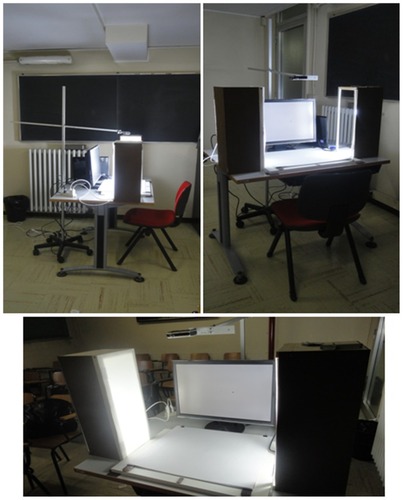
Each subject sat at a table covered with a non-reflective material to avoid reflections during the tests and to ensure clear video capture. A 22-inch LCD screen was used for the presentation of visual stimuli. This was placed on the table in front of the subject, at approximately 55–60 centimeters distance from the subject’s eyes. A special lighting system set at 100 watts was used and the point on which people had to rest the index finger of their right hand during projection of the stimulus and where they had to return at the end of each trial was clearly marked ().
Figure 1 The setting.
The subject could not see their own right hand during stimulus presentation because the working space was covered; the cover was removed 2 seconds after the stimulus disappeared from the screen. The subject, who was given unlimited time for visual analysis, controlled the disappearing of the stimulus.
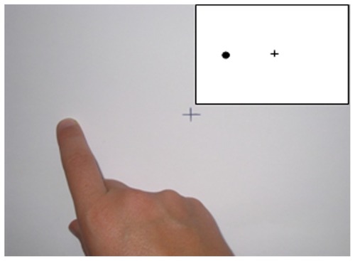
For each trial, the target stimulus consisted of a black circle (5 mm diameter) projected on a white background. This stimulus was positioned at distances of 3, 6, 9 cm and along rays at 45, 135, 180, 225, 270, and 315 degrees compared with a black cross (5 × 5 mm) located centrally in the field of projection.
Each subject was preliminarily allowed to undertake a number of trials of each test sufficient to make sure that they understood the task exactly. These trials were not recorded.
In the delayed pointing (P) task, the visual stimulus was presented and, after 2 seconds without visual input, the subject was asked to indicate with their index finger the target position with respect to the point marked on the shelf (the starting point; see ). In the delayed distance (D) task, after the presentation of the stimulus and a latency period of two seconds, the subject was asked to mimic with the first and second fingers the distance between the two elements.
Figure 2 Performance in delayed pointing task (stimulus represented in box).
The two tasks were administered in separate blocks following the ABBA order. The positions along the horizontal axis, 0 and 180 degrees, were presented three times because they were thought to be the most significant positions;Citation20 therefore, for each block there were 36 trials, 18 of which related to non-horizontal positions (45, 90, 135, 225, 270, or 315 degrees) and 18 related to horizontal positions (0, 180 degrees). Pseudo-randomized sequences were used to avoid the construction of sequences with identical consecutive stimuli and sequences with more than three consecutive stimuli on the horizontal axis.
In the horizontal delayed pointing (DP) task, we asked the subjects to indicate with their index finger the remembered position of the visual target; in the horizontal delayed anti-pointing (DAP) task, the subjects instead had to indicate the symmetrical (with respect to the starting point) position. Both tasks were performed 2 seconds after the stimulus disappeared. Since the horizontal axis is the most sensitive in the detection of dysfunction after brain injury, we presented stimuli at distances of 3, 6, or 9 cm from the central cross on the 0 and 180 degrees axis, for a total of 18 trials per task.
Assessment of the dorsal stream
The visual-motor skills relating to the dorsal stream were evaluated with tests in which the action (reaching or grasping) was executed without any latency and while the visual stimulus remained visible.
In the immediate pointing (IP) task, a black circle (5 mm diameter) and a central cross (5 × 5 mm) were printed on A4 white paper. The target stimulus (black circle) was presented at distances of 3, 6, or 9 cm along the rays of 45, 135, 180, 225, 270, and 315 degrees with respect to the central cross. We asked the subject to indicate the black circle. A total of 36 trials were administered, with the stimuli presented in random order.
In the grasping (G) task, gray rulers of 1 × 1 × 3 cm, 1 × 1 × 6 cm, and 1 × 1 × 9 cm size were inclined 45 or 90 degrees to the axis of symmetry of an A4 white sheet of paper. We asked the subject to grasp the ends of the rulers with their first and second fingers. A total of 36 trials were administered, with the different rulers and positions presented in random order.
Detection apparatus
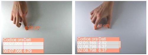
A camera orthogonally placed at about 50 cm from the plane filmed all movements. The videos were reworked with the video analysis software Dartfish ProSuite 5.0 (Dartfish, Fribourg, Switzerland). This software allowed measurement of the positions and distances pointed by the subjects (see ). Data obtained were then transferred to a spreadsheet for statistical analysis.
Figure 3 Data resulting from video analysis in a pointing and in a grasping task.
At the beginning of each new task administration, we calibrated our system by filming a ruler with millimeter increments marked on it placed on the shelf, to correct any possible distortion in the recorded video.
Statistical analysis
Data were analyzed with SPSS (v 19; IBM, Armonk, New York, USA).
For the P, D, IP, DP, and DAP tasks, we calculated the differences between the real distances and those indicated by each subject. For the G task, we calculated the differences between the maximum opening of the first and second finger before the ruler was grasped and the ruler’s length. Therefore, in the first group of mentioned tasks, we quantified measure errors, while, for the G task, we measured the excess of distance between the first phalanges of the first and second finger at about two-thirds of the flight phase compared with the real size of the object to be grasped.
For the IP task, we considered the number of errors (defined as pointing in a place different from the location of the stimulus).
Each task was analyzed with a general linear model (GLM), which can be described as a generalization of a multiple linear regression model to more than one dependent variable. A difference was considered significant if P ≤ 0.05.
Results
P and D tasks
The means and standard deviations of the systematic error in the P and D tasks are shown in .
Table 1 Systematic error in delayed pointing and delayed distance tasks (mean [standard deviation])
We divided data in groups according to the target position on the visual fields (both on the horizontal and vertical planes).
We first applied a GLM 2 (P or D) × 3 (spatial position: left, center, right). Data analysis revealed a significant interaction between the stimuli spatial position and the task [F(1.336, 0. 856) = 4.53; P = 0.017]. We highlighted a linear effect of stimuli position on the task [F(1, 1.625) = 5.914; P = 0.026] in the P task but not in the D task.
We also applied a GLM 2 (P or D) × 3 (spatial position: superior, middle, inferior). We found a significant interaction between the position of the stimulus and the task [F(1.71, 2.49) = 32.03; P < 0.001]. In both the P and D tasks, we highlighted a linear effect of stimuli position on the task: F(1, 0.477) = 21.32 (P < 0.001) and F(1, 0.377) = 11.01 (P = 0.004), respectively.
Means and standard deviations of the random error are shown in . No significant effects or interactions were found.
Table 2 Random error in delayed pointing and delayed distance tasks (mean [standard deviation])
P and DAP tasks
Means and standard deviations of the systematic error are shown in .
Table 3 Systematic error in delayed pointing and delayed anti-pointing tasks (mean [standard deviation])
We applied a GLM 2 (P or DAP) × 2 (reaction/response side: left or right) × 3 (distance: 3, 6, or 9 cm). The analysis revealed a significant effect of stimuli distance from the reference cross [F(1.07, 28.12) = 16.84; P = 0.001] on both tasks. The means and standard deviations of random error are shown in .
Table 4 Random error in delayed pointing and delayed anti-pointing tasks (mean [standard deviation])
We applied a GLM 2 (P or DAP) × 2 (reaction/response side: left or right) × 3 (distance: 3, 6, or 9 cm). Data analysis revealed a significant effect of the distance of the stimulus from the reference cross [F(1.78, 2.37) = 25.8; P < 0.001] on both tasks: subjects overestimated short distances and underestimated long distances. Presentation side of the stimulus did not seem to influence task performance.
G task
Means and standard deviations of the systematic error are shown in .
Table 5 Systematic error in grasping task (mean [standard deviation])
We applied a GLM 2 (ruler inclination: 45 or 90 degrees) × 3 (ruler length: 3, 6, or 9 cm). Data analysis showed a significant effect of both ruler length [F(1.53, 145.26) = 733.30; P < 0.001] and inclination [F(1, 238.58) = 1707.22; P < 0.001], without any statistically significant interaction between the two.
Subjects opened their fingers about 20% more than necessary according to ruler length, a percentage that tended to increase when the ruler was placed at 45 degrees.
IP task
Statistical analysis was not conducted because subjects made no errors during this task.
Discussion
For tasks studying the ventral stream, conditions were chosen to avoid the activation of the dorsal stream. We used three study conditions to achieve this goal. First, the answer to the task was given on a plane that was orthogonal to the plane where the stimulus had been seen. This was done to force subjects to activate the streams and not to rely on a simple memory of the position of the stimulus. Second, we used a latency of 2 seconds, as Milner and Goodale have suggested is a time that enhances activation of the dorsal stream over the ventral stream when performing a task.Citation18,Citation19 Third, a movable shelf was used to prevent the subject from watching their own hand while receiving the input in tasks meant to activate the ventral rather than the dorsal stream.
P and D tasks
In the P task, statistical analysis revealed that the systematic error tended to be higher for stimuli projected in the inferior part of the visual field. This could be explained by a predominance of the flexor tone occurring during arm pointing movements in proximal peri-personal space.
In contrast, in the D task, the error tended to be higher for stimuli projected in the superior part of the visual field. Brain lateralization could play a role in explaining this phenomenon: the right hemisphere of the brain is specialized in visuo-spatial representation and has been shown to perform a more careful analysis of stimuli projected in the superior left part of the visual field.Citation22 Moreover, all subjects we tested were from a Western culture: the habit of reading from left to right could contribute to the measured differences.
P and DAP tasks
In these tasks, the only factor influencing performance seems to have been target distance from the reference cross. We registered an overestimation (of about 0.75 cm) for a short distance (3 cm) and an underestimation (of about −0.20 cm) for a long distance (9 cm). To explain this, it could be hypothesized that an unconscious “obstacle avoidance behavior” intervened.Citation21 According to this hypothesis, the reference cross would be perceived as an “obstacle” and subjects would overestimate the target position when it was close to the reference cross to avoid it. This behavior would prevail on the tendency toward an underestimation of target distance, which emerged when the stimulus was further away from the reference cross.
Statistical analysis of random error highlighted a significant interaction between distance and subject’s performance: response variability was wider for longer distances, without a significant effect of the side of presentation of the stimulus.
G task
The excess of finger opening could be explained by neuro-motor constraints: the shoulder, elbow, and wrist drawing movement near the body involve a predominance of flexor tone. In healthy subjects, this is balanced by the activation of antagonist extensor muscles. The predominance of arm muscle flexor tone is greater when the ruler reaches an inclination of 45 degrees rather than 90 degrees; to counteract this, subjects tend to increase the activation of the extensor muscles, including those acting on the fingers.
Moreover, wrist position influences the finger extrinsic muscles: at an inclination of 45 degrees, the activation of the forearm extensor muscles is stronger and this leads to an increased contrast of the action of the flexor digitorum profundus, flexor digitorum superficialis and flexor pollicis longus. This could result in a relative reduction in the flexor tone acting on the fingers.
IP task
The IP task was designed to investigate visual-motor skills depending on the dorsal stream and to detect the presence of pathological conditions such as optic ataxia. As expected for a healthy sample, all performances were completely normal.
Conclusion
Our method provides findings in line with the published literature. Accuracy and precision increased when pointing targets were located in the superior left visual field.Citation22 A predominance of the flexor tone in proximal peri-personal space arm movements was evident.Citation23
The absence of significant random errors in any task and the accuracy in data collection are significant factors increasing the validity of our method. The innovative technique we explored allowed us to obtain easily precise measurement of finger position, distances, and aperture of finger grip. It is not an invasive procedure and does not require the use of fixed markers. Thus, we believe that our method could be useful in many settings, because it is as easy to reproduce as the commonly used paper and pencil tests and does not cost as much as more complex devices.Citation6
However, our study has some relevant limitations. The major limitation is that we recruited only healthy subjects; as such, we need to collect data from patients known to have a disruption of the dorsal and/or ventral stream due to a neurological disorder. This is the only way to confirm whether our tasks can discriminate between these conditions. Moreover, we will need to perform the same assessment before and after treatment to verify the tasks’ sensitivity to change.
Future studies need to be conducted with a comparison assessment technique to verify the reliability of our method.
Disclosure
The authors received no grants or funding for this paper and declare no conflicts of interest in this work.
References
- DesrosiersJHébertRBravoGDutilEUpper-extremity motor co-ordination of healthy elderly peopleAge Ageing19952421081127793331
- CrawfordJDMedendorpWPMarottaJJSpatial transformations for eye-hand coordinationJ Neurophysiol2004921101915212434
- JeannerodMMechanisms of visuomotor coordination: a study in normal and brain-damaged subjectsNeuropsychologia198624141783517680
- TurveyMTFonsecaSNature of motor control: perspective and issuesSternadDProgress in Motor Control: A Multidisciplinary PerspectiveNew York, NYSpringer Verlag200993123
- GottliebGLSongQAlmeidaGLHongDACorcosDDirectional control of planar human arm movementJ Neurophysiol1997786298529889405518
- ChiappediMDe BernardiEDalla ToffolaEBejorMChild visuomotor skills: preliminary findings using a new low-cost movement analysis methodFunct Neurol2010251454820626996
- UngerleiderLMishkinMTwo cortical visual systemsIngleDJGoodaleMAMansfieldRJWAnalysis of Visual BehaviourCambridge, MAMIT Press1982549586
- RizzolatiGSinigagliaCSo quel che fai. Il cervello che agisce e i neuroni specchio [I know what you’re doing:the acting brain and mirror neurons]MilanRaffaello Cortina Editore2006
- JamesTWCulhamJHumphreyGKMilnerADGoodaleMAVentral occipital lesions impair object recognition but not object-directed grasping: an fMRI studyBrain2003126Pt 112463247514506065
- MilnerADPerrettDIJohnstonRSPerception and action in ‘visual form agnosia’Brain1991114Pt 1B4054282004249
- DehaeneSChangeuxJPNaccacheLSackurJSergentCConscious, preconscious, and subliminal processing: a testable taxonomyTrends Cogn Sci200610520421116603406
- MilnerADGoodaleMAThe Visual Brain in ActionOxfordOxford University Press1995
- JeannerodMThe representing brain: neural correlates of motor intention and imageryBehav Brain Sci1994172187202
- MotterBCMountcastleVBThe functional properties of the light-sensitive neurons of the posterior parietal cortex studied in waking monkeys: foveal sparing and opponent vector organizationJ Neurosci1981113267346556
- DanckertJGoodaleMASuperior performance for visually guided pointing in the lower visual fieldExp Brain Res20011373–430330811355377
- GréaHPisellaLRossettiYA lesion of the posterior parietal cortex disrupts on-line adjustments during aiming movementsNeuropsychologia200240132471248012417474
- PisellaLGréaHTiliketeCAn ‘automatic pilot’ for the hand in human posterior parietal cortex: toward reinterpreting optic ataxiaNat Neurosci20003772973610862707
- WestwoodDAGoodaleMAPerceptual illusion and the real-time control of actionSpat Vis2003163–424325412858950
- MilnerADPaulignanYDijkermanHCMichelFJeannerodMA paradoxical improvement of misreaching in optic ataxia: new evidence for two separate neural systems for visual localizationProc Biol Sci199926614342225222910649637
- LepelleyMCThullierFBolmontBLestienneFGAge-related differences in sensorimotor representation of space in drawing by handClin Neurophysiol2010121111890189720483658
- MilnerADGoodaleMATwo visual systems re-viewedNeuropsychologia200846377478518037456
- de MontalembertMAuclairLMamassianP“Where is the sun” for hemi-neglect patients?Brain Cogn201072226427019850395
- KirschWHerbortOButzMVKundeWInfluence of motor planning on distance perception within the peripersonal spacePLoS One201274e3488022545092