Abstract
Background and methods
This study investigated the effects of sleep deprivation on total and partial (early and late) declarative memory and activation in the areas of the brain involved in these activities. The study included two experiments. Experiment 1 included 40 male residents of an orphanage aged 16–19 years, who were divided into four groups (n = 10 each) and subjected to total sleep deprivation, normal sleep, early-night sleep deprivation, or late-night sleep deprivation. Experiment 2 included eight students from the same institution who were divided into the same four groups (n = 2) as in experiment 1. Declarative memory was tested using lists of associated word pairs in both experiments, and activation of the relevant brain regions was measured before and after retrieval by single-photon emission computed tomography for subjects in experiment 2 only.
Results
Students subjected to normal sleep had significantly higher scores for declarative memory retrieval than those subjected to total sleep deprivation (P = 0.002), early-night sleep deprivation (P = 0.005), or late-night sleep deprivation (P = 0.02). The left temporal lobe showed the highest rate of activity during memory retrieval after normal sleep, whereas the frontal, parietal, and right temporal lobes were more active after sleep deprivation.
Conclusion
Both slow wave sleep and rapid eye movement sleep play an active role in consolidation of declarative memory, which in turn allows memory traces to be actively reprocessed and strengthened during sleep, leading to improved performance in memory recall.
Introduction
Consolidation of memories is widely believed to benefit from sleep.Citation1 The benefits of sleep-dependent performance in humans have been widely demonstrated, appearing in declarative,Citation2 spatial,Citation3 perceptual,Citation4 motor,Citation5 and sensorimotor tasks.Citation6 However, relating single sleep states, ie, rapid eye movement (REM) sleep and slow wave sleep to types of memory (ie, declarative and nondeclarative) is more complicated.Citation7–Citation9 It has been hypothesized that the effect of sleep state on memory processing is task-dependent, ie, procedural memory would benefit from REM sleep, whereas declarative memory would be linked with slow wave sleep.Citation10 This model is referred to as the “dual process” hypothesis.Citation11 However, much of the experimental evidence is inconsistent with this theory, suggests cooperation between slow wave sleep and REM is needed for memory consolidation,Citation12 and has led to the “sequential” hypothesis, according to which both slow wave sleep and REM sleep are involved in memory processing.Citation13 The best known model discussing the role of sleep in memory is that of replay.Citation1 However, at the neurophysiological level, there is evidence suggesting that sleep deprivation leads to a decrease in neurobehavioral functioning, including vigilance, memory, and impulse control.Citation14–Citation17 This deterioration is attributed to less activation in the brain regions which are responsible for these functions, ie, the prefrontal cortex and the temporal lobes. At the same time, the frontal and parietal regions show more activation in some sleep-deprived subjects when they perform specific tasks, and activation of these regions is correlated with thalamic activity.Citation9,Citation15 This study investigated the impact of total and partial (early and late) sleep deprivation on declarative memory retrieval and attempted to correlate scores for declarative memory retrieval with the degree of activation of different brain regions following normal sleep and after deprivation of the different sleep states.
Materials and methods
Written informed consent was obtained from all participating students and their guardians, with the approval of the orphanage, after giving a full explanation of the aim of the study and a detailed description of its protocol. The study was approved by the ethics committee at the Faculty of Medicine, Assiut University. Before embarking on this study, the principal investigator lived for about a year with subjects at the orphanage where the research took place, with the aim of exploring the living environment of the subjects and establishing relationships with them.
Experiment 1
A total of 40 male student residents, aged 16–19 (mean 18 ± 1.2) years, participated in this orphanage-based study. All participants had normal percentiles (range 0–95) for intelligence quotient, as measured by the standardized Arabic version of Raven’s progressive matrices.Citation18 Students were excluded if they suffered sleep deprivation for any reason or reported an abnormal sleep pattern prior to the study, or if their intelligence quotient was less than the 70th percentile.
The 40 subjects were randomly allocated to one of four groups (n = 10 each) according to hours and type of sleep, ie, slow wave sleep and REM sleep.Citation19 Students in group A were allowed approximately 8 hours of continuous night-time sleep from 11 pm to 7 am (normal sleep); students in group B were deprived of sleep throughout the night (total sleep deprivation); students in group C were deprived of sleep for the first half of the night from 11 pm to 3 am, during which slow wave sleep predominates, and were then allowed to sleep for the second half of the night (early-night sleep deprivation); and group D students were allowed to sleep for the first half of the night, and were then awakened after four hours of sleep and kept awake for the second half of night from 3 am to 7 pm, during which REM sleep predominates (late-night sleep deprivation).
Instrument used to measure declarative memory retrieval
A word-pair association task was used to test declarative memory, defined as “consciously accessible memories of fact-based information”.Citation14 This task consists of two lists of associated word pairs, each containing 18 associated word pairs of Arabic concrete nouns (eg, cat-animal). These lists of associated word pairs had been standardized for word frequency, length, emotionality, meaningfulness, and concreteness in a pilot study prior to undertaking this work.
In the learning condition, the word pairs were first presented to the subject (each pair for 5 seconds). A cued recall followed immediately thereafter, in which the subject had to write the matching word after being shown the first word of the pair. If the participant was not able to recall the correct word, the correct answer was displayed. Recall was repeated until the subject had achieved 100% correct responses, ie, for 36 word pairs.
In the retest condition, the lists were presented on paper with each pair having a missed word, and the subjects were asked to write the word that completed the pair. The number of correctly completed word pairs was recorded. In both the training condition and the retest condition, the subject was given unlimited time to respond to the cued recall. Performance was measured as the number of correctly completed word pairs on retesting.
In the normal sleep group, subjects learned the two lists of associated word pairs between 10 pm and 11 pm before going to bed at their usual time. After a night of normal sleep, retrieval was tested at 8 am. In the total sleep deprivation group, learning took place between 10 pm and 11 pm, with the subjects staying awake until morning and retrieval tested at 8 am. In the early-night sleep deprivation group, learning took place between 10 pm and 11 pm, with subjects staying awake from 11 pm to 3 am, during which slow wave sleep is more predominant,Citation1 going to sleep at 3 am and awakened at 7 am, with retrieval tested at 8 am. In the late-night sleep deprivation group, learning took place between 10 pm and 11 pm, with subjects going to bed from 3 am to 7 am, during which REM sleep is predominant,Citation1 and awakened at 7 am, with retrieval tested at 8 am.
Experiment 2
After repeated trials, eight right-handed healthy male students of mean age 18.2 ± 0.9 years and with a normal intelligence quotient agreed to participate in the second part of the study. Declarative memory was tested using the same protocol as in experiment 1, with the only difference being use of single-photon emission computed tomography (SPECT) to image the brain before and after declarative memory recall in all participants.
The participants continued in the same four groups to which they had been allocated in experiment 1, with each group now consisting of only two students. Each subject underwent baseline SPECT brain imaging at 8 am and post-retrieval imaging two hours later using Tc-99m ethyl cysteinate dimer (ECD). The subjects lay still and blindfolded in a dark room, with a catheter inserted into an antecubital vein for at least 15 minutes before injection. Imaging was done 5 minutes post injection using a dual-head Piker AXIS® gamma camera (MedWOW, Nicosia, Cyprus) equipped with a low energy all-purpose collimator interfaced to a dedicated computer.
At the baseline examination (before recall), the subjects received an injection of 222 MBq (6 mCi) Tc-99m ECD IV, and SPECT imaging was performed 10–30 minutes post injection. Two hours later, another dose (222 mBq [6 mCi]) was injected immediately before the volunteer started to retrieve the associated word pairs. Environmental conditions were kept constant during both scans.
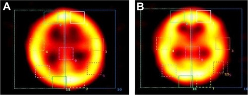
The subjects were imaged in the supine position with the neck hyperextended and a head strap used for fixation. A total of 32 projections was acquired over 180 degrees of rotation. The timing of acquisition was 30 seconds per frame, with the radius of rotation dependent on camera fixation in the noncircular programmed motion, ensuring that the detector heads remained very close to the subject’s head in each projection. Projection images in 64 × 64 matrices were smoothed and reconstructed using Hamming filtered back projections. No attenuation or scatter correction was performed. Transverse, sagittal, and coronal cuts 3.9 mm in thickness were computed. These parameters were fixed for both the baseline and post-retrieval scans. A comparison of the average count per pixel of 25 automated brain regions between both scans (baseline and after retrieval) was done using built-in computer software ().
Figure 1 Comparison between baseline (A) and post-retrieval (B) activation of brain region in a subject after total sleep deprivation.
Data analysis
Kruskal–Wallis one-way analysis of variance was used to measure differences in memory retrieval between the four study groups. The Mann–Whitney test was used to test for differences between each two types of sleep, ie, slow wave sleep and REM sleep.
Results
shows the means of correctly recalled word pairs for the declarative memory task in the four sleep groups. Using Kruskal–Wallis one-way analysis of variance, it was found that students in group 1 (normal sleep) had a significantly higher mean score for associated word pairs than the three sleep-deprived groups.
Table 1 Differences in recall between the study groups using the Kruskal–Wallis test
shows the differences in mean score for recalled memory word pairs between the study groups. Using the Mann–Whitney test, the normal sleep group recalled significantly more word pairs than the total sleep deprivation group (P = 0.002), the early-night sleep deprivation group (0.005), and the late-night sleep deprivation group (P = 0.02). However, the late-night sleep deprivation group scored significantly higher than the total sleep deprivation group for mean number of word pairs recalled (P < 0.03). shows the percentage activation in different brain regions using SPECT before and after recall of the memory task.
Table 2 Differences in recall between the study groups using the Mann–Whitney test
Table 3 Differences in activation of brain regions by single-photon emission computed tomography
Discussion
Sleep actively improves performance of recently acquired skills and tasks. The effect of sleep on recently acquired memory is obvious when people are sleep-deprived on the night following learning. The main finding of this study is that sleep facilitates consolidation of declarative memory and that being awake during the same period does not ( and ). These results are consistent with those of previous studies showing that retention of declarative memory benefits from a period of sleep in comparison with wakefulness after learning.Citation20–Citation22
The consolidation phase for declarative memory requires two types of sleep, ie, slow wave sleep and REM sleep, and sleep deprivation, whether total or partial, obstructs this process and impairs retrieval. Our results are consistent with those reported by Gais et al,Citation23 who showed that consolidation of declarative memory benefits from sleep, and that sleep is most effective when it follows within a few hours of learning, without longer periods of intervening wakefulness. Similarly, Axmacher et alCitation24 found that sleep has a vital role in consolidation of declarative memory. Our results are also in line with the sequential hypothesis, which suggests that both slow wave sleep and REM sleep are necessary for consolidation of memory.Citation25
From another point of view, our finding that students who had normal sleep showed significantly higher memory scores than those who only had slow wave sleep or REM sleep might provide further support for the model of replay which assumes that REM and slow wave sleep support system consolidation and synaptic consolidation, respectively. During slow wave sleep, slow oscillations, spindles, and ripples coordinate reactivation and redistribution of hippocampal-dependent memories to neocortical sites, whereas during REM sleep, local increases in plasticity-related immediate-early gene activity favor subsequent synaptic consolidation of memory in the cortex.Citation14
In contrast with our findings, Genzel et alCitation26 reported that deprivation of either REM sleep or slow wave sleep did not affect sleep-dependent memory consolidation, and that there was a significant correlation between declarative tasks and stage 2 sleep spindles. These conflicting results could be attributed to the fact that sleep, as a biological process, cannot be switched off at any one stage and be switched on and continued later on. However, awakening of subjects to deprive them of any stage of sleep would be followed by restarting their sleep cycle after reinitiation of sleep, without continuing on to the next stage of sleep.
With regard to the effect of order of sleep deprivation on recall of declarative memory tasks in the different study groups, we found that total sleep deprivation has the worst effect on recall, followed by early-night sleep deprivation, and then late-night sleep deprivation, while normal sleep had the best effect on recall. This deleterious effect of sleep deprivation on memory consolidation could be attributed to inhibition of the interplay of sleep stages within the ultradian cycle, which orchestrates a complex process of feed-forward and feed-backward mechanisms between the hippocampus and the neocortex that enhances memory consolidation by repeatedly shuffling information back and forth, in the form of “hippocampal-neocortical dialog”.Citation27
Functional neuroimaging using SPECT before and after recall of declarative memory tasks in students who participated in experiment 2 revealed a direct proportional relationship between the level of performance on recall and degree of activation of certain regions of the brain, particularly the thalamus. In all the study groups, students with better retrieval scores showed a higher level of thalamic activation, which reached a peak (104%) for students in the normal sleep group. The high activation in the thalamus spread to all regions of the brain, activating most areas to variable degrees, especially the temporal lobes (left 64.9% and right 55.7%) which are well known to be involved in memory. In accordance with the findings of Greenberg et alCitation28 and Bunge et al,Citation29 our results demonstrate involvement of the left temporal lobe in retrieval of declarative memory on a background of normal sleep. However, this role is not limited to the left temporal lobe, and is supported by activity in the right temporal, frontal, and parietal lobes.
In contrast, no notable activation of the temporal lobe was found in students who were totally sleep-deprived or in those subjected to early-night sleep deprivation. However, higher scores for word retrieval were associated with higher levels of activation in the left frontal lobe (59.4% in the total sleep deprivation subjects and 77.1% in the early-night sleep deprivation subjects), left parietal lobe (45.7% and 56.0%, respectively), and 74% in right frontal lobe only in early night sleep deprivation. These higher levels of activation are likely to compensate for the noticeable lack of temporal lobe activity (). This could suggest that declarative memory retrieval tasks on a background of total sleep deprivation or early-night sleep deprivation depend mainly on activation of the frontoparietal lobes. However, with late-night sleep deprivation, good performance in memory retrieval was associated with distinct activity in the right temporal lobe (101.8%), in addition to high activity in both the left and right frontal and parietal lobes (45.44% for right frontal, 56.2% for left frontal, 64.68% for left parietal, and 45.88% for right parietal). These results are in accordance with those of Tomasi et alCitation15 and Chee et al,Citation16 who found that high performance on retrieval tasks after sleep deprivation was associated with activation of the frontoparietal cortices.
In conclusion, our study demonstrates that both slow wave sleep and REM sleep must participate in the consolidation of memory, given that deprivation of sleep, whether slow wave sleep or REM, hinders the neural pathways involved in transferring recently acquired memories for further consolidation in the brain. Therefore, sleep-deprived subjects are likely to perform poorly on memory tasks. This low performance is related to failure of activation of the brain regions responsible for retrieving memory, namely the temporal lobes.
A limitation of this study is that only eight of 48 subjects who participated in the first part of this experiment went on to participate in the subsequent neuroradiological study. However, it is still reasonable on the basis of our results to recommend that students optimize their lifestyles regarding hours of study and obtain adequate sleep at night following learning to help with memory consolidation and recall of learned information later on.
Acknowledgments
We are sincerely grateful to the Assiut City orphanage, including its teachers, supervisors, and resident students, for their participation in our research. We would also like to thank the staff of the Nuclear Medicine Unit, Assiut University Hospital, for their involvement in this study.
Disclosure
The authors report no conflicts of interest in this work.
References
- WalkerMPA refined model of sleep and the time course of memory formationBehav Brain Sci200528516416047457
- FennKMGalloDAMargoliashDRoedigerHLNusbaumHCReduced false memory after sleepLearn Mem20091650951319706833
- TalarminiLMNieuwenhuisILCTakashimaAJensenOSleep directly following learning benefits consolidation of spatial associative memoryLearn Mem20081523323718391183
- FennKMNusbaumHCMargoliashDConsolidation during sleep of perceptual learning of spoken languageNature200342561461614534586
- KormanMDoyonJDoljanksyJCarrierJDaganYKarniADaytime sleep condenses the time course of motor memory consolidationNat Neurosci2007101206121317694051
- BrawnTPFennKMNusbaumHCMargoliashDConsolidation of sensorimotor learning during sleepLearn Mem20081581581918984561
- DiekelmannSWilhelmIBornJThe whats and whens of sleep-dependent memory consolidationSleep Med Rev20091330932119251443
- MednickSCAlaynickWAComparing models of sleep-dependent memory consolidationJ Exp Clin Med20102156164
- KopaszMLoesslBHornyakMSleep and memory in healthy children and adolescents — a critical reviewSleep Med Rev20101416717720093053
- TakashimaAPeterssonKMRuttersFDeclarative memory consolidation in humans: a prospective functional magnetic resonance imaging studyProc Natl Acad Sci USA200610375676116407110
- WalkerMPStickgoldRSleep-dependent learning and memory consolidationNeuron20064412113315450165
- FogelSMSmithCCoteKADissociable learning-dependent changes in REM and non REM sleep in declarative and procedural memory systemsBehav Brain Res2007180486117400305
- FiccaGSalzaruloPWhat in sleep is for memorySleep Med2004522523015165527
- DiekelmannSBornJThe memory function of sleepNat Rev Neurosci20101111412620046194
- TomasiDWangRLTelangFImpairment of attentional networks after 1 night of sleep deprivationCereb Cortex20091923324018483003
- CheeMWTanJCZhengHLapsing during sleep deprivation is associated with distributed changes in brain activationJ Neurosci2008285519552818495886
- WagnerUBornJMemory consolidation during sleep: interactive effects of sleep stages and HPA regulationStress200811284117853075
- TantawyAOThe effect of culture factor on culture-free tests in the light of standardizing the sequenced matrix on the Egyptian environmentJ Res Educ Psychol19883211243
- RauchsGBertranFGuilleryBConsolidation of strictly episodic memories mainly requires rapid eye movement sleepSleep20042739540115164890
- EllenbogenJMHulbertJCStickgoldRDingesDFThompson-SchillSLInterfering with theories of sleep and memory: sleep, declarative memory, and associative interferenceCurr Biol2006161290129416824917
- RobertsonEMCohenDAUnderstanding consolidation through the architecture of memoriesNeuroscientist20061226127116684970
- WilhelmIDiekelmannSBornJSleep in children improves memory performance on declarative but not procedural tasksLearn Mem20081537337718441295
- GaisSLucasBBornJSleep after learning aids memory recallLearn Mem20061325926216741280
- AxmacherNHauptSFernandezGElgerCEFellJThe role of sleep in declarative memory consolidation — direct evidence by intracranial EEGCereb Cortex20081850050717573370
- GiudittaAMandilePMontagnesePPiscopoSVesciaSThe role of sleep in memory processing: the sequential hypothesisMaquetPSmithCStickgoldRSleep and Brain PlasticityNew York, NYOxford University Press2003
- GenzelLMDreslerMWehrleRGrözingerMSteigerASlow wave sleep and REM sleep awakenings do not affect sleep dependent memory consolidationSleep20093230231019294950
- BuzsákiGThe hippocampo-neocortical dialogueCereb Cortex1996681928670641
- GreenbergDLRiceHJCooperJJCabezaRRubinDCLaBarKSCo-activation of the amygdala, hippocampus and inferior frontal gyrus during autobiographical memory retrievalNeuropsychologia20054365967415721179
- BungeSABurrowsBWagnerADPrefrontal and hippocampal contributions to visual associative recognition: interactions between cognitive control and episodic retrievalBrain Cogn20045614115215518931