
Abstract
Vagus nerve stimulation (VNS) is acknowledged as a highly effective therapy for various neurological conditions, including refractory epilepsy, depression, Alzheimer’s disease (AD), migraine, and stroke. Presently, there is an increasing focus on understanding the impact of VNS on cognitive aspects. Numerous studies suggest that VNS suppresses the body’s inflammatory response, leading to enhanced cognitive function in patients. Vascular cognitive impairment (VCI) is a severe cognitive dysfunction syndrome resulting from prolonged chronic cerebral hypoperfusion (CCH), where the primary pathogenesis is CCH-induced neuroinflammation. In this paper, we present a comprehensive overview of the research advancements in using VNS for treating VCI and discuss that VNS improves cognitive function in VCI patients by suppressing neuroinflammation, offering insights into a potential novel approach for addressing this condition.
Introduction
Vascular cognitive impairment (VCI) is a form of cognitive dysfunction resulting from cerebrovascular diseases.Citation1 It is classified into mild and severe categories, with the latter referred to as vascular dementia (VaD). The occurrence and prevalence of severe VCI, also known as VaD, show a substantial rise with age, particularly in populations aged over 75 in developed countries.Citation2 In the elderly population over 65 years old in China, the occurrence of VaD is 1.50%. VaD ranks as the second most prevalent form of dementia following Alzheimer’s disease (AD).Citation3 Moreover, it stands as the most common type of dementia after a stroke,Citation4 with nearly one in ten patients experiencing cognitive dysfunction within the initial year following a stroke.Citation2,Citation5 As the population ages, the prevalence of cerebrovascular disease has been steadily rising, leading to a corresponding increase in the number of individuals affected by VCI. This not only has significant health implications but also imposes a substantial economic burden on both families and society. Chronic cerebral hypoperfusion (CCH) is identified as a potential factor in the development of VaD, triggering neuroinflammatory responses and oxidative stress.Citation6 While the majority of studies concentrate on pharmaceutical interventions for VaD,Citation3 there is a limited array of evidence-based medical options for treating VCI using other therapeutic methods. Vagus nerve stimulation (VNS) stands out as a neural regulation technique, involving the application of electrical stimulation to the vagus nerve for the treatment of various brain diseases. Since the initial instance of VNS procedures for epilepsy in 1988, it has received subsequent approvals for the treatment of drug-refractory epilepsy from regulatory bodies such as the European Commission (1994), the US Food and Drug Administration (1997),Citation7,Citation8 and the National Medical Products Administration in China (2000).Citation9 Consequently, has been employed in clinical practice for over three decades. Presently, it is acknowledged as one of the most efficacious methods. VNS is categorized into invasive vagal nerve stimulation (iVNS) and non-invasive vagal nerve stimulation, also known as transcutaneous vagal nerve stimulation (tVNS). The latter can be further subcategorized into transcutaneous auricular vagus nerve stimulation (taVNS) and transcutaneous cervical vagal nerve stimulation (tcVNS). Given that iVNS is an invasive procedure requiring general anesthesia and carrying a risk of complications, taVNS has emerged as a safe, non-invasive, and well-tolerated alternative, activating vagal projections and pathways with effects comparable to those of iVNS. Through extensive research on VNS for epilepsy and AD, it has been discovered that VNS can enhance the cognitive function of patients,Citation9,Citation10 introducing innovative approaches to cognitive disorder treatment. Most studies have concluded that VNS improves patients’ cognitive function by suppressing the body’s inflammatory response. In contrast, VCI is a severe cognitive dysfunction syndrome caused by persistent CCH. The underlying pathogenic factor is mainly CCH-induced neuroinflammation. This review discusses that VNS improves cognitive function in patients with impaired VCI by inhibiting neuroinflammation, ie, the potential application value of VNS in adjunctive therapy for VCI.
VCI
Anatomical Structure--of the Cerebral Vascular Network
The brain relies significantly on a continuous supply of glucose and oxygen for its metabolism, making an uninterrupted and well-maintained blood supply crucial. The primary blood supply system is derived from the internal carotid artery and the vertebral artery, which collectively form the cerebral arterial circle, also known as the Circle of Willis, at the brain’s base. Surface arteries create an intricate network of cerebral microvessels, ensuring the delivery of nutrients and oxygen to the brain. In contrast, the perforating branches, originating from the Circle of Willis and proximal branches, ascend to supply the basal ganglion. Unlike leptomeningeal vessels and capillaries, perforating arteries have minimal collateral vessels. Consequently, the occlusion of a perforating artery is adequate to induce a small ischemic lesion, known as lacunar infarction. Additionally, the deep subcortical white matter (WM), nourished by long perforating arteries with low perfusion pressures, is particularly vulnerable to hemodynamic instability.Citation2
Potential Pathogenesis of VCI
The origins of VCI are a subject of debate. Initially, many scholars linked VCI to large vessel infarction.Citation1 However, as neuroimaging techniques have advanced, researchers have observed that diffuse cerebral white matter lesions (WMLs) are more prevalent than multiple strokes and are considered closely tied to the development of cognitive dysfunction.Citation1,Citation11,Citation12 In recent years, an increasing number of studies have proposed that chronic cerebral hypoperfusion resulting from cerebrovascular injury is a major contributing factor to VCI.Citation2,Citation6,Citation11,Citation13,Citation14 Neuroinflammation holds significance in numerous neurodegenerative conditions. Chronic cerebral hypoperfusion-induced ischemia and hypoxia can trigger neuroinflammation, which, in turn, may result in neuronal dysfunction or in severe cases neuronal death, ultimately contributing to cognitive impairment.Citation15 Neuroinflammation denotes a sequence of immune responses activated when immune cells in the central nervous system recognize signals of injury.Citation16 These processes encompass the activation of microglia, elevated levels of cytokines and chemokines, the mobilization of peripheral immune cells, and damage to local tissues.Citation17,Citation18 Within the context of inflammation, immunogenic molecules can trigger microglial activation, setting off subsequent immune responses and oxidative stress.Citation19 Neuroinflammation has the potential to cause harm to white matter and neuronal structures, resulting in learning and memory impairments, ultimately contributing to and expediting the progression of neurodegenerative disorders like dementia.Citation20 The neuropathological criteria for VCI remain complex due to the diverse causative factors and the complexity of neuropathology.
VNS Improves Cognitive Function
The precise mechanism by which VNS ameliorates cognitive dysfunction is not yet fully understood. Initially, researchers noted cognitive function improvement in patients with refractory epilepsy who underwent VNS. Similar positive outcomes were subsequently observed in studies exploring VNS for AD, depression, and schizophrenia.Citation9,Citation10,Citation21 Clinical investigations have even identified cognitive enhancement in healthy adults following VNS treatment ().Citation22 Presently, research into the mechanisms underlying the cognitive benefits of VNS primarily focuses on vagal afferent fibersCitation23–25 and the cholinergic anti-inflammatory pathway (CAP).Citation8
Table 1 Effect of VNS on Cognitive Memory Processes in Healthy Volunteers
Vagus
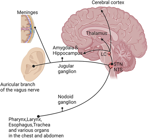
The vagus nerve (VN), constituting the tenth pair of cranial nerves, is the longest and most extensively distributed among the cerebral nerves. It comprises 80% sensory nerve fibers (afferent nerves) and 20% motor nerve fibers (efferent nerves). The efferent nerves regulate organs located below the neck, including the heart, lungs, and gastrointestinal tract. Afferent nerves project to various cortical and subcortical brain structures such as the hippocampus, thalamus, hypothalamus, insula, prefrontal cortex, and motor cortex. Originating in the medulla oblongata, the VN has four primary nuclei: Dorsal Motor Nucleus (DMN), Nucleus Ambiguous (NA), Spinal Trigeminal Nucleus (STN), and Nucleus of the Solitary Tract (NTS). Sensory nerve fibers of the VN extend into the brainstem, predominantly terminating in the NTS,Citation23 which serves as an integration center for sensory information. Subsequently, these fibers project directly to the dendrites of locus coeruleus (LC) norepinephrine-ergic neurons. Both the NTS and LC project to various brain regions, including the thalamus, amygdala, medial septum, hippocampal formation, and cerebral cortex (). Some of these regions play a significant role in memory storage. Specifically, VNS triggers the release of norepinephrine (NE) through the LC, NTS, and other cognitive-related structures like the thalamus, amygdala, hippocampus, and cerebral cortex, thereby enhancing memory.Citation23–28 Functional magnetic resonance imaging has revealed that VNS induces local blood flow changes in the brainstem, thalamus, hypothalamus, amygdala, and hippocampus. The observed changes indirectly imply that VNS may improve memory function by activating the aforementioned brain regions associated with cognition.Citation10 Moreover, animal experiments have demonstrated that VNS stimulation enhances spatial memory and fear memory in rats, with a correlation observed with the release of NE during stimulation.Citation29 Consequently, intermittent chronic electrical stimulation of VN afferent fibers activates the NTS-LC-NE pathway, ultimately improving cognitive function. These conclusions are predominantly affirmed in studies related to epilepsy, Alzheimer’s disease, depression, and cognitive function.Citation9,Citation10,Citation21,Citation23–25,Citation28,Citation30
Figure 1 Schematic diagram of vagus nerve afferent fibers. Thalamus; Amygdala; Hippocampus; Cerebral cortex; locus coeruleus (LC); Nodoid ganglion; Jugular ganglion; Pharynx, Larynx, Esophagus, Trachea and various organs in the chest and abdomen; Meninges; Auricular branch of the vagus nerve; nucleus of the solitary tract (NTS); spinal trigeminal nucleus (STN). Created with BioRender.com.
Cholinergic Anti-Inflammatory Pathway
Increasing evidence suggests a connection between inflammation and the onset of cognitive dysfunction, implicating various neuroinflammatory factors. Consequently, there is a belief that VNS may enhance cognitive function by triggering anti-inflammatory pathways. Findings from an animal experiment investigating postoperative cognitive dysfunction in aged rats support this notion, as VNS was shown to ameliorate cognitive dysfunction by suppressing the expression of postoperative inflammatory cytokines.Citation31 This provides evidence supporting the idea that VNS can enhance cognitive function by mitigating inflammatory responses. Furthermore, it has been demonstrated that VNS activates the cholinergic anti-inflammatory pathway (CAP).Citation8,Citation32 Specifically, vagal efferent fibers facilitate the release of acetylcholine (ACh), which binds to the α7 nicotinic acetylcholine receptor (α7nAChR)Citation33 on the surface of immune cells like macrophages and microglia. This process activates the intracellular JAK2/STAT3 signaling pathway, inhibiting the release of cytokines (inflammatory factors), such as tumor necrosis factor (TNF) α and interleukin-6 (IL-6), thus alleviating the inflammatory response.Citation34,Citation35 Moreover Wang et al indicated that by regulating the CAP improve cognitive impairment in CCH mice.Citation36 By activating CAP, VNS curtails the release of inflammation-associated cytokines, safeguarding and enhancing cognitive function. The anti-inflammatory mechanism of VNS positions it as a potential supplementary therapy for cognitive dysfunction.
Improvement of VCI by VNS
The precise mechanism through which VNS enhances VCI remains incompletely understood. Nevertheless, past research has indicated a connection between cerebrospinal fluid circulation (CSF) and cognitive function.Citation37,Citation38 Cheng et al observed that iVNS increased CSF circulation.Citation39 Additionally, animal experiments conducted by Choi et al demonstrated that taVNS promotes CSF circulationCitation40 and repetitive stimulation in animal models enhances cognitive function. In addition, Liu et al found that VNS improved cognitive function in cerebral ischemia-reperfusion rats, and the main mechanism was related to NE. Because the experiment mainly investigated the improvement of cognitive function within 30 minutes after ischemic stroke, it could not be directly equated with VCI.Citation29 Zhao et alCitation41 found that tcVNS attenuates cerebral ischemic injury and reduces apoptosis by promoting microglial neuron M2 polarization. We elaborated in the potential mechanism of VCI above that blocking microglia activation improves cognitive dysfunction, ie, suggesting that nVNS may play a neuroprotective role by inhibiting inflammatory responses. It is important to note that there is currently a lack of clinical trial data related to the use of VNS for improving VCI.
Summary
The initiation of neuroinflammation due to chronic cerebral hypoperfusion-induced ischemia and hypoxia is implicated in the progression of VCI. This review summarized the pathological mechanisms of VCI and the mechanisms by which VNS improves cognitive function, and provided rationale and ideas for the use of VNS in the treatment of VCI. However, the lack of extensive long-term, large-scale clinical trials poses a challenge in confirming its effectiveness, durability, and safety. This underscores the need for further validation through randomized controlled trials with an adequate sample size.
Abbreviations
VCI, Vascular Cognitive Impairment; VaD, Vascular Dementia; AD, Alzheimer’s disease; CCH, Chronic Cerebral Hypoperfusion; VNS, Vagus Nerve Stimulation; iVNS, invasive Vagal Nerve Stimulation; tVNS, transcutaneous Vagal Nerve Stimulation; taVNS, transcutaneous auricular Vagus Nerve Stimulation; tcVNS, transcutaneous cervical Vagal Nerve Stimulation; WM, White Matter; WMLs, White Matter Lesions; LC, Locus Coeruleus; NTS, Nucleus of the Solitary Tract; NE, Norepinephrine; CAP, Cholinergic Anti-Inflammatory Pathway; VN, Vagal Nerve; DMN, Dorsal Motor Nucleus; NA, Nucleus Ambiguous; STN, Spinal Trigeminal Nucleus; Ach, Acetylcholine; α7nAChR, α7 nicotine acetylcholine receptor α7; TNF, Tumor Necrosis Factor; IL-6, Interleukin-6; CSF, Cerebrospinal Fluid.
Disclosure
The authors declare that they have no competing interests in this work.
Additional information
Funding
References
- van der Flier WM, Skoog I, Schneider JA, et al. Vascular cognitive impairment. Nat Rev Dis Primers. 2018;4(1):18003. doi:10.1038/nrdp.2018.3
- Iadecola C, Duering M, Hachinski V, et al. Vascular cognitive impairment and dementia: JACC Scientific Expert Panel. J Am Coll Cardiol. 2019;73(25):3326–3344. doi:10.1016/j.jacc.2019.04.034
- Cognitive Disorders Professional Committee, Neurology Branch, Chinese Medical Doctor Association, Chinese Guidelines for Diagnosis and Treatment of vascular cognitive Disorders. Guidelines for diagnosis and treatment of vascular cognitive Impairment in China in 2019. Chin J Med. 2019;99(35):2737–2744. doi:10.3760/cma.j.issn.0376-2491.2019.35.005
- O’Brien JT, Thomas A. Vascular dementia. Lancet. 2015;386(10004):1698–1706. doi:10.1016/S0140-6736(15)00463-8
- Lu L, Yu X, Li LJ, et al. Diagnosis and treatment of vascular cognitive impairment (2020 edition). Chin Clin Educ Gen Pract. 2021;19(03):197–199.
- Hu Y, Zhang M, Liu B, et al. Honokiol prevents chronic cerebral hypoperfusion induced astrocyte A1 polarization to alleviate neurotoxicity by targeting SIRT3-STAT3 axis. Free Radic Biol Med. 2023;202:62–75. doi:10.1016/j.freeradbiomed.2023.03.018
- Vonck K, Raedt R, Naulaerts J, et al. Vagus nerve stimulation…25 years later! What do we know about the effects on cognition? Neurosci Biobehav Rev. 2014;45:63–71. doi:10.1016/j.neubiorev.2014.05.005
- Johnson RL, Wilson CG. A review of vagus nerve stimulation as a therapeutic intervention. J Inflamm Res. 2018;11:203–213. doi:10.2147/JIR.S163248
- Fan S, Wang K, Meng F. Research progress on the effects of vagus nerve stimulation on cognition. Chin J Neurosurg. 2019;35(8):855–858. doi:10.3760/cma.j.issn.1001-2346.2019.08.022
- Broncel A, Bocian R, Kłos-Wojtczak P, Kulbat-Warycha K, Konopacki J. Vagal nerve stimulation as a promising tool in the improvement of cognitive disorders. Brain Res Bull. 2020;155:37–47. doi:10.1016/j.brainresbull.2019.11.011
- Yu W, Li Y, Hu J, Wu J, Huang Y. A study on the pathogenesis of vascular cognitive impairment and dementia: the chronic cerebral hypoperfusion hypothesis. J Clin Med. 2022;11(16):4742. doi:10.3390/jcm11164742
- Inoue Y, Shue F, Bu G, Kanekiyo T. Pathophysiology and probable etiology of cerebral small vessel disease in vascular dementia and Alzheimer’s disease. Mol Neurodegener. 2023;18(1):46. doi:10.1186/s13024-023-00640-5
- Rajeev V, Chai YL, Poh L, et al. Chronic cerebral hypoperfusion: a critical feature in unravelling the etiology of vascular cognitive impairment. Acta Neuropathol Commun. 2023;11(1):93. doi:10.1186/s40478-023-01590-1
- Poh L, Sim WL, Jo DG, et al. The role of inflammasomes in vascular cognitive impairment. Mol Neurodegener. 2022;17(1):4. doi:10.1186/s13024-021-00506-8
- Tian Z, Ji X, Liu J. Neuroinflammation in vascular cognitive impairment and dementia: current evidence, advances, and prospects. Int J Mol Sci. 2022;23(11):6224. doi:10.3390/ijms23116224
- Woodburn SC, Bollinger JL, Wohleb ES. The semantics of microglia activation: neuroinflammation, homeostasis, and stress. J Neuroinflammation. 2021;18(1):258. doi:10.1186/s12974-021-02309-6
- Estes ML, McAllister AK. Alterations in immune cells and mediators in the brain: it’s not always neuroinflammation! Brain Pathol. 2014;24(6):623–630. doi:10.1111/bpa.12198
- Belarbi K, Cuvelier E, Bonte MA, et al. Glycosphingolipids and neuroinflammation in Parkinson’s disease. Mol Neurodegener. 2020;15(1):59. doi:10.1186/s13024-020-00408-1
- DiSabato DJ, Quan N, Godbout JP. Neuroinflammation: the devil is in the details. J Neurochem. 2016;139(Suppl 2):136–153. doi:10.1111/jnc.13607
- Lyman M, Lloyd DG, Ji X, Vizcaychipi MP, Ma D. Neuroinflammation: the role and consequences. Neurosci Res. 2014;79:1–12. doi:10.1016/j.neures.2013.10.004
- Liu L, Jiang ZL. Research progress of vagus nerve electrical stimulation in memory disorders. Chin J Rehabil Med. 2021;36(11):1460–1464. doi:10.3969/j.issn.1001-1242.2021.11.026
- Ridgewell C, Heaton KJ, Hildebrandt A, Couse J, Leeder T, Neumeier WH. The effects of transcutaneous auricular vagal nerve stimulation on cognition in healthy individuals: a meta-analysis. Neuropsychology. 2021;35(4):352–365. doi:10.1037/neu0000735
- Li L, Wang D, Pan H, et al. Non-invasive vagus nerve stimulation in cerebral stroke: current status and future perspectives. Front Neurosci. 2022;16:820665. doi:10.3389/fnins.2022.820665
- Colzato L, Beste C. A literature review on the neurophysiological underpinnings and cognitive effects of transcutaneous vagus nerve stimulation: challenges and future directions. J Neurophysiol. 2020;123(5):1739–1755. doi:10.1152/jn.00057.2020
- Li ZD, Qiu HJ, Wang XQ, Zhang CC, Zhang YJ. Transcutaneous auricular vagus nerve stimulation in poststroke cognitive impairment: protocol for a randomised controlled trial. BMJ Open. 2022;12(10):e063803. doi:10.1136/bmjopen-2022-063803
- Dolphin H, Dukelow T, Finucane C, Commins S, McElwaine P, Kennelly SP. “The Wandering Nerve Linking Heart and Mind” - the complementary role of transcutaneous vagus nerve stimulation in modulating neuro-cardiovascular and cognitive performance. Front Neurosci. 2022;16:897303. doi:10.3389/fnins.2022.897303
- Sommer A, Fischer R, Borges U, Laborde S, Achtzehn S, Liepelt R. The effect of transcutaneous auricular vagus nerve stimulation (taVNS) on cognitive control in multitasking. Neuropsychologia. 2023;187:108614. doi:10.1016/j.neuropsychologia.2023.108614
- Naparstek S, Yeh AK, Mills-Finnerty C. Transcutaneous Vagus Nerve Stimulation (tVNS) applications in cognitive aging: a review and commentary. Front Aging Neurosci. 2023;15:1145207. doi:10.3389/fnagi.2023.1145207
- Liu AF, Zhao FB, Wang J, et al. Effects of vagus nerve stimulation on cognitive functioning in rats with cerebral ischemia reperfusion. J Transl Med. 2016;14:101. doi:10.1186/s12967-016-0858-0
- Yap JYY, Keatch C, Lambert E, Woods W, Stoddart PR, Kameneva T. Critical review of transcutaneous vagus nerve stimulation: challenges for translation to clinical practice. Front Neurosci. 2020;14:284. doi:10.3389/fnins.2020.00284
- Cai L, Lu K, Chen X, Huang JY, Zhang BP, Zhang H. Auricular vagus nerve stimulation protects against postoperative cognitive dysfunction by attenuating neuroinflammation and neurodegeneration in aged rats. Neurosci Lett. 2019;703:104–110. doi:10.1016/j.neulet.2019.03.034
- Alen NV. The cholinergic anti-inflammatory pathway in humans: state-of-The-art review and future directions. Neurosci Biobehav Rev. 2022;136:104622. doi:10.1016/j.neubiorev.2022.104622
- Li J, Zhang Q, Li S, et al. α7nAchR mediates transcutaneous auricular vagus nerve stimulation-induced neuroprotection in a rat model of ischemic stroke by enhancing axonal plasticity. Neurosci Lett. 2020;730:135031. doi:10.1016/j.neulet.2020.135031
- Huffman WJ, Subramaniyan S, Rodriguiz RM, Wetsel WC, Grill WM, Terrando N. Modulation of neuroinflammation and memory dysfunction using percutaneous vagus nerve stimulation in mice. Brain Stimul. 2019;12(1):19–29. doi:10.1016/j.brs.2018.10.005
- Kalkman HO, Feuerbach D. Modulatory effects of α7 nAChRs on the immune system and its relevance for CNS disorders. Cell Mol Life Sci. 2016;73(13):2511–2530. doi:10.1007/s00018-016-2175-4
- Wang Z, Han B, Qi J, Cao X, Gu H, Sun J. Chuanzhitongluo capsule improves cognitive impairment in mice with chronic cerebral hypoperfusion via the cholinergic anti-inflammatory pathway. Exp Gerontol. 2024;189:112407. doi:10.1016/j.exger.2024.112407
- Li M, Kitamura A, Beverley J, et al. Impaired glymphatic function and pulsation alterations in a mouse model of vascular cognitive impairment. Front Aging Neurosci. 2022;13:788519. doi:10.3389/fnagi.2021.788519
- Attier-Zmudka J, Sérot JM, Valluy J, et al. Decreased cerebrospinal fluid flow is associated with cognitive deficit in elderly patients. Front Aging Neurosci. 2019;11:87. doi:10.3389/fnagi.2019.00087
- Cheng KP, Brodnick SK, Blanz SL, et al. Clinically-derived vagus nerve stimulation enhances cerebrospinal fluid penetrance. Brain Stimul. 2020;13(4):1024–1030. doi:10.1016/j.brs.2020.03.012
- Choi S, Jang DC, Chung G, Kim SK. Transcutaneous auricular vagus nerve stimulation enhances cerebrospinal fluid circulation and restores cognitive function in the rodent model of vascular cognitive impairment. Cells. 2022;11(19):3019. doi:10.3390/cells11193019
- Zhao XP, Zhao Y, Qin XY, Wan LY, Fan XX. Non-invasive vagus nerve stimulation protects against cerebral ischemia/reperfusion injury and promotes microglial M2 polarization via interleukin-17A inhibition. J Mol Neurosci. 2019;67(2):217–226. doi:10.1007/s12031-018-1227-7