
 ?Mathematical formulae have been encoded as MathML and are displayed in this HTML version using MathJax in order to improve their display. Uncheck the box to turn MathJax off. This feature requires Javascript. Click on a formula to zoom.
Abstract
Bovine serum albumin (BSA) is highly water soluble and binds drugs or inorganic substances noncovalently for their effective delivery to various affected areas of the body. Due to the well-defined structure of the protein, containing charged amino acids, albumin nanoparticles (NPs) may allow electrostatic adsorption of negatively or positively charged molecules, such that substantial amounts of drug can be incorporated within the particle, due to different albumin-binding sites. During the synthesis procedure, pH changes significantly. This variation modifies the net charge on the surface of the protein, varying the size and behavior of NPs as the drug delivery system. In this study, the synthesis of BSA NPs, by a desolvation process, was studied with salicylic acid (SA) as the active agent. SA and salicylates are components of various plants and have been used for medication with anti-inflammatory, antibacterial, and antifungal properties. However, when administered orally to adults (usual dose provided by the manufacturer), there is 50% decomposition of salicylates. Thus, there has been a search for some time to develop new systems to improve the bioavailability of SA and salicylates in the human body. Taking this into account, during synthesis, the pH was varied (5.4, 7.4, and 9) to evaluate its influence on the size and release of SA of the formed NPs. The samples were analyzed using field-emission scanning electron microscopy, transmission electron microscopy, Fourier transform infrared, zeta potential, and dynamic light scattering. Through fluorescence, it was possible to analyze the release of SA in vitro in phosphate-buffered saline solution. The results of chemical morphology characterization and in vitro release studies indicated the potential use of these NPs as drug carriers in biological systems requiring a fast release of SA.
Introduction
Nanoparticle (NP) and microparticle carriers present an important drug delivery potential for the administration of therapeutic drugs. Systems with controlled release offer numerous advantages over conventional dosage forms, since they have better efficiency and low toxicity and provide convenience to the patient.Citation1 The use of nanomaterials as pharmaceutical drug carriers to increase antitumor efficacy has been studied for more than 30 years.Citation2 Initial clinical studies with liposomal nanocarriers were conducted in 1970.Citation3 Currently, the use of nanoscale materials for drug delivery and diagnostics is in the forefront of medicine, since the encapsulation of a drug into NPs significantly improves its release profile in cells or tissues. By a proper synthesis, these nanomaterials can interact selectively with particular types of cells, passing through physiological barriers and penetrating deep into the tumor sites.Citation4,Citation5
In general, the use of nanocarriers can protect drugs from degradation and improve their function by facilitating the absorption and diffusion across the epithelium, modifying the distribution and pharmacokinetic profile of drugs in tissues and/or improving penetration and intracellular distribution.Citation1,Citation4–Citation6 Several drug nanocarrier systems have been investigated in cancer treatment to minimize side effects and improve antitumor efficacy.
The performance of NPs depends on dimensionality, surface charge, surface modification, and hydrophobicity, which can affect the reactivity and physical and chemical properties of the material.Citation1 Nanocarriers with optimized physical, chemical, and biological properties can penetrate the cells, where other larger materials would not be taken up or eliminated from the body. Smaller sizes (50–300 nm) enable interaction with biomolecules in the cell or on the cell surface, and thus nanocarriers can potentially affect cellular responses.Citation7
Systems based on proteins have been studied for medicinal drug delivery, nutrient bioactive peptides, and probiotic organisms.Citation7 Proteins are interesting materials since they possess the advantages of synthetic polymers, high adsorption capacity, low toxicity, biodegradability, and non-immunogenicity.
Human serum albumin (HSA) has a long history of pharmaceutical applications as a biodegradable carrier for the delivery of drugs. It is the most abundant protein in blood plasma, representing 52%–62% of total plasma proteins.Citation8 It is a globular protein with dimensions of 4 × 4 × 14 nm spheroids, containing ~585 amino acids with a molecular weight of ~66,500 Da. The isoelectric point (IEP) of albumin is 4.5–5.0 and its surface is not charged. The main physiological function of albumin is to maintain the osmotic pressure and pH of the blood and still play an important role in the transport of a variety of endogenous and exogenous compounds, including fatty acids, metals, amino acids, steroids, and therapeutic drugs.Citation9,Citation10 Among other physiological functions, HSA controls the transport of drugs and nutrients through the human body and is also responsible for the effective deposition of drugs. It is easily available, stable, and of low cost. Historically, albumin has been extensively used as a biodegradable carrier of anticancer drugs due to its excellent biocompatibility and high stability in the blood and can accumulate in malignant or inflamed tissues.Citation6
Bovine serum albumin (BSA) is highly water soluble and binds drugs and inorganic substances noncovalently. In addition, its structure is homologous to the three-dimensional structure of HSA. The main difference lies in the number of tryptophans (Trps). BSA has two Trps, while HSA has only one. This difference is useful in the context of its study by spectrofluorimetry, since this amino acid is the main one responsible for the intrinsic fluorescence of proteins.Citation11–Citation13
Microparticles and NPs of albumin can be obtained by the precipitation method in organic solvents, followed by a process of cross-linking with glutaraldehyde molecules (desolvation method).Citation14
Transportation systems based on albumin NPs represent an important strategy since significant amounts of drug can be incorporated within the particle, depending on the drug-binding sites.Citation12–Citation17 Due to the well-defined structure of the protein containing charged amino acids, albumin NPs could favor the electrostatic adsorption of negatively or positively charged molecules, inside or on the surface, and the presence of hydrophobic cavities may facilitate the incorporation of water-insoluble drugs.Citation18–Citation20 BSA particles are small compared to microparticles and generally have more controlled properties for drug delivery when compared to liposomal nanocarriers.Citation6,Citation21,Citation22
The first commercial product based on albumin NPs used in oncology was a 130 nm particle with bound paclitaxel.Citation23,Citation24 Currently, some studies have reported that particles of 20–200 nm are effective carriers for hydrophobic compounds.Citation14
Salicylic acid (SA) and salicylates are components of various plants and have been used for medications with anti-inflammatory, antibacterial, and antifungal properties. Among its properties, SA has been considered a promising drug for the prevention of cardiovascular disease and cancer.Citation25–Citation27 SA and salicylates are the active metabolite of aspirin (acetyl SA) and have been used since the fifth century. However, these salicylic-derived drugs have a 50% bioavailability if orally administered, as they lose this activity following the initial step of deacetylation to SA.Citation28–Citation30
Recent reports in the specialized literature demonstrated that SA may be encapsulated or entrapped in different polymer matrices, and its release can be controlled by degradation and subsequent reabsorption of the polymer matrix of the sample by the human body.Citation31–Citation38 Thus, sustained and slow releasing of SA may be more appropriate for reducing the risk of cancer and cardiovascular diseases. The rapid release of SA may be desired for antimicrobial and anti-inflammatory applications, such as treatment of infections. Whereas release is totally dependent on the encapsulating matrix, the bioaccessibility of SA can be modified in the encapsulation process since it is related to and dependent on the chemical interaction between SA and the matrix.Citation36,Citation39
In this context, it is important to study and optimize these nanocarrier systems to better understand how to control drug release and its dependence on functional and biological aspects. As the interaction between the drug and albumin is limited by the surface aspects of these NPs, such as charge, binding sites, and amino acids, it is important to have a high ratio of drug to albumin, which may increase the drug concentration in target organs or tissues by decreasing the dose of drugs and toxic side effects.Citation40 Furthermore, albumin has an unusual ability to bind various compounds, making it an excellent candidate for nanotechnology applications.
For the abovementioned reasons, the study of the synthesis and in vitro release of BSA NPs containing SA as the active agent was conducted. BSA NPs were synthesized by the desolvation method, where the size and surface charge of the NPs could be controlled by altering the pH in synthetic process. Therefore, the pH effect was evaluated on NP synthesis and the in vitro release of SA. The NPs were characterized by ultraviolet (UV)–visible, fluorescence and Fourier transform infrared (FTIR) spectroscopy. The analysis of zeta potential and dynamic light scattering (DLS) was used to investigate the physiochemical properties, namely size and charge for the SA–BSA NPs synthesized at different pHs. The structural surface morphology of BSA NPs and SA–BSA NPs was examined by field-emission scanning electron microscopy (FE-SEM) and transmission electron microscopy (TEM) showing a spherical smooth surface and SA entrapped in BSA NPs. The amount of unloaded SA and the release experiments were evaluated by fluorescence spectroscopy. The chemical and morphological characterization results and the in vitro release studies indicated the potential use of these NPs as drug carriers in biological systems requiring a fast release of SA.
Materials and methods
Reagent and chemicals
BSA (lyophilized powder, 66,000 kDa), 8% aqueous glutaraldehyde and SA were purchased from Sigma-Aldrich. Absolute ethanol was obtained from Synth. All chemicals were of analytical grade and were used without any further purification. All aqueous solutions were prepared in Milli-Q water. The pH of aqueous solutions was adjusted with 0.1 M HCl or 0.1 M NaOH using an Orion PerpHecT pH meter.
Preparation of BSA NPs and SA–BSA NPs
BSA NPs and SA–BSA NPs were prepared by the desolvation process.Citation34,Citation35
BSA NPs
BSA (200 mg) in 2 mL Milli-Q water (approximately pH 7.4) was stirred at 500 rpm at room temperature (25°C) for 10 minutes. The dissolved protein was transformed into NPs by the continuous addition of ethanol (1 mL/min), forming a turbid suspension. After 5 minutes, 0.16 mL of 8% (v/v) aqueous glutaraldehyde was added to cross-link the desolvated BSA NPs, and the reaction was kept under constant magnetic stirring for 18 hours. Next, the NP suspension was purified by three cycles of centrifugation at 12,000 rpm (Hermle Labortechnik GmbH) for 20 minutes to remove non-desolvated BSA, glutaraldehyde and ethanol. For each centrifugation step, NPs were redispersed in the same volume of deionized water (10 mL) by using an ultrasonication bath for 5 minutes. The obtained samples were then dried with nitrogen obtaining a nanosized powder.
SA–BSA NPs
In this procedure, 20 mg SA was dissolved in 2 mL Milli-Q water with the pH adjusted (5.4; 7.4; or 9), and 200 mg BSA were then added, followed by stirring for 10 minutes. Subsequently, the desolvating agent (absolute ethanol, 8 mL) was continuously added dropwise (1 mL/min) in solution with constant magnetic stirring at 500 rpm, resulting in the formation of a spontaneous opalescent suspension. After the addition of ethanol, 0.16 mL of 8% (v/v) aqueous glutaraldehyde was added to cross-link the desolvated BSA NPs, and the reaction was kept under constant magnetic stirring (500 rpm) for 18 hours at room temperature.
The resulting colloidal suspension was centrifuged at 12,000 rpm (Hermle Labortechnik GmbH) for 20 minutes. The supernatant was removed to obtain the particles. The samples obtained were purified by three cycles of centrifugation at 12,000 rpm for 20 minutes to remove non-desolvated BSA, free drug, glutaraldehyde, and ethanol.
For each centrifugation step, NP samples were redispersed in the small volume of deionized water (10 mL) by using an ultrasonication bath for 5 minutes. The samples were dried by solvent evaporation, resulting in an NP powder.
Synthesis yield and entrapment efficiency (EE%)
To determine the percent yield of synthesis, the NP powder sample obtained was weighed, and the percent yield was calculated using the following Equationequation 1(1) :
(1)
The amount of drug adsorbed or encapsulated in the albumin NPs was determined from the amount of free SA (weight [W]) in the supernatant after the centrifugation process, determined by fluorescence spectroscopy.
A standard calibration curve of peak maximum of fluorescence versus concentration, using known concentrations of SA in water, was plotted to determine free SA. The amount of SA loaded was determined using Equationequation 2(2) :
(2)
“In vitro” drug release experiments
To evaluate the release behavior of SA from the NPs, drug release experiments were performed in vitro. NPs (12 mg) were re-dispersed in 7 mL of phosphate-buffered saline (PBS, pH 7.4) incubated at 37±0.5°C under stirring at 100 rpm. At different times, 0.5 mL of the release medium was removed and another 0.5 mL of fresh PBS was supplied to maintain the total volume of the original solution at 7 mL. The aliquots removed were centrifuged, and the supernatant containing the drug (SA) was analyzed by fluorescence spectroscopy (excitation 330 nm), and all measurements were performed in triplicate. The cumulative drug release was plotted against time.
Characterization techniques
Physical characterization was performed using DLS and zeta potential, which measured size and surface charge of BSA NPs, respectively, using a Zetasizer Nano (Malvern, ZS 10, Malvern, UK) ZEN3601, with He–Ne laser, 633 nm. For measurements, BSA NP powder was suspended in absolute ethanol and Milli-Q water. Both dispersions were sonicated for 10 minutes to obtain uniformly dispersed NPs. The measurements were performed in triplicate at room temperature.
The structural morphology of BSA NPs was determined by FE-SEM (JSM-7500F; JEOL, Tokyo, Japan) and TEM (CM 200 Philips; Eindhoven, the Netherlands). For SEM measurements, NP solutions in ethanol (20 µL) were dripped and converted into powder on the surface of silicon substrates. Prior to analysis, samples were gold coated to make them electrically conductive and suitable for SEM.
For TEM analysis, 20–30 µL of the NP solutions in ethanol was dripped on a 200-mesh copper grid coated with carbon. The copper grid was allowed to dry for 2 hours at room temperature before observation.
UV–visible spectroscopy was used to analyze the NPs in aqueous solution (0.05 mg/mL), and the maximum absorption peak was measured by scanning 190–800 nm with a quartz cuvette of 1 cm path length, using a Shimadzu UV–visible spectrophotometer (UVmini-1240; Shimadzu, Kyoto, Japan).
Fluorescence spectroscopy was used to determine the presence and amount of free drug in solution to evaluate drug release. The fluorescence spectra were obtained using a microplate spectrofluorometer (Synergy H1 Hybrid reader; BioTek, Winooski, VT, USA) at room temperature, with excitation at 330 nm and emission in the range of 350–450 nm and with a fixed slit width of 5 nm. A standard calibration curve, fluorescence intensity at 405 nm versus concentration, was plotted to correlate fluorescence and concentration of free SA. The molecular structure and conformational changes in BSA due to interaction with SA were analyzed by FTIR spectroscopy. These measurements were performed using a Bruker Vertex 70 (Bruker; Ettlingen, Germany) in the region between 4000 and 400 cm−1, at room temperature in attenuated total reflectance (ATR) mode.Citation19
Results and discussion
As discussed earlier, protein NPs have recently been the focus of research involving the interaction between albumin and other hydrophobic drug substances. The drug-binding properties of albumin NPs are clearly important for the understanding of reaction mechanisms, which provide a path for pharmacokinetic and pharmacodynamic mechanisms of these substances in various tissues. Studies conducted by Weber et alCitation41 demonstrated that the process of protein NP formation depends on the amount of added desolvation agent, pH of the starting solution and amount of cross-linking agent (glutaraldehyde). The ethanol amount and pH of the solution directly alter NP size. Changing the amount of ethanol or rate of its addition in the process modifies the solubility of proteins, consequently altering the size of the NPs formed. Studies by Langer et alCitation42 indicated that varying size of the NPs can be attributed mainly to the manual and dropwise addition of ethanol. Burns and ZydneyCitation43 investigated the effect of solution pH and showed that the loading surface has a strong effect on protein adsorption.
Changing pH significantly alters the net charge on the protein surface, since it consists of hydrogens of amino acids that interact with different ions in solution. The IEP of albumin is around 4.9, and at this pH the net charge on the protein surface is zero.Citation44–Citation46 At pH 4.9, there is a lack of electrostatic repulsion, and thus amorphous aggregates are readily formed through nonspecific interactions, mainly hydrophobic in nature.
At other pHs, positive charges may be generated by protonation of primary amino groups (particularly lysine), while negative charges are generated from deprotonation of carboxylic acid groups (glutamate and aspartate).Citation44 Charges are generated on the whole surface of the protein, leading to greater electrostatic repulsion between molecules and decrease in hydrophobic interactions, thereby reducing aggregation and favoring a structural reorganization of the protein. Moreover, they can significantly alter the interaction and/or binding of compounds (drugs).
At pH above the IEP, negative charges and electrostatic forces dominate over hydrophobic interactions.Citation45,Citation46 Another important component in the process of NP formation is the amount of cross-linking agent (glutaraldehyde) added. Glutaraldehyde does not change the size of the NPs but significantly alters the charge on the surface of the NPs obtained.Citation42 Glutaraldehyde is a low-cost, water-soluble bifunctional reagent with high reactivity. The cross-linking of proteins or between protein molecules occurs by nucleophilic attack of the ε-amino groups of lysine and arginine residues in the protein by the two carbonyl groups of glutaraldehyde, forming Schiff bases in solution.Citation47,Citation48 The Schiff bases are unstable under acidic conditions but very stable at basic pH, aiding in the NP formation process. In addition, the lysine residues generally are not involved in the binding site, resulting in moderate preservation of the conformation of the protein and thereby the biological activity of the protein. As the amount of glutaraldehyde is linked to the number of free amino groups on the surface, it was kept constant during the study, since the amount of charge on the surface is associated with the efficacy of the interaction with the drug.Citation48–Citation51
For comparison purposes, the synthesis of drug-free NP albumin was also carried out at pH 7.4, according to the procedure described in the “Materials and methods” section. The protein NPs were obtained by the known process of desolvation. To do that, initially, BSA was placed in aqueous solution with the desired pH. In the case of the synthesis of NPs containing drugs, SA was also added to the albumin solution and allowed to interact using a magnetic stirring for 10 minutes. In this step, the charges formed on the surface of proteins by changing the pH could now interact differently with SA added. Ethanol (8 mL) was then added as desolvation agent at a controlled rate of 1 mL/min, which gradually decreases the solubility of proteins in solution leading to agglomeration. In this step, it was already possible to observe the formation of NPs, where some were partially soluble and stabilized in solution. To obtain NPs stabilized in solution with definite shape, glutaraldehyde was added, acting as a cross-linking agent. In this process, through covalent bonds, cross-linking agent was part of the protein surface NPs. The glutaraldehyde inserted into the protein, during the cross-linking process, has no toxicity, and the obtained NPs are nontoxic.Citation45,Citation47,Citation48 However, the free compound in solution may exhibit toxicity. So, unreacted glutaraldehyde remaining in the solution was removed by centrifugation and purification of the NPs obtained.Citation45,Citation47,Citation48,Citation52
The purified NPs were dried by solvent evaporation under nitrogen flow. The yield of NPs and the entrapment efficiency (EE%) obtained were determined according to EquationEquations 1(1) and Equation2
(2) , respectively, and are shown in .
Table 1 Percent yield and EE% of synthesis of BSA NPs and SA–BSA NPs
The low yield obtained for the reactions at pH 5.4 indicated that charges generated on the protein surface (close to BSA IEP) hampered drug entrapment into NPs.
Physical, chemical, and morphological characterization of NPs
After drying, pure BSA, BSA NPs, and SA–BSA NPs were analyzed by UV–visible absorption and fluorescence spectroscopy. These techniques provided physical characterization of the formation of the synthesized albumin NPs and presence of SA.
The UV–visible spectra of pure BSA, BSA NPs, and SA–BSA NPs were significantly different () taking into account the position of maximum absorption peaks, when a spectral scan was evaluated between 190 and 800 nm. shows the fluorescence spectra, using an excitation wavelength of 280 nm and emission range of 310–450 nm during evaluation.Citation53
Figure 1 UV–visible absorption and fluorescence emission spectra of pure BSA, BSA NPs, SA-BSA NPs and salicylic acid.
Abbreviations: BSA, bovine serum albumin; NPs, nanoparticles; SA, salicylic acid; UV, ultraviolet.
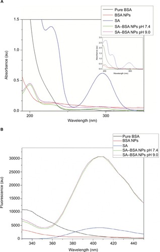
shows that pure BSA had two absorption peaks at 192 and 278 nm. The intense peak at 192 nm is associated with the BSA backbone absorption, while the peak at 278 nm is associated with weak absorption of the aromatic amino acids phenylalanine (Phe), tyrosine (Tyr), and tryptophan (Trp).Citation53,Citation54 SA exhibited a strong absorption bands at 205, 228, and 296 nm. With the formation of BSA NPs, there was a strong decrease in the intensity and a blue shift for the absorption peak of 192 nm, and a slight increase for the peak at 278 nm, revealing that the changes related to the formation of BSA NPs mainly occurred in the BSA backbone bonds. It is noteworthy that the decrease in intensity in the SA may be linked to the fact that for concentration pure SA (0.05 mg/mL) and even SA–BSA NPs at a concentration of 0.05 mg/mL do not have the same amount of SA available. These results indicated that, in the formation of BSA NPs, glutaraldehyde modified mainly the BSA backbone amino acids, maintaining the aromatic amino acids Phe, Tyr, and Trp for drug interaction.Citation53,Citation54
According to chemical analysis, in the process of drug interaction with the NPs, the following can be considered: 1) SA interaction with BSA initially occurs in water at pH adjusted and 2) in the next step, cross-linking with glutaraldehyde occurs.
It is known that the main drug-binding region of BSA is localized in hydrophobic cavities of the IIA and IIIA subdomains, which exhibit similar chemical properties and are called sites I and II. These sites are mainly composed of amino acids to which molecules may bind on hydrophobic or positively charged surfaces. Thus, these two subdomains can specifically interact with negatively charged molecules or delocalized negative charges, such as heterocyclic ligands or carboxylic acids.Citation53,Citation54
Initially, when SA was added to the reaction medium, there was interaction at sites I and II, mainly with Tyr, Trp, and Phe, which absorb in the region of 278 nm.
SA–BSA NPs exhibited significant spectroscopic changes. They showed absorption in the regions of 202, 230, and 295 nm, while absorption at 278 nm decreased substantially, indicating the binding of SA to BSA in the NPs obtained.
As shown in , the fluorescence data corroborated the UV–visible results. It is known that BSA shows an optimal excitation wavelength at 278 nm, with maximum of emission around 350 nm, attributed to the amino acid residues of Tyr, Trp, and Phe. In contrast, some studies indicate that the intrinsic fluorescence can be attributed to Trp in BSA chain when the excitation is between 285 and 290 nm.Citation55 In the BSA chain, there are two different types of Trp (134 and 212), localized in sites I and II, which can interact or bind with ligands. Trp-134 is localized on the protein surface and Trp-212 in the hydrophobic cavity of subdomain II. Thus, for pure BSA, BSA NPs, and SA–BSA NPs,Citation56 the fluorescence wavelength changes were indicative of conformational and chemical changes in the BSA structure.Citation57,Citation58
The intrinsic fluorescence of BSA and SA, when excited at 280 nm, was obtained at 335 and 405 nm, respectively. Thus, according to the fluorescence spectra, the formation of BSA NPs leads to a decrease in the fluorescence intensity at 335 nm with a slight associated blueshift, indicating a conformational modification related to changes in the protein surface chain due to interaction with glutaraldehyde. The addition of SA at the beginning of the synthesis process leads to a strong decrease in the fluorescence intensity around 334 nm and an increase at 408 nm, characteristic emission of SA.Citation11,Citation59–Citation63
The BSA NPs and SA–BSA NPs obtained were analyzed by FTIR spectroscopy. This technique has been used to evaluate the chemical and conformational changes that occur when NPs are formed or when they interact with other compounds through the slight shift in characteristic bands in the spectral regions of amide I and amide II.Citation39 shows the FTIR spectra of pure BSA, BSA NPs, and SA–BSA NPs synthesized at pH 7.4 and 9.0.
Figure 2 Pure BSA, BSA NPs, and SA–BSA NPs FTIR spectra.
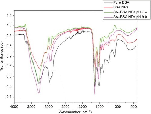
shows the FTIR spectrum analysis, where it was possible to observe the major bands of pure BSA at 3280 cm−1 (amide A, related to N–H stretching), 2970 cm−1 (amide B, N–H stretching of
free ion), 1643 cm−1 (amide I, C=O stretching), 1515 cm−1 (amide II, related to C–N stretching and N–H bending vibrations), 1392 cm−1 (CH2 bending groups) and ~1260 cm−1 (amide III, related to C–N stretching and N–H bending). The most intense bands are associated with the secondary structure and conformation of proteins. The spectra of BSA NPs and SA–BSA NPs exhibited these characteristic bands of the protein and SA structure shifted slightly as shown in .Citation45,Citation64,Citation65
Table 2 Bands of amides A, B, I, II, and III for the samples, according to the FTIR spectra
A small shift, of the absorption bands, was observed when compared to pure BSA with BSA NPs and SA–BSA NPs. The related changes in the amide I, II, and III bands confirm the formation of NP albumin and SA-loaded NP. In addition, some bands showed intensity differences (), since it was possible to note a strong decrease intensities in the amide B (59% decrease) and amide III (40% decrease) bands, indicating changes in the C–N and/or NH bonds, due to interactions of different groups on BSA.
As discussed earlier, the pH change and the glutaraldehyde addition modify the surface charge of NPs, changing the electrostatic potential and colloidal stability of the protein NPs in solution.Citation18,Citation56,Citation65 Thus, the electrostatic potential and the particle size were evaluated by zeta potential and DLS measurements by dispersion of the NPs in water and absolute ethanol. SA–BSA NPs synthesized at pH 7.4 showed respective zeta potential and DLS values of −6.45±1.23 mV and 182.20±12.20 d·nm for NPs dispersed in water and −33.2±1.90 mV and 81.48±0.9 d·nm for NPs dispersed in ethanol. For the SA–BSA NPs synthesized at pH 9, these values were 9.25±1.63 mV and 125.25±1.75 d·nm for NPs in water, and 32.8±4.3 mV and 76.54±0.46 d·nm for NPs in ethanol.Citation18,Citation56
The type of solvent used for dispersion strongly changes the colloidal stability of NPs. The NP solution in absolute ethanol exhibited a greater zeta potential and lower DLS value than the aqueous NP solution. The water pH changes the NP charge surface leading to the agglomeration of BSA NPs. A higher zeta potential value causes the particles to become stable by preventing their aggregation.
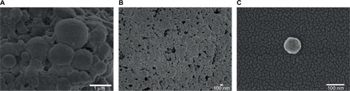
Morphological analysis of BSA NPs and SA–BSA NPs was carried out with FE-SEM, and the images obtained are shown in . Both types of synthesized NPs had a spherical morphology and an average size on a micrometric scale for BSA NPs (600±60 nm) and nanometric average, 110±7 nm and 138±6 nm, for SA–BSA NPs synthesized at pH 7.4 and 9.0, respectively. Furthermore, BSA NPs showed a greater nonuniform distribution, widely differing in size when compared to SA–BSA NPs.
Figure 3 Representative images for (A) BSA NPs (magnification =25,000×), (B) SA–BSA NPs at pH 7.4 (magnification = 30,000×), and (C) SA–BSA NPs at pH 7.4 in detail (magnification =190,000×).
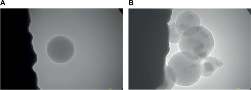
Drug encapsulation by the protein during the synthesis process was also investigated by TEM, and the result obtained is shown in . SA–BSA NPs showed a spherical morphology, as demonstrated by FE-SEM. Furthermore, it was possible to note the presence of structures with irregular surfaces inside these NPs, believed to be the encapsulated SA.
Figure 4 TEM image of (A) BSA NPs and (B) SA–BSA NPs synthesized at pH 7.4. Magnification =26,500×.
“In vitro” release studies
SA release from the protein NPs was analyzed by fluorescence measurement of suspensions of SA–BSA NPs in PBS, simulating a physiological environment at pH 7.4, and the results are shown in .
Figure 5 In vitro SA release from SA BSA NPs in PBS.
Abbreviations: BSA, bovine serum albumin; NPs, nanoparticles; PBS, phosphate-buffered saline; SA, salicylic acid.
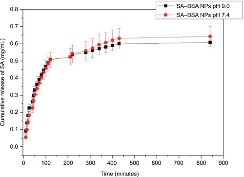
The values of the release rate of NPs synthesized at pH 7.4 and 9.0 are provided in .
Table 3 Release rate of NPs (mg/mL·min)
A similar release behavior was seen for the samples obtained with the two synthesis procedures used, where there was an immediate release of SA, followed by a high release rate up to 120 minutes. Later, the release rate decreased and remained approximately constant from 400 minutes.
Conclusion
SA-loaded BSA NPs were synthesized with the desolvation process using glutaraldehyde cross-linking at different pHs. The pH change suggests that the process is associated with protein surface charges, generated at the beginning of the synthesis, and that this directly influences the entrapment process of SA, since synthesis was ineffective at pH 5.4. However, the pH slightly alters the release of SA from the protein NP. The release of SA occurs immediately, progressing to ~120 minutes. From this time, the release making constant is greatly reduced making constant starting 400 minutes, and these NPs may be applied in biological systems that require a rapid anti-inflammatory response.
Acknowledgments
The authors thank the Brazilian agencies CAPES, FAPESP, and CNPq for financial support, under contracts FAPESP 2014/204710 and CNPq 304810/2010-0. They are also grateful to Professor Marcelo Ornaghi Orlandi of the Department of Physical Chemistry, Instituto de Química, UNESP – Universidade Estadual Paulista, Araraquara, 14800-900, Brazil, for the FE-SEM and TEM analyses and to Dr Elaine Cristina Paris from EMBRAPA Instrumentação – Rua XV de Novembro, 1452, CP 741, CEP 13560-970, São Carlos, SP, Brazil, for zeta potential and DLS measurements. Dr A Leyva helped with English editing of the manuscript. Some parts of the results were presented as a poster in the 2015 MRS Fall Meeting and Exhibit, November 29, 2015, to December 4, 2015, Boston, MA, USA, and XIV Brazil MRS Meeting, September 27, 2015, to October 1, 2015, Rio de Janeiro, Brazil.
Disclosure
The authors report no conflicts of interest in this work.
References
- MehravarRJahanshahiMSaghatoleslamiNFabrication and evaluation of human serum albumin (HSA) nanoparticles for drug delivery applicationInt J Nanosci200983319322
- BrasseurFCouvreurPKanteBActinomycin D absorbed on polymethylcyanoacrylate nanoparticles: increased efficiency against an experimental tumorEur J Cancer19801611144114457227421
- GregoriadisGNeerunjunEDTreatment of tumour bearing mice with liponsome-entrapped actinomycin D prolongs their survivalRes Commun Chem Pathol Pharmacol19751023513621153840
- MohamedFVan der WalleCFEngineering biodegradable polyester particles with specific drug targeting and drug release propertiesJ Pharm Sci2008971718717722085
- RasmussenJWMartinezELoukaPWingettDGZinc oxide nanoparticles for selective destruction of tumor cells and potential for drug delivery applicationsExpert Opin Drug Deliv2010791063107720716019
- ElzoghbyAOSamyWMElgindyNAAlbumin-based nanoparticles as potential controlled release drug delivery systemsJ Control Release2012157216818221839127
- YanaSZhangaHPiaoJStudies on the preparation, characterization and intracellular kinetics of JD27-loaded human serum albumin nanoparticlesProcedia Eng2015102590601
- KopacTBozgeyikKYenerJEffect of pH and temperature on the adsorption of bovine serum albumin onto titanium oxideColloids Surf A20083221928
- BrandesNWelzelPBWernerCKrohLWAdsorption-induced conformational changes of proteins onto ceramic particles: differential scanning calorimetry and FTIR analysisJ Colloid Interface Sci20062991566916500671
- HuangBXKimHYDassCProbing three-dimensional structure of bovine serum albumin by chemical cross-linking and mass spectrometryJ Am Soc Mass Spectrom20041581237124715276171
- TianJLiuJTianXHuZChenXStudy of the interaction of kaempferol with bovine serum albuminJ Mol Struct20046911–3197202
- TrnkováLBoušováIKubíčekVDršataJBinding of naturally occurring hydroxycinnamic acids to bovine serum albuminNat Sci201026563570
- XuHYaoNXuHWangTLiGLiZCharacterization of the interaction between eupatorin and bovine serum albumin by spectroscopic and molecular modeling methodsInt J Mol Sci2013147141851420323839090
- XuRFisherMJulianoRLTargeted albumin-based nanoparticles for delivery of amphipathic drugsBioconjug Chem201122587087821452893
- YangZGongWWangZA novel drug-polyethylene glycol liquid compound method to prepare 10-hydroxycamptothecin loaded human serum albumin nanoparticleInt J Pharm20154901–241242826027489
- PatilGVBiopolymer albumin for diagnosis and in drug deliveryDrug Dev Res2003583219247
- IracheJMMerodioMArnedoACamapaneroMAMirshahiMEspuelasSAlbumin nanoparticles for the intravitreal delivery of anticytomegaloviral drugsMini Rev Med Chem20055329330515777263
- MohantaVMadrasGPatilSLayer-by-layer assembled thin film of albumin nanoparticles for delivery of doxorubicinJ Phys Chem C2012116953335341
- WanXZhengXPangXThe potential use of lapatinib-loaded human serum albumin nanoparticles in the treatment of triple-negative breast cancerInt J Pharm20154841–2162825700543
- SubiaBKunduSCDrug loading and release on tumor cells using silk fibroin-albumin nanoparticles as carriersNanotechnology201324303510323262833
- TuHLuYWuYFabrication of rectorite-contained nanoparticles for drug delivery with a green and one-step synthesis methodInt J Pharm20154931–242643326231105
- WeiYLiLXiYQianSGaoYZhangJSustained release and enhanced bioavailability of injectable scutellarin-loaded bovine serum albumin nanoparticlesInt J Pharm20144761–214214825269007
- NieSXingYKimGJSimonsJWNanotechnology applications in cancerAnnu Rev Biomed Eng2007925728817439359
- KratzFAlbumin as a drug carrier: design of prodrugs, drug conjugates and nanoparticlesJ Control Release2008132317118318582981
- GuptaRADuboisRNColorectal cancer prevention and treatment by inhibition of cyclooxygenase-2Nat Rev Cancer200111112111900248
- PatersonJRLawrenceJRSalicylic acid: a link between aspirin, diet and the prevention of colorectal cancerQJM200194844544811493722
- AmannRPeskarBAAnti-inflammatory effects of aspirin and sodium salicylateEur J Pharmacol200244711912106797
- ErdmannLMacedoBUhrichKEDegradable poly(anhydride ester) implants: effects of localized salicylic acid release on boneBiomaterials200021242507251211071600
- ErdmannLUhrichKESynthesis and degradation characteristics of salicylic acid-derived poly(anhydride-esters)Biomaterials200021191941194610941915
- LevyGComparative pharmacokinetics of aspirin and acetaminophenArch Intern Med198114132792817469620
- Whitaker-BrothersKUhrichKInvestigation into the erosion mechanism of salicylate-based poly(anhydride-esters)J Biomed Mater Res A200676347047916315189
- TangSZJuneSMHowellBAChaiMSynthesis of salicylate dendritic prodrugsTetrahedron Lett2006474476717675
- JiJHaoSWuDHuangRXuYPreparation, characterization and in vitro release of chitosan nanoparticles loaded with gentamicin and salicylic acidCarbohydr Polym2011854803808
- MengMFengYGuanWLiuYXiYYanYSelective separation of salicylic acid from aqueous solutions using molecularly imprinted nano-polymer on wollastonite synthesized by oil-in-water microemulsion methodJ Ind Eng Chem201420639753983
- NowatzkiPJKoepselRRStoodleyPSalicylic acid-releasing polyurethane acrylate polymers as anti-biofilm urological catheter coatingsActa Biomater2012851869188022342353
- ChandorkarYBhagatRKMadrasGBasuBCross-linked, biodegradable, cytocompatible salicylic acid based polyesters for localized, sustained delivery of salicylic acid: an in vitro studyBiomacromolecules201415386387524517727
- DasguptaQChatterjeeKMadrasGCombinatorial approach to develop tailored biodegradable poly(xylitol dicarboxylate) polyestersBiomacromolecules201415114302431325322446
- DasguptaQChatterjeeKMadrasGControlled release of salicylic acid from biodegradable cross-linked polyestersMol Pharm20151293479348926284981
- RogersMAYanYFBen-ElazarKSalicylic acid (SA) bioaccessibility from SA-based poly (anhydride-ester)Biomacromolecules20141593406341125082798
- XieLTongWYuDXuJLiJGaoCBovine serum albumin nanoparticles modified with multilayers and aptamers for pH-responsive and targeted anti-cancer drug deliveryJ Mater Chem2012221360536060
- WeberCKreuterJLangerKDesolvation process and surface characteristics of HSA-nanoparticlesInt J Pharm2000196219720010699717
- LangerKBalthasarSVogelVDinauerNVon BriesenHSchubertDOptimization of the preparation process for human serum albumin (HSA) nanoparticlesInt J Pharm20032571–216918012711172
- BurnsDBZydneyALEffect of solution pH on protein transport through ultrafiltration membranesBiotechnol Bioeng1999641273710397836
- EiseleKGropeanuRAZehendnerCMFine-tuning DNA/albumin polyelectrolyte interactions to produce the efficient transfection agent cBSA-147Biomaterials201031338789880120817248
- RohiwalSSSatvekarRKTiwariAPRautAVKumbharSGPawarSHInvestigating the influence of effective parameters on molecular characteristics of bovine serum albumin nanoparticlesAppl Surf Sci2015334157164
- RohiwalSSPawarSHSynthesis and characterization of bovine serum albumin nanoparticles as a drug delivery vehicleInt J Pharm Bio Sci2014545157
- BhushanBDubeyPKumarSUSachdevAMataiIGopinathPBionanotherapeutics: niclosamide encapsulated albumin nanoparticles as a novel drug delivery system for cancer therapyRSC Adv201551207812086
- MigneaultIDartiguenaveCBertrandMJWaldronKCGlutaraldehyde: behavior in aqueous solution, reaction with proteins, and application to enzyme crosslinkingBiotechniques2004375798802
- OkudaKUrabeIYamadaYOkadaHReaction of glutaraldehyde with amino and thiol compoundsJ Ferment Bioeng1991712100105
- WeetallHHImmobilized enzymes: analytical applicationsAnal Chem1974467602A615A
- GuisánJMAldehyde-agarose gels as activated supports for immobilization-stabilization of enzymesEnzyme Microb Technol1988106375382
- BhushanBGopinathPAntioxidant nanozyme: a facile synthesis and evaluation of the reactive oxygen species scavenging potential of nanoceria encapsulated albumin nanoparticlesJ Mater Chem B201532448434852
- TianZYSongLNZhaoYSpectroscopic Study on the Interaction between Naphthalimide-Polyamine Conjugates and Bovine Serum Albumin (BSA)Molecules201520164911652326378511
- NaveenrajSAnandanSBinding of serum albumins with bioactive substances – nanoparticles to drugsJ Photochem Photobiol C2013145371
- NiYSuSKokotSSpectrofluorimetric studies on the binding of salicylic acid to bovine serum albumin using warfarin and ibuprofen as site markers with the aid of parallel factor analysisAnal Chim Acta2006580220621517723775
- RohiwalSSTiwariAPVermaGPawarSHPreparation and evaluation of bovine serum albumin nanoparticles for ex vivo colloidal stability in biological mediaColloids Surf A Physicochem Eng Asp20154802837
- GentiliPLOrticaFFavaroGStatic and dynamic interaction of a naturally occurring photochromic molecule with bovine serum albumin studied by UV−visible absorption and fluorescence spectroscopyJ Phys Chem B200811251167931680119367911
- JoshiPChakrabortySDeySBinding of chloroquine – conjugated gold nanoparticles with bovine serum albuminJ Colloid Interface Sci2011355240240921216410
- JuPFanHLiuTCuiLAiSProbing the interaction of flower-like CdSe nanostructure particles targeted to bovine serum albumin using spectroscopic techniquesJ Lumin2011131817241730
- LiuXHXiPXChenFJXuZHZengZZSpectroscopic studies on binding of 1-phenyl-3-(coumarin-6-yl)sulfonylurea to bovine serum albuminJ Photochem Photobiol B20089229810218571426
- WangYQTangBPZhangHMZhouQHZhangGCStudies on the interaction between imidacloprid and human serum albumin: spectroscopic approachJ Photochem Photobiol B200994318319019126446
- HuYJLiuYWangJBXiaoXHQuSSStudy of the interaction between monoammonium glycyrrhizinate and bovine serum albuminJ Pharm Biomed Anal200436491591915533690
- GrdadolnikJA FTIR investigation of protein conformationBull Chem Technol Maced2002212334
- KongJYuSFourier transform infrared spectroscopic analysis of protein secondary structuresActa Biochim Biophys Sin200739854955917687489
- LiCZhangDGuoHPreparation and characterization of galactosylated bovine serum albumin nanoparticles for liver-targeted delivery of oridoninInt J Pharm20134817986