
Abstract
Synovial chondromatosis is a rare and benign proliferative disorder of the synovial membrane in joints and bursae. Herein, we present the case of a 34-year-old male with synovial chondromatosis that caused limitation in the elbow joint in terms of mechanical function.
Introduction
Synovial chondromatosis (SC) resulting from the metaplasia of synovial tissue is a rare proliferative disorder with monoarticular involvement that is found in the joints and bursae. Although the etiology is unclear, it is considered as a metaplastic formation. SC can develop secondary to osteoarthritis, although it is generally a primary condition. It is most commonly seen between 30 and 50 years of age, and the prevalence in men is double of that in women. The knee joint is the most frequently involved site, but it may also involve other joints in a lesser degree.Citation1 Pain and progressive limitations of movements occur with effusion and occasional snapping of the joint.Citation2 SC expands the joint and can occur in extra-articular locations, but if it invades beyond the joint capsule, one would suspect a different entity.Citation3
Case report
A 34-year-old male presented to our clinic with pain, mechanical limitation, and swelling in the right elbow, that had become increasingly severe over the previous 3 years. In physical examination, there was limitation in extension by 30° and in flexion by 20° when compared to the contralateral elbow. It was detected that there was a palpable, painful, rigid, immobile mass at the anterior and posterior aspects of the elbow joint.
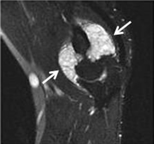
In radiological evaluation, it was seen that there was narrowing in the joint space and cortical irregularity in the anterior region (). On computerized tomography (CT) scan, it was seen that there was soft tissue swelling at the right elbow joint, and cortical erosive changes in the distal humerus (). On magnetic resonance (MR) imaging, hyperintense, well-defined, nodular mass lesions were seen in fat suppression sequences ().
Figure 1 Anterioposterior (A) and lateral (B) radiographs of elbow.
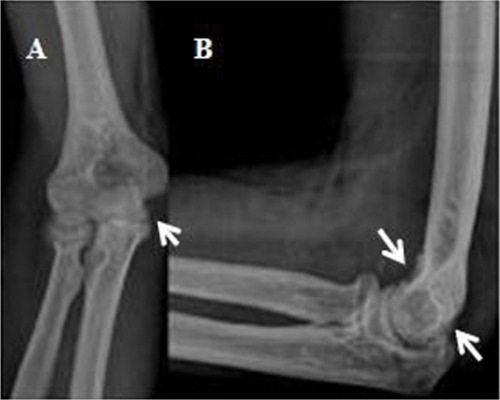
Figure 2 On CT scan, chondral erosions can be seen posterior to the elbow joint in axial sections with arrow.
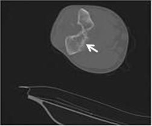
Figure 3 On MR imaging, hyperintense, well-defined, nodular mass lesions can be seen in fat suppression sequences (arrows).
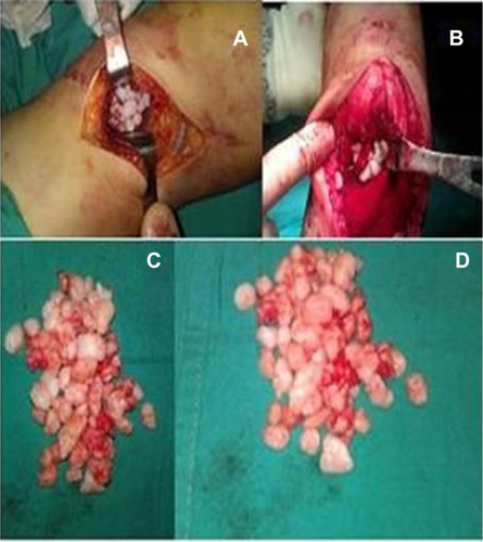
Surgery was scheduled for the patient with an initial diagnosis of SC. First, olecranon fossa was exposed through the lateral and medial tricep muscles following a midline incision at the posterior aspect of the elbow. Numerous chondral loose bodies, which resulted in limitation of extension of the elbow and filled the olecranon fossa leading to enlargement of the joint capsule, were removed after capsulotomy. A second incision was made at the anterior aspect of the elbow for lesion in the anterior region of the elbow. The anterior capsule was exposed by preserving the vascular and neural components. Again, numerous rigid, free chondral loose bodies, which extended to the radius head and a process inside the joint, were removed following capsulotomy (). Both anterior and posterior synovial excisions were made. After excision, the range of motion was assessed intraoperatively. It was noted that preoperative flexion and extension limitations were relieved.
Figure 4 Chondral components removed during (A and B) and after (C and D) surgery can be seen.
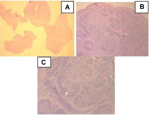
In the histopathological evaluation of the chondral loose bodies that were removed, SC was confirmed by observation of well-defined chondrocytes and mature cartilage tissues with lobulation (). No recurrence was detected during 24-month follow-up.
Figure 5 (A) Lesion consisting of mature, lobulated cartilage tissue of varying sizes (H&e, 10×). (B) Chondrocytes forming lobular structures within well-defined chondroid matrix (H&e, 40×). (C) Chondrocytes displaying polygonal nuclei with clear cytoplasm.
Abbreviation: H&E, hematoxylin and eosin.
Discussion
SC is an extremely rare proliferative disorder resulting from the metaplasia of cartilage tissue, which has a mesenchymal subintimal layer of synovium. The disease is characterized by the formation of multiple intrasynovial and osteochondral cartilaginous nodules. SC is generally associated with monoarticular involvement, while it is more frequently seen in large joints. The etiology is unclear.Citation4,Citation5 It is extremely rare for it to involve the elbow joint.Citation6 In a study by Mueller et al and Kamineni et al, only 20 cases and 12 cases of elbow involvement were reported in recent years, respectively.Citation7,Citation8
The HLA-DR and CD68 gene expression seen in previous studies supports the view that a reactive condition may play a role in the etiopathogenesis of SC; also many other abnormalities have been reported as non-diploid karyotypes, rearrangements, losses, or gains of chromosomes.Citation9,Citation10 However, no cytogenetic analysis was performed in our patient.
Malign transformation was reported in a few cases during long-term follow-up although spontaneous regression and transformation to chondrosarcoma are extremely rare.Citation11 Anract et al suggested that malign transformation should be suspected in the presence of osseous involvement and progressive clinical findings.Citation12 In our case, no apparent osseous involvement was observed despite occasional cortical erosions on CT scan and MR imaging. No changes indicating malign transformation were observed during 24-month follow-up.
In SC, the most common symptoms include pain, swelling, limitation, and snapping.Citation6 Clinical evaluation, plain radiographs, sonography, CT scan, and MR imaging are helpful in diagnosis. In addition, histopathological evaluation is warranted. In plain radiographs, chondral loose bodies inside the joint can be visualized depending on the degree of calcification or ossification. However, lack of radiopacity makes diagnosis challenging. In our case, CT scan and MR imaging were used for further evaluation due to the lack of radiopacity on plain radiographs. MR imaging is the most effective modality in early diagnosis.
In SC, the differential diagnosis includes pigmented villonodular synovitis, rheumatoid arthritis, synovial hemangioma, and chondrosarcoma.Citation13 It can also develop secondary to other articular disorders, including osteochondritis dissecans, tuberculosis arthritis, degenerative joint disease, and osteochondral fractures.Citation4 No mitotic changes, pleomorphism, or atypia suggesting malignant transformation were observed; in addition, no underlying condition was identified in our case.
The treatment involves the removal of loose bodies via pen or arthroscopic synovectomy. Arthroscopic treatment has been generally preferred in recent years due to early rehabilitation and less morbidity.Citation5,Citation14 However, in our case, open synovectomy was preferred as arthroscopy did not seem to be possible due to numerous loose bodies spreading to the anterior and posterior of the elbow region.
Conclusion
In conclusion, SC with monoarticular involvement is an extremely rare, proliferative disorder. As it is difficult to make a diagnosis based solely on clinical evaluation, the diagnosis should be supported by consistent radiological and histopathological findings. After establishing diagnosis, it is treated with complete synovial excision and the removal of loose bodies. Long-term follow-up should be performed due to the likelihood of recurrence.
Disclosure
The authors report no conflicts of interest in this work.
References
- TriasAQuintanaOSynovial chondrometaplasia: review of world literature and a study of 18 Canadian casesCan J Surg19761921511581260555
- KuralCAkyıldızMFErtürkHBayraktarKSynovial chondromatosis of the ankle joint and elbow in two casesActa Orthop Traumatol Turc200539544144416531704
- HermannGAbdelwahabIFKleinMKenanSLewisMSynovial chondromatosisSkeletal Radiol19952442983007644946
- ChillemiCMarinelliMde CupisVPrimary synovial chondromatosis of the shoulder: clinical, arthroscopic and histopathological aspectsKnee Surg Sports Traumatol Arthrosc200513648348815726326
- BruggemanNBSperlingJWShivesTCArthroscopic technique for treatment of synovial chondromatosis of the glenohumeral jointArthroscopy200521563315891738
- JazrawiLMOngBJazrawiAJRoseDSynovial chondromatosis of the elbowAm J Orthop200130322322411300131
- MuellerTBarthelTCramerAWernerAGohlkeFPrimary synovial chondromatosis of the elbowJ Shoulder Elbow Surg20009431932210979529
- KamineniSSO’DriscollWMorreyBFSynovial osteochondromatosis of the elbowJ Bone Joint Surg Br200284-B96196612358386
- ApteSSAthanasouNAAn immunohistological study of cartilage and synovium in primary synovial chondromatosisJ Pathol199216632772811381426
- BuddinghEPKrallmanPNeffJRNelsonMLiuJBridgeJAChromosome 6 abnormalities are recurrent in synovial chondromatosisCancer Genet Cytogenet20031401182212550753
- HallamPAshwoodNCobbJFazalAHeatleyWMalignant transformation in synovial chondromatosis of the knee?Knee20018323924211706733
- AnractPKatabiMForestMBenoitJWitvoëtJTomenoBSynovial chondromatosis and chondrosarcoma. A study of the relationship between these two diseasesRev Chir Orthop Reparatrice Appar Mot19968232162249005459
- GoelACullenCPaulASFreemontAJMultiple giant synovial chondromatosis of the kneeKnee20018324324511706734
- GriesserMJHarrisJDLikesRLJonesGLSynovial chondromatosis of the elbow causing a mechanical block to range of motion: a case report and review of the literatureAm J Orthop201140525325621734934