
Abstract
Purpose
The purpose of this study was to investigate the serum hypercoagulability state and common viral and protozoan infections in Coats’ disease versus a normal control group.
Materials and methods
In this comparative case series, 22 consecutive patients with Coats’ disease and 19 non-Coats’ patients undergoing lensectomy for congenital, traumatic, or senile cataract between January 2011 and June 2014 were included. Laboratory data for hypercoagulability states and common viral and protozoan infections were investigated.
Results
The mean age for the Coats’ group was 14.5 years (median 8 years, range: 2 months to 59 years), and for the control group it was 30.6 years (median 17 years, range: 2–82 years). In patients aged 10 years or younger, anticytomegalovirus immunoglobulin G (IgG) (P≤0.01), homocysteine (P=0.03), and serum beta globulin (P<0.001) were associated with Coats’ disease. In those older than 10 years, higher serum protein S (P=0.04), beta globulin (P=0.05), and gamma globulin (P=0.04) were related to Coats’ diagnosis. After adjusting for sex and age as confounding factors, only beta globulin was found to be associated with Coats’ disease in logistic regression analysis (odds ratio: 1.8, 95% confidence interval: 1.0–3.1, P=0.02).
Conclusion
Serum beta globulin levels appear to be elevated in patients with Coats’ disease.
Introduction
Coats’ disease first described in 1908,Citation1 is an idiopathic, typically unilateral, retinal vasculopathy that manifests with retinal telangiectasia, exudation, and retinal detachment.Citation2–Citation4 Coats’ disease shows a male predominance, occurs more often in early childhood, and can lead to vision loss. Less commonly, this condition presents in teenagers and young adults often with less aggressive features.Citation4 Coats’ disease is a major simulator of retinoblastoma, a life-threatening pediatric ocular malignancy.Citation1–Citation4
In a study of angiographic findings of patients with Coats’ disease, we noticed that occlusion of the retinal microvasculature with peripheral nonperfusion and shunt or microshunt formation between arterioles and venules in the retina was a prominent finding.Citation5 This led to a speculation regarding serum factors that could lead to the microocclusions, such as a hypercoagulability state. Realizing that such a state would more likely lead to bilateral manifestations, we continued to explore serum features in Coats’ disease. In 2012, we found a significantly high level of anti-cytomegalovirus (CMV) immunoglobulin G (IgG) in 92% of Coats’ patients, higher serum proteins C and S, anti-herpes simplex virus (HSV) IgG I/II, alpha-2 globulin, and homocysteinemia in patients with Coats’ disease.Citation6 In the second step of the analysis, it was planned to compare these factors in patients with Coats’ disease with a control group. Herein, we evaluated serum hypercoagulable factors and viral and protozoan diseases in patients with Coats’ disease versus normal controls.
Materials and methods
A prospective, single-center, comparative, consecutive cross-sectional study was conducted for evaluating blood hypercoagulability state and infectious diseases, including CMV, herpes simplex, Epstein-Barr virus (EBV), toxoplasma and toxocara infections, in all patients with Coats’ disease from February 2011 to December 2013. The research adhered to the tenets of the Declaration of Helsinki and the study was approved by Tehran University of Medical Sciences Institutional Review Board. Each patient or parents were carefully informed about the purpose of the research, and oral consent for laboratory examinations was obtained. Each patient or parents provided their written informed consent for this study.
Coats’ disease was defined as unilateral or bilateral retinal vasculopathy characterized by retinal telangiectasia, capillary non-perfusion, multiple aneurysmal formation, exudation, and exudative retinal detachment.Citation1–Citation9 Patients with Coats’ disease were grouped into those aged 10 years or less versus those older than 10 years. Clinical factors were compared to a control group, which consisted of patients with cataract undergoing lensectomy for congenital, traumatic, or senile cataract, with no evidence of retinal vascular disease.
Patients were evaluated with best-corrected visual acuity, indirect ophthalmoscopy for fundus features and color fundus photography, fluorescein angiography, and B-scan echography as needed. Fluorescein angiography was performed using a scanning laser ophthalmoscope (HRA, Heidelberg, Germany) or RetCam 120 (Clarity Medical Systems, Inc., Pleasanton, CA, USA).
The blood sample for serum studies was obtained from patients immediately before the treatment of Coats’ disease. Treatment options included cryotherapy, photocoagulation, and intravitreal antivascular endothelial growth factor and/or subtenon triamcinolone depending on the patient’s condition. Serum studies included hemoglobinopathies by hemoglobin electrophoresis; serum protein electrophoresis; coagulable states by serum protein C, protein S level, antiphospholipid antibody, anticardiolipine antibody, antithrombin III, homocysteine level, and lipid profile; and infectious states by anti-HSV IgG, anti-CMV IgG, anti-EPV, anti-toxoplasma and anti-toxocara serum antibodies were measured with a enzyme-linked immunosorbent assay (ELISA) technique according to the manufacturer’s protocol.
Statistical analysis
Statistical analyses were performed using Statistical Package for Social Sciences (v 17.0; SPSS, Chicago, IL, USA). All data are presented as mean ± standard deviation (SD) or median (range of data). A Mann–Whitney test was used for quantitative or skewed distributed parameters. To assess the relationship between the qualitative variable and Coats’ diagnosis, Fisher’s exact test was applied. Logistic regression models were used to assess the relation of target exposure with Coats’ diseases adjusting for possible confounding factors. A P-value of less than 0.05 was considered to be statistically significant.
Results
During the study period, there were 22 patients with Coats’ disease () and 19 control patients. Of those with Coats’ disease, 13 were 10 years or younger and 9 were older. (). The mean age for Coats’ patients was 14.5 years (median 8 years, range: 2 month to 59 years), and for the control group it was 30 years (median 17 years, range: 2–82 years) ().
Figure 1 Clinical appearance of a patient with Coats’ disease. (A and B) The fundus image of a patient with Coats’ disease with exudative total retinal detachment. There is central arteriolar mild dilation and tortuosity. Peripheral telangiectatic vessels along aneurysmal changes are visible. Many microvascular shunts nearly in 360° of periphery and midperiphery of retina are evident. (C and D) Angiographic finding as telangiectatic vessels, microaneurysms, sacular aneurysms, shunt vessels and peripheral avascular area in temporal and inferior retina.
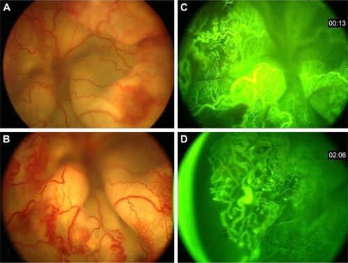
Table 1 Coats’ disease based on the age category: demographic features
Table 2 Coats’ disease based on the age category: comparison of serum values with normal controls
For those 22 patients with Coats’ disease, the mean time from the first notice of the disease by patients (or parents) to diagnosis was 7.1±5.4 months in the younger age group (≤10 years old) and 7.8±9.9 months in the older age group (>10 years old). There were three (13.6%) with bilateral involvement. Using the Coats’ disease classification,Citation4 the conditions of the eyes were classified as stage 1 (n=0), stage 2 (n=7), stage 3 (n=12), stage 4 (n=3), and stage 5 (n=0). Total retinal detachment was seen in six (28%) cases, whereas macular detachment was seen only in three (14%) cases. In two (9%) cases, there was no retinal detachment. The temporal and inferior regions were the most common sites for retinal detachment. Using fluorescein angiography, avascular non-perfused areas were found in 18 (82%) cases, located in the periphery region in 10 (46%) and in the macular region in 2 (9%) ().
The first step of analysis was a comparison of the two age groups of Coats’ disease (≤10 years vs >10 years). This revealed no significant difference in the lipid profile, serum antithrombin III, anticardiolipine and antiphospholipid antibody serum antibody titer as well as polymerase chain reaction test for toxoplasma, toxocara, or EBV in the two groups. There was statistical difference (Mann–Whitney test-skewed deviation) in the two groups with regard to hemoglobin (P=0.02), serum anti-HSV IgG (P=0.05), anti-CMV IgG (P=0.05), protein C (P=0.07), homocysteine (P<0.01), serum alpha-2 globulin (P<0.01), and serum gamma globulin (P=0.05). Due to the small sample size in each age group, the logistic regression analysis was inconclusive.
The second step of analysis involved comparison of laboratory findings in Coats’ disease versus the normal control group (). The serum levels in both Coats’ and control patients were compared using the Mann–Whitney test. There was no significant difference in the lipid profile, serum antithrombin III, anticardiolipine, and antiphospholipid antibody serum antibody titer as well as polymerase chain reaction for toxoplasma, toxocara, or EBV. However, in the control group, the serum titer of anti-HSV IgG was higher and that of anti-CMV IgG was lower. In Coats’ group, serum beta globulin was significantly higher in both young (≤10 years, P<0.001) and old (>10 years, P=0.05) subgroups compared to controls. Applying Bonferroni correction did not change the results of serum beta globulin in the group less than 10 years old.
Logistic regression analysis for the main serologic findings in Coats’ patients (irrespective of age) and control group was performed. After adjustment for age and sex, a significant association persisted for the presence of higher titer of serum beta globulin in Coats’ disease compared to controls (odds ratio [OR]: 1.8, 95% confidence interval [CI]: 1.0–3.1, P=0.02) (). Anti-HSV showed a mild negative impact (P=0.01, OR =−0.94) on the diagnosis of Coats’ disease. After adjusting for age and sex, in those less than 10 years, the serum beta globulin was found to be significantly associated with the diagnosis of Coats’ disease (OR: 6.3, 95% CI: 1.2–32.6, P=0.02). In patients greater than 10 years with Coats’ disease, there was a non-significant association with the diagnosis of Coats’ disease (OR: 1.3, 95% CI: 0.8–2.0, P=0.24). For the older age, anti-HSV antibody was borderline significant (OR: 0.98, 95% CI: 0.97–1.00, P=0.05). The small sample size precluded more conclusive results in the logistic regression analysis, adjusting for all other covariates.
Table 3 Coats’ disease comparison with controls: adjusted to age and sex as confounding factors (logistic regression)
Discussion
In this study, we could not find any association between hypercoagulability state and Coats’ disease (compared to controls). The only factor associated with Coats’ disease was elevated serum beta globulin mainly in the younger age group. We also recognized the negative impact of anti-HSV antibody on Coats’ disease in the older age group.
Serum beta globulins are grossly measured by electrophoresis. Serum protein electrophoresis is a technique to assess the two major fractions of protein in blood, including albumin and globulins.Citation10 Albumin is a transport protein that plays a significant role in fat solubility.Citation11 The globulin fraction of blood includes hundreds of serum proteins, including carrier proteins, enzymes, complement, and immunoglobulins. Globulins are divided into four groups by electrophoresis: alpha, alpha-2, beta, and gamma, depending on their migratory pattern between the anode and the cathode of the electrophoresis test.Citation11 The beta fraction of serum globulins is known to have two peaks, named as beta 1 and beta 2. Beta 1 is composed of mostly transferrin, and beta 2 contains beta-lipoprotein.Citation12 IgA, IgM, and sometimes IgG, along with complement proteins (C3), beta-2 microglobulin, plasminogen, angiostatins, properdin, sex hormone-binding globulin, transferrin, hemopexin, and factor H, can also be identified in the beta fraction.Citation10–Citation12 These proteins carry out numerous biological functions in the human body, including iron transport and monitoring immune response.Citation10,Citation11
Beta globulin protein can be elevated or depressed in various diseases. Beta globulin is found to be elevated in iron deficiency anemia, hypercholesterolemia, pregnancy, estrogen therapy, and also substantially increased in liver disease.Citation9,Citation10,Citation13,Citation14 It is found to be decreased in malnutrition, cirrhosis, and immune deficiency due to decreased synthesis, and in nephrotic syndrome due to protein loss in the kidney.Citation11
In this series of Coats’ disease, the beta globulin fraction was elevated, which is particularly evident in patients lesser than 10 years. This age-related difference could correlate with milder manifestations of Coats’ disease in older patients. The relationship of this fraction with Coats’ disease is unclear, but could correlate to higher C3 (complement protein 3) level or other factors. Complement 3 is an important protein in the immune system, which plays a vital role in the acute phase response. It controls several biological processes, including cell lysis, chemotaxis, anaphylaxis, vascular permeability, and cellular membrane adhesion. This protein is categorized as a beta globulin and might represent the elevation of beta globulin in Coats’ disease in this report.Citation12,Citation15–Citation18
Chronic low-grade inflammation has been correlated with elevated C-reactive protein, as well as complement C3 plasma levels, and these can be predictive of arterial thrombotic events.Citation16–Citation20 Elevated C3 is also associated with prolonged fibrinolysis.Citation18,Citation21–Citation23 Both of these roles could relate to the vascular pathology of Coats’ disease.
The discrepancy found in the younger and older age groups could suggest a difference in the main pathophysiological basis of Coats’ disease in children and adults. More studies are needed for the confirmation of this observation. There are limitations of this study, including the small sample size and unclear relevance of beta-globulin relationship to the condition studied. This relationship could be further explored with larger cohort and more precise fractionation of the beta-globulin subgroup to better understand the exact protein(s) that contribute to this finding and its relationship to Coats’ disease.
Conclusion
This analysis demonstrated that children with Coats’ disease showed elevated serum levels of beta globulin. We speculate that this finding could reflect the known fluorescein angiographic-evident ischemic component and possibly now a low-grade inflammatory component.
Disclosure
The authors report no conflicts of interest in this work.
References
- Coats’GForms of retinal diseases with massive exudationRoyal London Ophthalmic Hosp Rep190817440525
- SmithenLMBrownGCBruckerAJYannuzziLAKlaisCMSpaideRFCoats’ disease diagnosed in adulthoodOphthalmology200511261072107815882905
- CahillMO’KeefeMAchesonRMulvihillAWallaceDMooneyDClassification of the spectrum of Coats’ disease as subtypes of idiopathic retinal telangiectasis with exudationActa Ophthalmol Scand200179659660211782226
- ShieldsJAShieldsCLHonavarSGDemirciHCaterJClassification and management of Coats disease: the 2000 Proctor LectureAm J Ophthalmol2001131557258311336931
- GhassemiFShieldsCLMashayekhiAFluorescein angiographic findings in Coats’ diseaseOral presentation in World Ophthalmology Congress 2012February 16–20, 2012Abu Dhabi, United Arab Emirates
- GhassemiFSabourSCoats disease and cytomegalovirus infectionIran J Ophthalmol20122427578 Available from: http://irjo.org/browse.php?a_id=669&slc_lang=en&sid=1&ftxt=1Accessed December 20, 2016
- JonesJHKrollAJLouPLRyanEACoats’ diseaseInt Ophthalmol Clin200141418919811698747
- EgbertPRChanCCWinterFCFlat preparations of the retinal vessels in Coats’ diseaseJ Pediatr Ophthalmol1976136336339798026
- ChangMMMcLeanIWMerrittJCCoats’ disease: a study of 62 histologically confirmed casesJ Pediatr Ophthalmol Strabismus19842151631686502405
- McPherson PincusRAHenryMRBernardJHenry’s Clinical Diagnosis and Management by Laboratory Methods22nd edPhiladelphia, PAElsevier Saunders2011231244
- BusherJTSerum albumin and globulinWalkerHKHallWDHurstJWClinical Methods: The History, Physical, and Laboratory Examinations3rd edBoston, MAButterworths1990497499
- O’ConnellTXHoritaTJKasraviBUnderstanding and interpreting serum protein electrophoresisAm Fam Physician200571110511215663032
- GraySJBarronESThe electrophoretic analyses of the serum proteins in diseases of the liverJ Clin Invest194322219120016694993
- RavelRClinical Laboratory Medicine: Clinical Application of Laboratory Data6th edSt. LouisMosby1995343350
- KilicarslanAUysalARoachECAcute phase reactantsActa Medica201321227 Available from: http://www.tip.hacettepe.edu.tr/actamedica/2013/Acta13(2).pdfAccessed December 20, 2016
- AjjanRGrantPJFutersTSComplement C3 and C-reactive protein levels in patients with stable coronary artery diseaseThromb Haemost20059451048105316363249
- CojocaruIMCojocaruMMuşuroiCMuşuroiCDruţăABăcanuMStudy of some markers of inflammation in atherothrombotic pathogenesis of acute ischemic strokeRom J Intern Med2002401–410311615526546
- RidkerPMSilvertownJDInflammation, C-reactive protein, and atherothrombosisJ Periodontol2008798 Suppl1544155118673009
- D’AmbrosioALPinskyDJConnollyESThe role of the complement cascade in ischemia/reperfusion injury: implications for neuroprotectionMol Med20017636738211474130
- MunshiNCLongoDLAndersonKCPlasma Cell Disorders: Harrison’s Principles of Internal Medicine118th edNew York, NYThe McGraw-Hill Companies2012936944 Available from: http://accessmedicine.mhmedical.com/content.aspx?bookid=331§ionid=40726850Accessed December 20, 2016
- HowesJMRichardsonVRSmithKAComplement C3 is a novel plasma clot component with anti-fibrinolytic propertiesDiab Vasc Dis Res20129321622522253322
- SchroederVCarterAMDunneJMansfieldMWGrantPJProinflammatory and hypofibrinolytic phenotype in healthy first-degree relatives of patients with type 2 diabetesJ Thromb Haemost2010892080208220586918
- SeyaTNagasawaSMatsukuraMHasegawaHAtkinsonJPGeneration of C3d, g and C3d by urokinase-treated plasma in association with fibrinolysisComplement198522–31651742935360