
Abstract
Objective
To evaluate the role of serum apelin as a diagnostic tool in retinopathy of prematurity (ROP) disease.
Patients and methods
Thirty-eight preterm infants (60% male) with gestational age ranging from 30 to 36 weeks admitted to the neonatal intensive care unit, KJO Hospital, Saudi Arabia with proven diagnosis of ROP were included in the study. In addition, 27 preterm infants without ROP served as controls. All newborn infants in the study were subjected to adequate history taking, full clinical examination, and fundus examination by indirect ophthalmoscope (at 4–6 weeks) as well as determination of serum apelin at birth and at 4–6 weeks of age.
Results
The study revealed that oxygen therapy longer than 7 days’ duration, cesarean section (as a mode of delivery), sepsis, mechanical ventilation, blood transfusion, premature rupture of membranes, pneumothorax, perinatal asphyxia, cardiac problems, and neonatal jaundice were considered as risk factors related to development of ROP. Serum apelin levels were significantly lower in patients than controls (P<0.001) at time of diagnosis of the disease (4–6 weeks) while no significant differences were observed in levels at birth.
Conclusion
Serum apelin was found to be of significant diagnostic value in the occurrence of ROP.
Introduction
Major improvements in neonatal survival have been observed in the last ten years, particularly at low gestational age.Citation1 Retinopathy of prematurity (ROP) is a proliferative disorder of immature retinal vasculature with multifactorial etiology.Citation2 ROP develops in 16% of all premature births, the figure rising to around 60% of infants weighing <1,500 g at birth and 65% for infants <1,250 g. Some studies suggest that as smaller and younger babies are surviving, its incidence is increasing. However, better understanding of screening and management of these babies have resulted in a decrease in its incidence.Citation3 Both oxygen-regulated and non-oxygen-regulated factors contribute to normal vascular development and retinal neovascularization. Vascular endothelial growth factor (VEGF) is an important oxygen-regulated factor, while non-oxygen-regulated growth factor is insulin-like growth factor-I (IGF-I).Citation4 The initial phase of retinal vessel growth retardation occurs from birth to postmenstrual age of ~30–32 weeks. Oxygen plays a critical role in this process. Hypoxia and hyperoxia, by the high levels of supplemental oxygen the infant may receive in the neonatal intensive care unit (NICU), inhibit production of growth factors, such as VEGF, which causes cessation of normal retinal growth, and vessels constriction with a potential for vaso-obliteration of new immature vessels, resulting in tissue hypoxia that releases factors to stimulate new retinal blood vessel growth, a process called neovascularization (Phase II).Citation5
Apelin is a pro-inflammatory adipocyte-derived factor that participates in vascular wall inflammation. It was identified in 1998 as an endogenous ligand of the human orphan G protein-coupled receptor, APJ.Citation6 Apelin receptor is expressed in several cell types.Citation7 The angiogenic activity is the consequence of apelin action on the proliferation and migration of the endothelial cells. Apelin expression is upregulated during new vessels formation and downregulated following vessels stabilization.Citation8
The aim of this study is to evaluate the role of serum apelin in ROP.
Patients and methods
This is a prospective observational case–control study. It included 82 infants with a gestational age <32 weeks and a birth weight of ≤1,500 g. The infants were screened for ROP at NICU, KJO Hospital, Saudi Arabia, during January 2014–March 2016.
Thirty-eight (20 males and 18 females) with classified ROP completed our study group. Twelve preterm babies who died before first ophthalmological examination and five preterm babies with multiple congenital anomalies were excluded from this study. Twenty-seven preterm neonates who did not develop ROP (16 males and 11 females) were studied as control group.
Ethical approval was obtained from the institutional review board of KJO Hospital, Saudi Arabia, and a written informed consent was provided by parents of all children prior to the study.
All the studied groups were subjected to detailed history taking, including identification, cause of admission, neonatal history, maternal history, and family history. Full clinical examinations include:
Assessment – gestational age was assessed using the date of the mother’s last menstrual period and modified Ballard Score.Citation9
Ophthalmological examination – fundus examination was performed after pharmacological mydriasis starting 2 hours before the examination with cyclophrine eyedrops (combination of 2.5% phenylephrine and 1% cyclopentolate), applied three times at 30-minute intervals. Under topical anesthesia with boxinate eyedrops, an infantile eyelid speculum was applied. Using indirect binocular ophthalmoscope (Keeler Vantage®, Tampa, FL, USA), 20-diopter double aspheric Volk lens, the entire retina was carefully examined in all directions of gaze and with gentle pressure on the eyeball, using a scleral indenter for better visualization of the peripheral retina. The first examination was performed between 4 and 6 weeks of age or between 31 and 33 weeks of postmenstrual age. Follow-up examinations were made every week until the retinal vascularization was well into zone III. The presence or absence of the ROP plus disease, the zone, stage, and the extent of the ROP were recorded according to the international classification of ROP (stages 1–5).Citation10
Laboratory tests
Routine laboratory investigations: Complete blood count, blood culture, C-reactive protein, liver and kidney functions, serum electrolytes, and arterial blood gases were monitored.
Measurement of serum apelin peptide: Blood samples were obtained from studied 82 preterm babies twice: at birth (cord blood) and after 4–6 weeks. Two-milliliter venous blood sample was collected on ethylenediaminetetraacetic acid, centrifuged for 10 minutes at 5,000 rpm, and the plasma was separated. The serum was stored at −80°C in polypropylene tubes until the time of assay. Serum level of apelin peptide was measured using enzyme-linked immunosorbent assay kits (RayBiotech, Inc, Norcross, GA, USA. EIA Kit).Citation11
Statistical analysis
Quantitative data were expressed as mean ± standard deviation (X ± SD). Qualitative data were expressed as frequency and percentage (%). Receiver operating characteristic (ROC) curve was used to define the best of serum apelin cutoff value.
Data were analyzed using Statistical Package for the Social Sciences (SPSS) version 18.0 (SPSS Inc., Chicago, IL, USA). A P-value <0.05 was considered significant.
Results
The demographic characteristics of the 38 ROP and 27 non-ROP infants were analyzed. The patient group included 38 neonates of gestational age ranging from 30 to 36 weeks (X ± SD: 34.37±1.29 weeks) and mean birth weight of 2.42±0.54 kg, compared with 27 age- and sex-matched preterm infants without ROP as control group ().
Table 1 Demographic characteristics of studied groups
Our study showed a highly significant negative correlation between stages of severity of ROP and Apgar score at 5 minutes of life in the patient group, while no correlation was found as regards mode of delivery, premature rupture of membranes, mechanical ventilation, pneumothorax, sepsis, blood transfusion, neonatal jaundice, and perinatal asphyxia. While, a significant positive correlation between the stages of ROP and oxygen therapy for >7 days was observed (P<0.05) ().
Table 2 Correlation between ROP and some risk factors among patients group
A comparison of mean levels of serum apelin between the groups of patients and controls was undertaken. At the sampling time at birth, there were no significant differences between respective mean values in the two groups (P>0.05). Measurements made at 4–6 weeks after birth showed higher mean concentrations of serum apelin in patients (P<0.05) ().
Table 3 Serum apelin levels at birth and 4–6 weeks among the studied groups
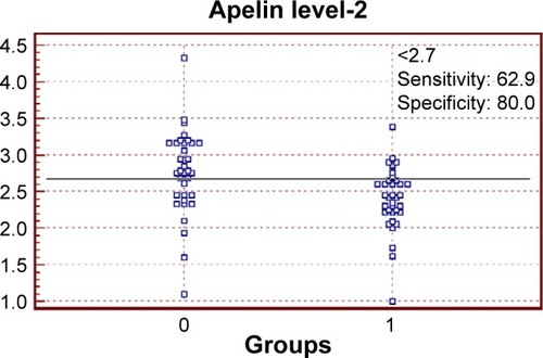
Serum apelin level cutoff value in samples taken at birth was 2.18 ng/mL, with sensitivity of 42.9%, specificity of 80%, positive predictive value of 68.2%, negative predictive value of 58.3%, and diagnostic accuracy of 59.1% ( and ). While the cutoff value of serum apelin levels taken at 4–6 weeks age was 2.7 ng/mL, with sensitivity of 62.9%, specificity of 80%, positive predictive value of 75.9%, negative predictive value of 68.3%, and diagnostic accuracy of 71% ( and ).
Figure 1 ROC curve between patients and control groups as regards serum apelin levels at birth.
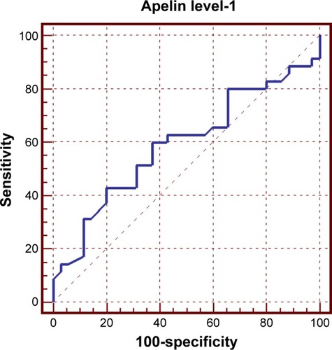
Figure 2 Interactive dot diagram between patients and control groups as regards serum apelin levels at birth.
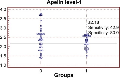
Figure 3 ROC curve between patients and control groups as regards serum apelin levels, at 4–6 weeks of age.
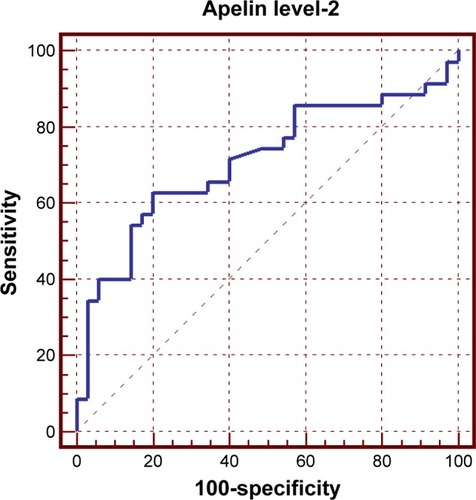
Figure 4 Interactive dot diagram between patients and control groups as regards serum apelin at 4–6 weeks of age.
Abbreviation: ROP, retinopathy of prematurity.
Discussion
ROP is a disorder of the developing retina of low-birth-weight preterm infants that potentially leads to blindness in a small but significant percentage of those infants. In almost all term infants, the retina and retinal vasculature are fully developed, and ROP cannot occur; however, in preterm infants, the development of the retina, which proceeds from the optic nerve head anteriorly during the course of gestation, is incomplete, with the extent of the immaturity of the retina depending mainly on the degree of prematurity at birth.Citation12 The rate of ROP in most premature infants has gone down greatly in developed countries over the past few decades due to better care in the NICU. However, more babies born very early are now able to survive, and these very premature infants are at the highest risk for ROP.Citation13 Studies have shown that apelin was involved in vascular pathophysiologyCitation6 and act as an angiogenic factor stimulating retinal endothelial cells’ proliferation, migration, and vascular tube formation.Citation14,Citation15
In the current study, 65 infants born with a gestational age before 32 weeks were screened for development of ROP, 38 infants developed ROP (58%), while only 41.5% were free. ROP reported by Taquin et alCitation16 study (32.4%) involved only low-birth-weight infants, while Chen et alCitation17 (10.8%) study involved infants with higher gestational age and birth weight (up to 2 kg and or 34 weeks’ gestational age).
In the present study, we investigate the most significant risk factors related to the development of ROP; we found that the most important risk factors included O2 therapy >7 days, cesarean section (as a mode of delivery), sepsis, prolonged mechanical ventilation, blood transfusion, premature rupture of membranes (PROM), development of hypoxia due to pneumothorax, hypoxic ischemic encephalopathy, cardiac problems, and newborn jaundice. These findings were going with many other studies as Chen et al,Citation18 while in Hadi and HamdyCitation19 cohort study, the risk factors for ROP were sepsis, continuous positive airway pressure, mechanical ventilation, neonatal indirect hyperbilirubinemia, intra ventricular hemorrhage, patent ductus arteriosis, and red blood cell and/or plasma transfusion. All these factors were significantly related to the occurrence of ROP and development of severe form of the disease except for indirect hyperbilirubinemia.
In our study, Apgar score in the first minute of life was mainly 4–6 (91.4%) (moderate Apgar score) and at 5 minutes was mainly >6 (7–9) (high Apgar score); there was a high significant negative correlation between severity of ROP and Apgar score at 1 and 5 minutes of life in the patients group. This comes in agreement with Martínez-Cruz et al’sCitation20 study.
In our study, serum apelin was detectable in all the studied groups, which come in concordance with the study of Malamitsi-Puchner et al,Citation21 who found that apelin was detectable in the serum of all studied neonates. We also evaluate the serum apelin level-1 (at birth) and level-2 (after 4–6 weeks) in both patients and control groups.
The average serum apelin level-1 is nearly equal in both patients and control groups; this comes in disagreement with Cekmez et al’sCitation22 study where the non-ROP (control) group had higher serum apelin level-1 than ROP (patients) group.
After 4–6 weeks, apelin level-2 was higher in non-ROP group than the ROP group, and this comes in agreement with Cekmez et al’sCitation22 study.
ROC curve was used to define the best cutoff value of apelin level-1 in our patients, which was 2.18, with sensitivity of 42.9%, specificity of 80%, positive predictive value of 68.2%, negative predictive value of 58.3%, and diagnostic accuracy of 59.1%. Another (ROC) curve was used to define the best cutoff value of apelin level-2 which was 2.7, with sensitivity of 62.9%, specificity of 80%, positive predictive value of 75.9%, negative predictive value of 68.3%, and diagnostic accuracy of 71%.Citation23
High positive predictive value of serum apelin level-2 test in our study prove that apelin test can predict the ROP early and prevent loss of vision if not controlled early enough. Sensitivity might be attributed to small number of the studied patients group (sensitivity of 62.9% and specificity of 80%), yet it suggests that the role of serum apelin in ROP should be further investigated.
Geiger et alCitation24 investigated the role of hypoxia on the expression of the adipokine apelin. Hypoxia in adipose tissue leads to a dysregulation of the expression of adipokines. He improved that apelin expression and secretion by human adipocytes are strongly induced under hypoxic conditions and that the early response on hypoxia with apelin induction is dependent on hypoxia-inducible factor 1-α (HIF-1α). Eyries et alCitation25 also reported that apelin is a hypoxia-inducible gene by both in vitro and in vivo studies. They found that hypoxia-induced apelin expression was mediated by HIF through a hypoxia-response element located within the first intron of the gene. Moreover, they determined that hypoxic induction of apelin expression regulated endothelial proliferation. Concerning the relationship between apelin and VEGF, they reported that apelin could enhance endothelial cell proliferation and aggregation in the presence of VEGF.Citation26,Citation27
Lim et al’sCitation28 study reported that human preterm and term labor significantly decreased apelin gene expression, but had no effect on APJ expression. They suggested an important role for apelin and APJ in the regulation of placental flow and vasculogenesis. The result of Zhang et alCitation29 showed that both apelin and APJ were positively expressed in the fibrovascular membrane of the ROP. The results also showed that apelin stimulates migration, proliferation, and capillary-like tube formation of RF/6A cells but not human umbilical venous endothelial cells and that apelin promotes angiogenesis in vivo. Therefore, they conclude that apelin/APJ signals seem to serve as a stimulatory factor in angiogenesis, probably in retina. Cekmez et alCitation22 used other tests (with serum apelin level test) to detect serum IGF-1 to aid early detection of ROP – it improved that circulating apelin concentrations in preterm babies may be linked with ROP like IGF-1 levels – while Lofqvist et alCitation30 used postnatal weight gain and serum IGF-1 for early detection of ROP.
Conclusion
Serum apelin was found to be of significant diagnostic value in the occurrence of ROP. Serum apelin values were significantly lower in patients group compared with controls group as regards ROP. There is also a significant positive correlation between the use of oxygen therapy >7 days and the severity of the ROP. The best cutoff value of serum apelin to detect ROP (after 4–6 weeks) was (2.7 ng/mL) with a sensitivity of 62.9% and specificity of 80%. Further studies are needed to 1) declare if serum apelin levels get affected by other diseases, such as respiratory distress syndrome, hemorrhagic diseases, or surgical conditions, and in neonates 2) consolidate the value of serum apelin in management of neonates admitted to NICU with ROP as a predictive marker.
Disclosure
The authors report no conflicts of interest in this work.
References
- EfferSBMoutquinJMFarineDNeonatal survival rates in 860 singleton live births at 24 and 25 weeks gestational age. A Canadian multicentre studyBJOG200210974074512135208
- ChawlaDAgarwalRDeorariAKPaulVKRetinopathy of prematurityIndian J Pediatr200875737618245940
- ZinAGoleGARetinopathy of prematurity-incidence todayClin Perinatol20134018520023719304
- SmithLEPathogenesis of retinopathy of prematurityGrowth Horm IGF Res200414Suppl AS140S14415135797
- HartnettMPennJMechanisms and management of retinopathy of prematurityN Engl J Med20123672515252623268666
- TatemotoKHosoyaMHabataYIsolation and characterization of a novel endogenous peptide ligand for the human APJ receptorBiochem Biophys Res Commun19982514714769792798
- DrayCKnaufCDaviaudDApelin stimulates glucose utilization in normal and obese insulin-resistant miceCell Metab2008843744519046574
- KidoyaHTakakuraNBiology of the apelin-APJ axis in vascular formationJ Biochem201215212513122745157
- BallardJLKhouryJCWedigKWangLEilers-WalsmanBLLippRNew Ballard score, expanded to include extremely premature infantsJ Pediatr19911194174231880657
- International Committee for the Classification of Retinopathy of PrematurityThe International Classification of Retinopathy of Prematurity revisitedArch Ophthalmol200512399199916009843
- ThanAChengYFohLCApelin inhibits adipogenesis and lipolysis through distinct molecular pathwaysMol Cell Endocrinol201236222724122842084
- FiersonWMAmerican Academy of Pediatrics Section on OphthalmologyAmerican Academy of OphthalmologyAmerican Association for Pediatric Ophthalmology and StrabismusAmerican Association of Certified OrthoptistsScreening examination of premature infants for retinopathy of prematurityPediatrics201313118919523277315
- YeSHellstromASmithLEHRetinopathy of prematurityMartinRJFanaroffAAFanaroff Martin’s Neonatal-Perinatal Medicine10th edPhiladelphia, PAElsevier Saunders2015104
- WangZNakayamaTInflammation, a link between obesity and cardiovascular diseaseMediators Inflamm2010201053591820847813
- KasaiAShintaniNOdaMApelin is a novel angiogenic factor in retinal endothelial cellsBiochem Biophys Res Commun200432539540015530405
- TaquinAMSyedRChadryTARetinopathy of prematurity: frequency and risk factors in a tertiary care hospital in Karachi, PakistanJ Pak Med Assoc20085818619018655427
- ChenJConnorKMAdermanCMSmithLEErythropoietin deficiency decreases vascular stability in miceJ Clin Invest200811852653318219389
- ChenJStahlAHellstromASmithLECurrent update on retinopathy of prematurity: screening and treatmentCurr Opin Pediatr20112317317821150442
- HadiAMHamdyISCorrelation between risk factors during the neonatal period and appearance of retinopathy of prematurity in preterm infants in neonatal intensive care units in Alexandria, EgyptClin Ophthalmol2013783183723674885
- Martínez-CruzCFSalgado-ValladaresMPoblanoATrinidad-PérezMCRisk factors associated with retinopathy of prematurity and visual alterations in infants with extremely low birth weightRev Invest Clin20126413614322991775
- Malamitsi-PuchnerAGourgiotisDBoutsikouMBakaSHassiakosDBrianaDDCirculating apelin concentrations in mother/infant pairs at term, Acta PaediatricaActa Paediatr2007961751175418001333
- CekmezFCanpolatFEPirgonOApelin, vaspin, visfatin and adiponectin in large for gestational age infants with insulin resistanceCytokine20115638739121742512
- Hajian-TilakiKReceiver Operating Characteristic (ROC) curve analysis for medical diagnostic test evaluationCaspian J Intern Med20134262763524009950
- GeigerKMuendleinAStarkNHypoxia induces apelin expression in human adipocytesHorm Metab Res20114338038521448846
- EyriesMSiegfriedGCiumasMHypoxia-induced apelin expression regulates endothelial cell proliferation and regenerative angiogenesisCirc Res200810343244018617693
- KidoyaHUenoMYamadaYSpatial and temporal role of the apelin/APJ system in the caliber size regulation of blood vessels during angiogenesisEMBO J20082752253418200044
- LuQJiangYRQianJTaoYApelin-13 regulates proliferation, migration and survival of retinal Müller cells under hypoxiaDiabetes Res Clin Pract20139915816723332048
- LimRBarkerGRileyCLappasMApelin is decreased with human preterm and term labor and regulates prolabor mediators in human primary amnion cellsReprod Sci20132095796723314958
- ZhangYJiangYRLuQYinHTaoYApelin in epiretinal fibrovascular membranes of patients with retinopathy of prematurity and the changes after intravitreal bevacizumabRetina20133361362023296045
- LöfqvistCNiklassonAEngströmEA pharmacokinetic and dosing study of intravenous insulin-like growth factor-I and IGF-binding protein-3 complex to preterm infantsPediatr Res20096557457919190540