
Abstract
Purpose
To describe various types of anterior capsular tears and an early diagnostic, flap motility, as a sign of posterior capsular rupture following posterior extension of radial tears.
Design
This was a prospective study carried out in 4,331 eyes that underwent phacoemulsification in a private practice setting from April 2015 to February 2016. Twenty six consecutive cases of anterior capsular tears were included. Morphological features of anterior capsular tears and resultant complications were evaluated. Parameters studied were surgical step during which the tear occurred, shape of tear, its extension in relation to the equator, and flap nature and motility in tear extending up to equator.
Main outcome measures
The main outcome measures were motility and nature of flaps in anterior capsular radial tears and the relation to posterior capsule rupture.
Results
Based on shape, extent, and angulation, anterior capsular tears were categorized into 5 types: Type I, pre-equatorial radial tear (26.92%); Type II, post-equatorial radial tear (3.85%); Type III, Argentinean flag sign pre-equatorial tear (57.69%); Type IV, Argentinean flag sign post-equatorial tear (7.69%), and Type V, mini punch (3.85%). Flaps were either seen to be everted and fluttering or inverted and non-fluttering. In all cases with everted fluttering flaps no posterior capsular rupture (PCR) was observed, while in cases with inverted non-fluttering flaps a PCR was observed (p<0.05).
Conclusion
Everted and fluttering flaps of the anterior capsular tears indicate pre-equatorial tear, while inverted and non-fluttering flaps indicate posterior capsule rupture following tear extension beyond the equator.
Video abstract
Point your SmartPhone at the code above. If you have a QR code reader the video abstract will appear. Or use:
Introduction
Radial tears in capsulorhexis margin are not an uncommon complication and can occur during any stage of phacoemulsification surgery.Citation1 Once a tear occurs in the capsulorhexis margin, it can easily extend to the equator and sometimes beyond it due to the irrigating fluid and the inter-capsular pressure created by lens material. If it extends to the posterior capsule causing a posterior capsule rent (PCR), then the surgery can get complicated by vitreous loss or nucleus drop.Citation2 At present, there is no known intraoperative clue that can ascertain the extent of intraoperative radial tear of the capsulorhexis margin. Consequently, the surgeon remains in a dilemma whether to continue or abort phacoemulsification.
In the present study, we describe various types of anterior capsular tears and for the first time an intraoperative sign, flap motility, that can help to ascertain the extent of the anterior capsular tear in relation to lens equator.
Materials and methods
This prospective study comprised 4,331 eyes with a clinical diagnosis of cataract that underwent phacoemulsification in a private practice setting from April 2015 to February 2016. The study analyzed anterior capsular tears in 26 consecutive cases, which occurred while performing phacoemulsification. The protocol adhered to the tenets of the Declaration of Helsinki and received approval from the ethics committee of Dr Om Parkash Eye Hospital. Prior to the surgical procedure all of the patients provided a written informed consent for the surgery and for inclusion in this study. The anterior capsular tears in patients with subluxated cataracts and maculoleucomatous corneal opacities were excluded from the study. Patients aged 20–70 years (mean 56.40±8.63 years) with variable grades of cataract were included in the study. Morphological features of anterior capsular tears and the resultant complications, if any, were studied. The parameters studied were anterior chamber depth (ACD), type and density of cataract, step of surgery during which tear occurred, shape of tear, and its extension in relation to the equator of lens. Additionally, the nature and motility of the flaps of the capsular tears were closely observed in relation to the extension of the tear up to the equator or beyond it. The nature of the flap was noted as everted or inverted/flat. The motility of the flap was noted as fluttering or non-fluttering.
The hypothesis was that the everted and fluttering flaps do not extend beyond the equator and the inverted and non-fluttering flaps indicate the extension of the flap beyond the equator onto the posterior capsule with associated complications. This is explained as follows:
Fluttering and everted flap in the pre-equatorial anterior capsular tear: When the edge of the anterior capsular tear does not extend beyond the equator, the capsular flap traverses a short distance prior to the equator. The flap thus formed remains everted and free. The end point of the extension of the flap functions as the pivotal point for the flap. The flap, being short, is responsive to the pressure gradient dynamics of the anterior chamber and thus flutters freely.
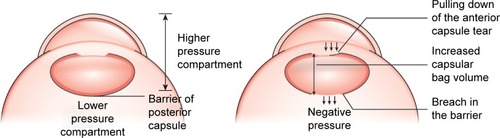
Non-fluttering and inverted flap in the post equatorial tear: During phacoemulsification, the intact posterior capsule compartmentalizes the eye into the highly pressurized anterior segment and relatively less-pressurized posterior segment. The inflated bag in the anterior compartment has the weight of the lenticular content and a high pressure due to the fluidics involved in the procedure. When the extension of the flap goes beyond the equator, there is no more division into the anterior and posterior compartments. The weight of the lenticular content, the fluid dynamics in the bag, and the high intraocular pressure in the anterior chamber cause the bag contents to move posteriorly. Simultaneously, there is extension of the tear (). Furthermore, the negative pressure in the posterior segment makes the bag contents to move posteriorly. The posterior displacement of the tear moves the corresponding anterior part of the tear along with it posteriorly. The continuous posterior pulling of the whole flap makes it flat and eventually inverted. Subsequently, the taut flap becomes totally non-responsive to the pressure gradient dynamics of the anterior chamber and stops fluttering.
Figure 1 Posterior capsule barrier and hypothesis of the non-fluttering flap.
Statistical analysis
Statistical analysis was performed by STATA 11.2 (College Station, TX, USA). Fisher exact test was used to measure the association between the posterior capsule rupture with everted flap, inverted flap, fluttering flap, and non-fluttering flap, respectively. These parameters were expressed as frequency and percentage. p<0.05 was considered as statistically significant.
Results
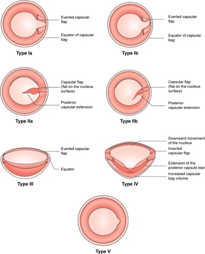
In this study, of the 26 cases, 14 were males and 12 were females with an age range of 29–67 years (56.4±8.63 years). Of the 26 cases, 17 (65.38%) had white mature cataract. Mean ACD was 2.92±0.34 mm. Based on the shape, extent, and angulation, the anterior capsular tears were categorized into five types ():
Type I – pre-equatorial radial tear
Type Ia – perpendicular pre-equatorial radial tear (11.54%)
Type Ib – acute-angled pre-equatorial radial tear (15.38%)
Type II – post-equatorial radial tear
Type IIa – perpendicular post-equatorial radial tear (3.85%)
Type IIb – acute angled post-equatorial radial tear (0%)
Type III – Argentinean flag sign pre-equatorial tear (57.69%)
Type IV – Argentinean flag sign post-equatorial tear (7.69%)
Type V – mini punch (3.85%)
Figure 2 Types of capsular tears.
The most common radial tears were the perpendicular tears not extending beyond the equator. The acute-angled tears were caused in cases of zonulopathy associated with pseudoexfoliation. Type V (mini punch) in the capsulorhexis was observed during phacoemulsification in one case by the phaco probe. Since the Type V (mini punch) did not extend to the periphery, it was excluded from the study for statistical analysis. Most of the anterior capsular tears (84%) occurred during the step of capsulorhexis ().
Table 1 Stage at which the anterior capsular tear occurred
In 21 out of 25 cases the flap was seen to be everted, while in 4 out of 25 cases the flap was inverted. Flaps of the tear in all the cases that had everted flaps were seen to be fluttering during phacoemulsification procedure. In these cases, no PCR was observed to occur (p-value <0.05; and ). The cases that had inverted flaps, no flap fluttering was observed and all these cases developed a PCR (p-value <0.05; and ).
Table 2 Relation of posterior capsule rupture with everted flap
Table 3 Relation of posterior capsule rupture with fluttering flap
Table 4 Relation of posterior capsule rupture with inverted flap
Table 5 Relation of posterior capsule rupture with non-fluttering flap
In all 4 cases with posterior extension of the tear with associated posterior capsule rupture, there is a correlation with the non-fluttering and everted nature of the flaps. Additionally, in all 21 cases of pre-equatorial tears with intact posterior capsule, the flaps are everted and fluttering. Nevertheless, the absence of inverted and non-fluttering flaps in these 21 cases indirectly validates the clinical correlation between the flap motility and posterior capsule rupture.
We provide videos of 3 cases of extended anterior capsular tears and the flap motility sign to ascertain its relationship to the extent of equatorial anterior capsular tears (Video S1).
Case 1
Capsulorhexis was completed with forceps. The nucleus was phacoemulsified by direct vertical chop technique. The nuclear core was separated and emulsified to debulk the nucleus. During the emulsification of the nuclear core, an extension in the rhexis was seen in the sub-incisional area. On careful evaluation, the flaps thus created by the tear in the anterior capsule were seen to be everted and fluttering with the fluidics in the anterior chamber. Phacoemulsification was continued taking care that the tear does not extend further. Maneuvers like nucleus rotation and chopping were carried out gently to avoid tear extension. During the procedure, a close vigil was kept on the nature of the flap. Nucleus emulsification was completed using safe phacoemulsification technique by dividing nuclear pieces into fully separated small pieces. The remaining cortex was removed with coaxial irrigation and aspiration handpiece. The flap continued to flutter and remained everted during the entire procedure. No extension of the tear to the posterior capsule was observed. A foldable intraocular lens (IOL) was implanted in the bag.
Case 2
This was a case of white intumescent cataract, and the presence of Argentinian flag sign was noted as soon as capsulorhexis was initiated. The capsulorhexis forceps was used to fashion a hemi-rhexis on both sides of capsular tears. The motility of flaps of tear in the anterior capsule was carefully observed and they were seen to be everted and fluttering with the fluidics in the anterior chamber. Phacoemulsification was continued and a close vigil was kept on the nature of the flap. Nucleus emulsification was completed using safe phacoemulsification technique by dividing nuclear pieces into completely separated small pieces with minimal force of separation. The cortex was removed with coaxial irrigation and aspiration handpiece. The flap continued to flutter and remained everted during the entire procedure. No extension of the tear onto the posterior capsule was observed.
Case 3
This was a case in which anterior capsular tear extended in to the posterior capsule resulting in posterior capsular rent. A difficulty was faced by the surgeon during capsulorhexis, and Vanna’s scissors and forceps were used for its completion. This caused an irregularity in the capsulorhexis margin at one place. During nuclear trenching, a radial anterior capsular tear occurred and soon it extended reaching close to equatorial area. The flaps created by the tear in the anterior capsule were seen to be everted and fluttering with the fluid-ics in the anterior chamber. However, fluttering of the flaps stopped as the nucleus was being divided into quadrants. Also, they had become inverted. At this stage, according to our hypothesis, a conversion to extracapsular extraction should have been done since the flap had extended beyond equator. However, phacoemulsification was continued. Further maneuvers led to the nucleus quadrant dropping into the vitreous. The PCR was visible at this stage suggestive of an extension onto the posterior capsule.
Discussion
Radial tears in the anterior capsular tears can occur during any stage of surgery such as during viscoelastic injection by the cannula, capsulorhexis, hydrodissection, phacoemulsification, irrigation/aspiration, movement of the second instrument, and IOL/iris prosthesis implantation.Citation1 Marques et al observed capsulorhexis as the most frequent step during a surgery to have radial anterior capsular tears.Citation1 The capsular tears occurred during capsulorhexis in 84% of our cases making it the most common step during surgery in our study.
Radial tears in capsulorhexis can be predisposed by various factors including shallow anterior chamber, weak zonules such as in coexisting pseudoexfoliation, previous inflammatory conditions, positive vitreous pressure, squeezing of eye intraoperatively, requirement of local anesthesia, intumescent cataracts, cataracts in pediatric age group with highly elastic anterior capsules, and handling of capsulorhexis by novice surgeons.Citation3–Citation14 In our study, majority of the cases with cap-sulorhexis tears had an ACD of <3 mm and were mature intumescent cataracts.
Peripheral extension of the radial tear in anterior capsulorhexis margin can cause serious complications if it travels beyond lens equator and into the posterior capsule. Although there are measures to rescue the tear extension including Little’s rescue technique, cystitome retrieval technique, rhexis restarting from the other side, tangential flap creation, and anterior localized zonulysis with flap rescue, the radial tear may still extend to the peripheral part of lens mostly due to an increase in intracapsular pressure.Citation15–Citation19
Radial tears do not cross the equator immediately in a significant number of cases because of the thickened mid periphery of anterior capsule and the barrier created by the zonular attachment to the anterior capsule.Citation20 Nevertheless, radial tears can breach the barrier of equator during the course of emulsification or thereafter, and subsequently have complications such as posterior capsular tear, vitreous loss, insufficient capsular support for IOL implantation, and nuclear drop. Phaco-surgeons employ different maneuvers such as creating additional radial tears, performing supra-capsular phacoemulsification, using stable chamber settings, avoiding deep or shallow anterior chamber, and employing techniques that put minimal stress to the extended capsular flapsCitation21–Citation25 to prevent the extension of the tear beyond the equator onto the posterior capsule.Citation26 Subsequently, the posterior capsular rupture rate varies depending upon the experience and the technique of the surgeon. In a Moorfields Eye Hospital study of anterior capsular tear extension, a high rate (24%) of posterior capsular rupture, with 5% nuclear drop and 11% requiring a secondary procedure, was reported.Citation27 In a study by Marques et al, the incidence of extension of the anterior capsule to the posterior capsule through the equator was noted as ~52%.Citation1
The intraoperative signs of posterior capsular rupture are the sudden deepening of the anterior chamber, pupillary snap sign, increased red glow, decreased followability, and nuclear tilt.Citation28 However, these signs of posterior capsular rupture do not appear instantly after the extension of the tear onto the posterior capsule. Additionally, the time lag for appreciating the posterior capsular rupture may vary regardless of the experience of the surgeon. These deficiencies in recognizing the signs of radial tear of anterior capsule traveling beyond lens equator and causing a rent in the posterior capsule can lead to nucleus drop and its sequels.
We describe for the first time a surgical sign – flap motility sign – which clearly helps to ascertain the extent of a flap extension.
While performing phacoemulsification, if the extended flap of the capsulorhexis is everted and fluttering, it indicates that the flap has not extended beyond the equator. In such cases, phacoemulsification can be safely continued without a dilemma. Nevertheless, safe phacoemulsification techniques should be employed, as described earlier, and a close vigil has to be kept on the nature and motility of the flap as the tear can extend to the posterior capsule. If the extended flap becomes inverted on the nucleus and stops fluttering, it indicates the end point of safe phacoemulsification and denotes the extension of the flap beyond the equator onto the posterior capsule. In such situations, an instant decision to abort phacoemulsification and adopt extra-capsular cataract surgery or small incision cataract surgery helps to decrease the incidence of posterior capsular rupture.
The flap motility sign helps in deciding whether an IOL implantation is required. If the flaps are fluttering at the end of phacoemulsification surgery, IOL implantation can be done safely in the bag over the intact posterior capsule.
Conclusion
Based on our definite results, peripherally extended everted and fluttering flaps of the tears indicate that the radial tears have not extended beyond equator. Additionally, inverted and non-fluttering tears symbolize the extension of tear beyond equator.
Acknowledgments
This paper was presented as an abstract at the 74th Annual Conference (AIOC) of All India Ophthalmological Society, Kolkata, India; February 25–28, 2016. Dr Tushya Om Parkash now has a degree in MS Ophthalmology and is currently affiliated with Dr Om Parkash Eye Institute, Amritsar, India (as of July 11, 2017).
Supplementary material
Video S1 Flap motility sign.
Disclosure
The authors report no conflicts of interest in this work.
References
- MarquesFFMarquesDMVOsherRHOsherJMFate of anterior capsule tears during cataract surgeryJ Cataract Refract Surg200632101638164217010860
- MohammadpourMErfanianRKarimiNCapsulorhexis: pearls and pitfallsSaudi J Ophthalmol2012261334023960966
- CoreyRPOlsonRJSurgical outcomes of cataract extractions performed by residents using phacoemulsificationJ Cataract Refract Surg199824166729494901
- SaberHButlerTJCottrellDGResistance of the human posterior lens capsule and zonules to disruptionJ Cataract Refract Surg19982445365419584252
- AssiaEIAppleDJTsaiJCLimESThe elastic properties of the lens capsule in capsulorrhexisAm J Ophthalmol199111156286322021174
- PrasadSPhacoemulsification learning curve: experience of two junior trainer ophthalmologistsJ Cataract Refract Surg19981207377
- SewardHCDaltonRDavisAPhacoemulsification during learning curve: risk/benefit analysisEye (Lond)19937Pt 11641688325410
- PingreeMFCrandallASOlsonRJCataract surgery complication in 1 year at an academic institutionJ Cataract Refract Surg199925570570810330649
- BrowiningOJCoboLMEarly experience in extracapsular cataract surgery by residentsOphthalmology19859212164716534088614
- BianchiEScarinciFRipandelliGRetinal pigment epithelium, age-related macular degeneration and neurotrophic keratouveitisInt J Mol Med201331123224223128960
- TauroneSRipandelliGPacellaEPotential regulatory molecules in the human trabecular meshwork of patients with glaucoma: immunohistochemical profile of a number of inflammatory cytokinesMol Med Rep201411121384139025351602
- FehérJKovácsIPacellaERadákZA mikrofl óra és a bélnyálkahártya kölcsönhatása az irritábilis bél, irritábilis szem és irritábilis elme szindróma kórtanában és kezelésében [Correlation of the microbiota and intestinal mucosa in the pathophysiology and treatment of irritable bowel, irritable eye, and irritable mind syndrome]Orv Hetil20141553714541460 Hungarian25194867
- PacellaEColliniSPacellaFPirainoDCSantamariaVDe BlasiRALevobupivacaine vs racemic bupivacaine in peribulbar anaesthesia: a randomized double blind study in ophthalmic surgeryEur Rev Med Pharmacol Sci201014653954420712261
- PacellaEPacellaFTroisiFEfficacy and safety of 0.5% levobupivacaine versus 0.5% bupivacaine for peribulbar anesthesiaClin Ophthalmol2013792793223723684
- LittleBCSmithJHPackerMLittle capsulorhexis tear-out rescueJ Cataract Refract Surg20063291420142216931249
- KarimSMROngCTMiahMRSleepTHanifudinAA novel technique of rescuing capsulorhexis radial tear-out using a cystotomeJ Vis Exp2011472317
- FranchiniAThe rhexis: errant, compromised, or smaller or larger than plannedCataract & Refractive Surgery Today201233538
- MohammadpourMRescue of an extending capsulorrhexis by creating a midway tangential anterior capsular flap: a novel technique in 22 eyesCan J Ophthalmol201045325625820436546
- PageTPAnterior zonulotomy: rescue technique for capsulorhexis tear-outJ Cataract Refract Surg201541102036203926703276
- AssiaEIAppleDJTsaiJCMorganRCMechanism of radial tear formation and extension after anterior capsulectomyOphthalmology19919844324372052296
- ColvardDMRadial anterior capsular tear causes and managementCataract & Refractive Surgery Today2009108384
- AlioJLMuletMEShalabyAMAttiaWHPhacoemulsification in the anterior chamberJ Cataract Refract Surg2002281677511777712
- OsherRHBarrosMGMarquesDMMarquesFFOsherJMEarly uncorrected visual acuity as a measurement of the visual outcomes of contemporary cataract surgeryJ Cataract Refract Surg20043091917192015342055
- Ocular Surgery News U.S Edition. Complications from anterior capsule tears may require secondary intervention Available from: http://www.healio.com/ophthalmology/cataract-surgery/news/print/ocular-surgery-news/%7B13f27d61-f984-4a1e-a4de-a917065a208b%7D/complications-from-anterior-capsule-tears-may-require-secondary-interventionAccessed January 21, 2017
- ReusNJManagement of anterior and posterior capsular tearsCataract & Refractive Surgery Today201173840
- VajpayeeRBSharmaNDadaTGuptaVKumarADadaVKManagement of posterior capsule tearsSurv Ophthalmol200145647348811425354
- CarifiGMillerMHPitsasCComplications and outcomes of phacoemulsification cataract surgery complicated by anterior capsule tearAm J Ophthalmol2015159346346925461300
- RaniNGuptaPPosterior capsular rent: risk factors, diagnosis and managementSurg Sci201455224226