
Abstract
Purpose
To investigate the characteristics in spontaneous release of epiretinal membrane (ERM) during watchful waiting and to introduce a possible mechanism of pathogenesis as a photo essay.
Methods
Records from all patients with ERM were obtained from Osaka Medical College Hospital from January 2001 to October 2012. Visual acuity (VA), fundus photo, and optical coherence tomography (OCT) were reviewed using the medical records. For statistical analysis, VA measured with a Landolt chart was converted to the logarithm of the minimum angle of resolution (logMAR). To investigate the pathogenesis of ERM, tryptase activity in vitreous, which plays a role in tissue fibrosis and remodeling, was measured in patients that underwent a vitrectomy for ERM, macular hole, and proliferative diabetic retinopathy (PDR).
Results
ERM was observed in 604 patients and spontaneous release of the ERM was observed in 13 patients with 14 eyes (four males and nine females, aged 33–78 years). Among the 14 eyes, mean VA did not change significantly through the release of the ERMs (0.17±0.18 before and 0.24±0.40 after release, P=0.544). Nine eyes showed posterior vitreous detachment or vitreomacular traction on OCT images and five eyes did not. ERM was released in five eyes with no accompanying vitreous traction by OCT during watchful waiting and seems to have peeled off by itself by contracting and rolling from the inferior side. Three eyes with deteriorated VA underwent vitrectomy due to macular hole or pseudomacular hole. Vitreal tryptase activity was significantly higher in patients with ERM compared to those with PDR (P<0.05).
Conclusion
Fundus photos of ERM auto-peeling were taken during long-term follow-up. Spontaneous release of ERM is possibly involved in vitreous traction or membrane contraction. In addition, tryptase may be involved in the development and contraction of ERM.
Epiretinal membrane (ERM) is caused by a fibroglial proliferation on the surface of the retina. Patients with vitreomacular traction syndrome are treated with ocriplasmin; however, it usually has little effect on the treatment of ERM.Citation1,Citation2 Whereas, some patients do show auto-peeling and spontaneous release of ERM without posterior vitreous detachment during watchful waiting, which involves the contraction of the ERM ( and ). However, this phenomenon usually takes a long time. Therefore, to investigate the characteristics in spontaneous release of ERM during watchful waiting, visual acuity (VA), fundus photo, and optical coherence tomography (OCT) were reviewed using our medical records obtained from Osaka Medical College Hospital from January 2001 to October 2012. Patient consent to review their medical records was not required by the institutional review board, and patient data were covered confidentially. For statistical analysis, VA measured with a Landolt chart was converted to the logarithm of the minimum angle of resolution (logMAR). ERM was observed in 604 patients and spontaneous release of ERM was observed in 13 patients with 14 eyes (four males and nine females, aged 33–78 years). Among the 14 eyes, mean VA did not change significantly through release of the ERMs (0.17±0.18 before and 0.24±0.40 after release, P=0.544). In five eyes, the released ERM seemed to have peeled off by itself by contracting and rolling from the inferior side without accompanying vitreous traction by OCT during watchful waiting. Three eyes with deteriorated VA underwent vitrectomy due to macular hole or pseudomacular hole.
Figure 1 Auto-peeling of idiopathic ERM can be seen in these fundus photos and macular OCT from a 33-year-old female with idiopathic ERM. These photos and OCT were taken in (A) July 2010 and (B) October 2012. Visual acuity was improved from 10/20 to 14/20.
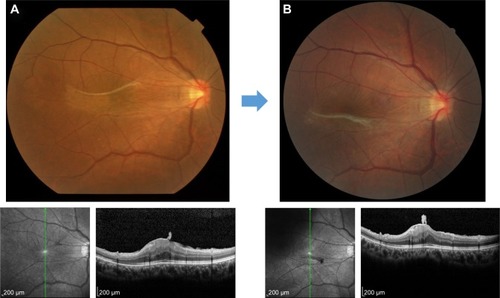
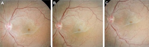
Figure 2 Auto-peeling of idiopathic ERM can be seen in these fundus photos from a 76-year-old female with idiopathic ERM. These photos were taken in (A) August 2008, (B) November 2009, and (C) February 2012.
Although interesting papers have been published regarding the pathohistology of ERMsCitation3,Citation4 and the remodeling of the extracellular matrix at the vitreoretinal interface,Citation2 the pathomechanisms leading to ERM still remains unclear, which raises the following questions:
Why does ERM occur in the macula of healthy subjects?
Why does ERM sometimes show auto-peeling and spontaneous release without posterior vitreous detachment?
What causes the recurrence of ERM in the macula again after successful removal of idiopathic ERM by vitrectomy? ()
Why does ERM reoccur despite the spontaneous release of ERM?
Figure 3 Recurrence of ERM in the macula after successful removal of idiopathic ERM by vitrectomy. Vitrectomy for idiopathic ERM was performed and visual acuity improved from 20/30 (A) to 20/20 (B). However, it reoccurred 1 year after the vitrectomy and the visual acuity decreased to 20/30 (C).
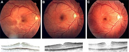
Based on the recurrent ERM depicted in , it is also necessary to think about the presence of internal limiting membrane (ILM). The removal of ILM is the key for ERM recurrence, and ILM peeling in vitrectomy of ERM could result in a significantly lower ERM recurrence rate by preventing reproliferation and ensuring complete removal of ERM fragments.Citation5 In addition, ERM tears and scrolling of tear edges include torn ILM (ILM ripping) histologically,Citation6,Citation7 and the presence of a postoperative ERM edge was recently reported to be associated with a higher risk of ERM recurrence.Citation8
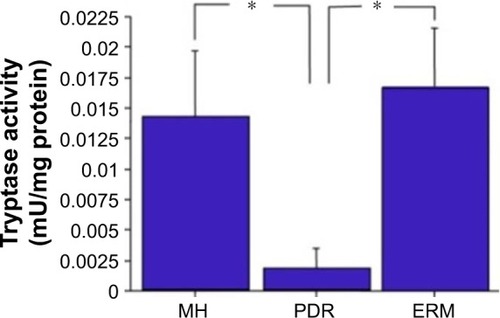
We add an interesting finding that may answer these questions by showing the possible involvement of tryptase in the development of idiopathic ERM (). Tryptase activity in the vitreous was significantly higher in patients with ERM (n=14) compared to those with PDR (n=13) (P<0.05). The Ethics Committee of Osaka Medical College, Takatsuki City, Osaka, Japan, approved this study. Tryptase is a serine protease produced in mast cells. Mast cells are located in the eye such as in choroid, ciliary body, conjunctiva, and sclera and play a role in tissue fibrosis, apoptosis, and remodeling. We have previously reported that tryptase activity in the vitreous increased in idiopathic ERM.Citation9 It has also been reported that ERM shows immunoreactivity for collagen types 1 and 2 histopathologically,Citation10 and tryptase stimulates fibroblast-mediated contraction of collagen type 1 gels,Citation11 Thus, the pathogenesis of ERM is possibly related to ERM auto-peeling by contracting the ERM, which results in spontaneous release of ERM. Here, we would like to add the possibility that tryptase affects the development of ERM and contraction of ERM, and mast cells play a key role in the pharmacological treatment of ERM, although further research is needed to determine where tryptase in the vitreous comes from exactly.
Figure 4 Tryptase activity in the vitreous. Tryptase activity in the vitreous was significantly higher in patients with ERM (n=14) and MH (n=14) compared to PDR (n=13); *P<0.05.
Disclosure
The authors report no conflicts of interest in this work.
References
- SpaideRFVitreomacular traction syndromeRetina201232Suppl 2S187S190 discussion S190–S18322929319
- BuSCKuijerRLiXRHooymansJMLosLIIdiopathic epiretinal membraneRetina201434122317233525360790
- HiscottPSGriersonIMcLeodDNatural history of fibrocellular epiretinal membranes: a quantitative, autoradiographic, and immunohistochemical studyBr J Ophthalmol198569118108234063247
- KampikAPathology of epiretinal membrane, idiopathic macular hole, and vitreomacular traction syndromeRetina201232Suppl 2S194S198 discussion S198–S19922929320
- SchechetSADeVienceEThompsonJTThe effect of internal limiting membrane peeling on idiopathic epiretinal membrane surgery, with a review of the literatureRetina201737587388027617536
- BoveyEHUfferSTearing and folding of the retinal internal limiting membrane associated with macular epiretinal membraneRetina200828343344018327135
- SiglerEJRandolphJCCalzadaJIIncidence, morphology, and classification of epimacular membrane ripRetina20133361158116523348863
- GaberRYouQSMuftuogluIKCharacteristics of epiretinal membrane remnant edge by optical coherence tomography after pars plana vitrectomyRetina Epub 201711110.1097/IAE.0000000000001466
- IkedaTNakamuraKOkuHThe role of tryptase and anti-type II collagen antibodies in the pathogenesis of idiopathic epiretinal membranesClin Ophthalmol201591181118626170611
- KritzenbergerMJunglasBFrammeCDifferent collagen types define two types of idiopathic epiretinal membranesHistopathology201158695396521480957
- SkoldCMOhkuniYLiuXDNumerofRRennardSICo-cultured human mast cells stimulate fibroblast-mediated contraction of collagen gelsInflammation2001251475111293665