
Abstract
Background
We have developed a new compact lightweight 8K ultra-high-definition (UHD; 7,680×4,320 pixels) camera and started medical application with an ophthalmic surgical microscope which is interchangeable with the conventional high-definition (1,920×1,080 pixels)/4K UHD (3,840×2,160 pixels) microscopic camera.
Methods
We did a feasibility study to apply our 8K UHD microscope in cataract surgery, glaucoma surgery and vitreous surgery using pig cadaver eyes. The 8K UHD microscope comprises a surgical microscope, a camera adaptor with relay lenses, an 8K UHD camera and an 8K UHD LCD to share the 8K UHD images with all surgical staff in real time.
Results
In ophthalmic surgeries, higher resolution images than conventional microscopic cameras were obtained with 8K UHD LCD equivalent to the observation through the microscopic eye pieces.
Conclusion
Based on the results of this feasibility study, clinical trials on human ophthalmic surgery using the new 8K UHD microscopic camera should be conducted in the near future.
Introduction
Japan Broadcasting Corporation (NHK) developed 8K ultra-high-definition (UHD) broadcasting technologies and achieved a series of devices specific for 8K UHD imaging in 2002. 8K UHD broadcasting system offers panoramic images of a resolution 16-fold higher than the images obtained with the high-definition (HD) technology.Citation1–Citation4 There are heightening expectations in recent years for important contributions of 8K UHD technology to innovative medical imaging in advanced image-guided diagnosis and treatment.
In ocular microsurgery, high-performance microscopes have been strongly demanded for the highest precision observation and operation. Ophthalmology microscopes attached with HD/4K UHD camera have been already commercialized. However, the image quality of the HD/4K UHD camera is inferior to that of the surgeons’ eyes’ view through the microscopic optical system. The first experiment in the world to apply 8K UHD in microscopic surgeries of cataract, glaucoma, and vitreoretinal was successfully conducted by the Medical Imaging Consortium in Miyake Eye Hospital (Nagoya, Japan) in December 2014 using our prototype 8K UHD camera, which was originally developed for an 8K UHD endoscope.Citation5 Subsequently, we have developed a new compact lightweight 8K UHD camera (Kairos Co., Ltd., KairoScope-E, Tokyo, Japan) in 2017Citation6 and started medical application of ophthalmic surgical microscope which is interchangeable with the conventional HD/4K camera.
In this paper, we report the results of the experiment to apply the new 8K UHD microscopic camera in ophthalmic surgeries using pig cadaver eyes.
Materials and methods
8K UHD microscopic camera for ophthalmic surgery
shows our new compact lightweight 8K UHD microscopic camera for ophthalmic surgery. The 8K UHD camera is interchangeable with the conventional HD/4K camera mounted on the ophthalmology microscopes. The dimensions of the 8K UHD camera are 75 mm (W)×75 mm (H)×170 mm (L) and the weight is 370 g. The 8K UHD camera has a super-35-mm complementary metal oxide semiconductor imaging sensor and its specifications are shown in . Especially, sensitivity of the imaging sensor is about four times superior to that of our prototype 8K UHD camera.
Table 1 8K UHD CMOS imaging sensor specifications
Figure 1 Our new 8K UHD camera mounted on a surgical microscope with a relay lens adapter.
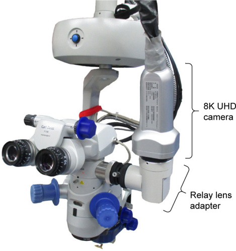
The prototype 8K UHD camera was too large to be mounted on a surgical microscope. On the contrary, our new 8K UHD camera size is nearly one fifth of the prototype one and can be attached to the surgical microscope without any mechanical interference on the microscope. Furthermore, as our compact 8K UHD camera is light, it can be attached to both table top and stand type microscopes without losing weight balance.
The optical system of an 8K UHD microscope comprises a surgical microscope, a camera adaptor with relay lenses, and an 8K UHD camera. Half quantity of the light from one ray path for the right eye is guided to the 8K UHD camera through the adaptor optics. The adaptor optics images the magnified surgical sites on the pixels of the 8K UHD camera with an optimal focal length of 289 mm.
Feasibility study using pig cadaver eyes
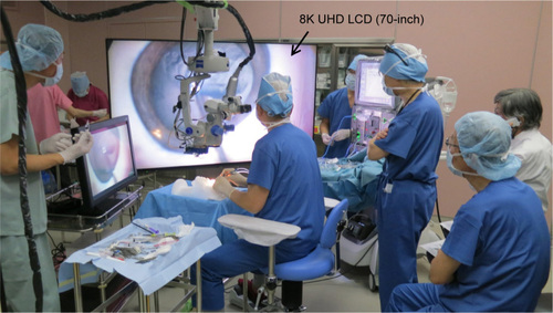
We tried cataract surgery (continuous circular capsulorhexis [CCC], phacoemulsification, and intraocular lens insertion), glaucoma surgery (observation through a gonioscope and sclera flap creating), and vitreous surgery (guillotine cut and peeling) using pig cadaver eyes in Miyake Eye Hospital. Our new 8K UHD camera was attached to an assistant microscope installation part of a commercialized ophthalmology microscope (Carl Zeiss, Zeiss Lumera T, Oberkochen, Germany) suspended from the ceiling. The intensity of microscopic illumination was between 40,000 and 47,000 lux in cataract and glaucoma surgery. In vitreous surgery, the intensity of endo-illumination and chandelier light was about 33,000 and 50,000 lux, respectively. A 70-inch 8K UHD monitor (LV-70002; Sharp Corporation, Osaka, Japan) was placed in front of a surgeon. shows the operating room site using an 8K UHD microscopic camera for ophthalmic surgeries.
Figure 2 8K UHD microscopic camera for ophthalmic surgery in the operating room site.
Results
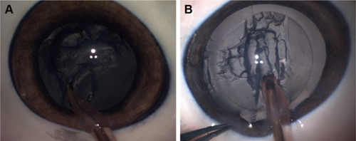
shows examples of the anterior eye segment – a comparison between the surgical field images of pig cadaver eyes in cataract surgery (phacoemulsification) obtained with the prototype 8K UHD microscopic camera () and that by the new 8K UHD microscopic camera (). In , since the brightness of the image was insufficient with the open aperture, a manual gain-up was added up to +15 dB by digital processing. The bright image of was obtained without an automatic digital gain-up and there was a margin of 1/2 to 1/3 of the aperture scale. Therefore, effectively ten times more sensitive image was obtained, so that the automatic gain control is available, which allows constant brightness even when changing light and dark areas. In addition, even aspirated groove texture of crystalline lens was highly clear with a fine difference like a shading change in .
Figure 3 The comparison of surgical field images of pig cadaver eye in cataract surgery (phacoemulsification). (A) Image obtained with the prototype 8K microscopic camera and (B) image obtained with the new 8K UHD microscopic camera.
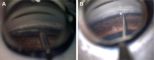
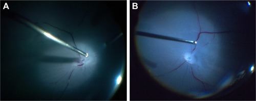
shows a comparison between the surgical field images of pig cadaver eyes in glaucoma surgery (observation through a gonioscope and creating scleral flap) obtained with the prototype 8K UHD microscopic camera () and that with the new 8K UHD microscopic camera (). Similar to the 8K UHD images in cataract surgery (), the images obtained with the new 8K UHD microscopic camera in glaucoma surgery () were brighter than those obtained with the prototype 8K UHD microscopic camera (). In , the fine microstructures of trabecular meshwork and ciliary body can be clearly observed with brighter 8K UHD resolution, and the quality was sufficiently high and equivalent to the microscopic direct observation through eyepieces.
Figure 4 The comparison of surgical field images of pig cadaver eye in glaucoma surgery. (A) Images obtained with the prototype 8K microscopic camera and (B) images obtained with the new 8K UHD microscopic camera through a gonioscope.
shows examples of posterior eye segment – a comparison between surgical field images of pig cadaver eyes in vitreoretinal surgery obtained with the prototype 8K UHD microscopic camera () and that with the new 8K UHD microscopic camera (). Posterior eye segment was illuminated by a chandelier endoillumination fiber, and quantity of illumination light for posterior eye segment was quite limited. High sensitivity of the new 8K UHD microscopic camera caused illumination of larger range () than the prototype 8K UHD microscopic camera ().
Figure 5 Comparison of surgical field images of pig cadaver eye in vitreoretinal surgery. (A) Images obtained with the prototype 8K microscopic camera and (B) images obtained with the new 8K UHD microscopic camera. Retinal capillary vessels were illuminated by a chandelier endo-illumination fiber.
Supplementary Video S1 shows detail of – with HD resolution down-converted from an original 8K UHD video.
Discussion
We have developed a new compact and lightweight 8K UHD microscopic camera for ophthalmic surgery, which is interchangeable with the conventional HD/4K microscopic camera. We did the feasibility study to apply our microscopic camera in ophthalmic surgeries using pig cadaver eyes, and images of sufficiently high quality could be obtained with 8K UHD LCD equivalent to the microscopic observation through the eye pieces for not only a surgeon but also other surgical staffs in real time.
Surgeons and other surgical staffs reported the following positive evaluations of the new 8K UHD microscopic camera for ophthalmic surgery:
The new compact and lightweight 8K UHD camera with higher sensitivity deserves a sufficient evaluation from the viewpoint of surgical suitability.
The new 8K UHD camera can increase the quantity and quality of microscopic digital images on the monitor, and the 8K UHD images are equivalent to real microscopic observation through eyepieces.
Brightness of the image approached practical use for 8K UHD microscopic camera.
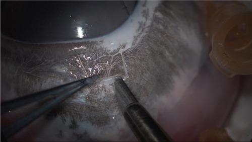
In glaucoma surgery, creating a sclera flap might not require stereoscopic vision so much because an 8K UHD image is easier to be seen than an image through a microscope with natural three-dimensional effect in a wide range, which might be possible for near-future “heads-up surgery” without observation through microscopic optical system ().
In cataract CCC, texture, thickness, and wrinkles of the capsule can be recognized easily.
In phacoemulsification, switching from normal illumination method to transillumination method causes easier recognition of the aspirated groove depth of crystalline lens.
In vitreous surgery, information about fine blood vessels and flow, membrane, and bleeding fiber tissue in the fundus might be obtained.
Figure 6 8K UHD image in creating a sclera flap of pig cadaver eye.
On the other hand, surgeons also noted the following challenges because of insufficient sensitivity of the 8K UHD image sensor:
Because enough light did not reach from the light source of the microscope, it was difficult to observe the posterior portion (vitreous body) of the eye, followed by the anterior portion (cataract) and surface portion (sclera), due to the structure of the eye ball with several optical systems that create aberrations.
Improvement of sensitivity of the new 8K UHD camera made it easier to focus on the operative field with suitable adjustment of the diaphragm for the adaptor optics lens and deeper depth of field; however, focusing is still more difficult than observing through the microscopic eye pieces.
Conclusion
The simulated surgeries using pig cadaver eye and comparison with the prototype 8K UHD microscopic camera have demonstrated the practicality of the new compact and lightweight 8K UHD microscopic camera for ophthalmic surgery in clinical practice and confirmed new possible use of the 8K UHD microscopic camera images. Based on the results of feasibility study (glaucoma surgery, cataract surgery, and vitreous surgery), clinical trials on human ophthalmic surgery using the new 8K UHD microscopic camera might be conducted in the near future.
Author contributions
All authors contributed toward data analysis, drafting and critically revising the paper, gave final approval of the version to be published, and agree to be accountable for all aspects of the work. Hiromasa Yamashita was in charge of optimal combination of the 8K UHD camera and a surgical microscope with a relay lens adapter and collection and assembly of the 8K UHD microscopic images. Kenkichi Tanioka was in charge of collection and assembly of the 8K UHD microscopic images and adjustment of aperture of the relay lens adapter during surgical operations. Goichiro Miyake was in charge of surgical operations during cataract surgery, glaucoma surgery, and vitreous surgery using pig cadaver eyes in Miyake Eye Hospital. Ichiro Ota was in charge of the planning, setting, and evaluation of this feasibility study. Toru Noda was in charge of an optimal design of the relay lens adapter to connect between microscopic optical system and the 8K UHD camera. Kensaku Miyake was in charge of final approval of the feasibility study in Miyake Eye Hospital. Toshio Chiba was the principal investigator of this feasibility study and was in charge of final approval of the article.
Acknowledgments
A part of this work was supported by the Japan Agency for Medical Research and Development (AMED) grant “Developing smart treatment centers with balanced safety and enhanced medical efficiency (8K UHD endoscope systems)”.
Supplementary material
Video S1 Detail of – with HD resolution down-converted from an original 8K UHD video.
Abbreviations: HD, high-definition; UHD, ultra-high-definition.
Disclosure
Hiromasa Yamashita is the Chief R&D Executive of Kairos Co., Ltd. The authors report no other conflicts of interest in this work.
References
- ShimamotoHYasueTKitamuraKA Compact 120 Frames/sec UHDTV2 Camera with 35 mm PL Mount LensSMPTE Motion Imaging J201412342128
- SugawaraMKanazawaMMitaniKShimamotoHYamashitaTOkanoFUltrahigh-Definition Video System with 4000 Scanning LinesSMPTE Motion Imaging J200311210–11339346
- SugawaraMEmotoMMasaokaKNishidaYShishikuiYSuper hi-vision for the next generation televisionITE Trans MTA2013112733
- YamashitaTMasaokaKOhmuraKEmotoMNishidaYSugawaraMSuper Hi-Vision Video Parameters for Next-Generation TelevisionSMPTE Motion Imaging J201212146368
- YamashitaHAokiHTaniokaKMoriTChibaTUltra-high definition (8K UHD) endoscope: our first clinical successSpringerplus201651144527652021
- YamashitaHTaniokaKChibaTA historical game-changer: The world’s smallest 8K UHD endoscope: current state of the artProc SPIE201810557105570A