Abstract
Purpose
To report a rare case of racemose hemangioma which developed spontaneous macular ischemia.
Methods
A 32-year-old healthy Caucasian lady presented complaining of recent deterioration of vision in her left eye. At presentation, her best corrected visual acuity (BCVA) was 20/20 in her right eye and counting fingers in her left eye (LE). Fundus examination and fluorescein angiography were performed. The patient had regular follow-up appointments over a period of 8 years.
Results
Fundus examination and fluorescein angiography revealed findings consistent with arteriovenous communications of the retina or racemose hemangioma, in the posterior pole of the LE with the presence of macular ischemia. Complete and systemic examination was unremarkable, excluding the possibility of Wyburn-Mason syndrome. Eight years after presentation, findings and BCVA in the LE have remained stable, with no extension of the retinal ischemia or development of neovascularization.
Conclusion
Although extensive retinal ischemia has been reported to result in complications such as retinal or iris neovascularization, in our case the macular ischemia has not expanded further over a period of 8 years. However, due to this macular ischemia the patient unfortunately lost her central vision.
Introduction
Racemose hemangioma is rare. The development of extensive retinal ischemia including macular ischemia resulting in rubeosis has been reported. We describe a case of racemose hemangioma which spontaneously developed macular ischemia alone, resulting in poor visual acuity. This finding has remained stable over a follow-up period of 8 years with no further complication.
Case report
A 32-year-old healthy Caucasian lady presented complaining of recent deterioration of vision in her left eye (LE). At presentation, her best corrected visual acuity (BCVA) was 20/20 in her right eye and counting fingers in her LE. The anterior segment and intraocular pressures were normal in both eyes. Fundus examination and fluorescein angiography revealed findings consistent with arteriovenous communications of the retina or racemose hemangioma, in the posterior pole of the LE with the presence of macular ischemia ( and ). There were no other signs of peripheral ischemia. Complete and systemic examination including MRI scan was unremarkable, excluding the possibility of Wyburn-Mason syndrome with arteriovenous malformations of the optic nerve and midbrain. There was no family history of note. Eight years after initial presentation, findings and BCVA in the LE have remained stable, with no extension of the retinal ischemia or development of neovascularization.
Figure 1 Color fundus picture of the left eye at presentation showing the retinal racemose angioma. The optic disc is obscured by vessels. The retinal vessels in the macular area are white indicating ischemia. Findings remained stable over the 8 year follow-up period.
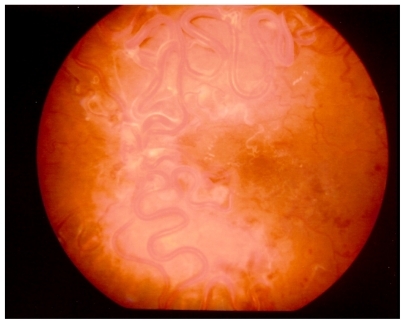
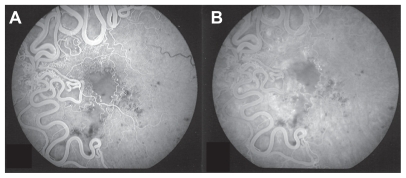
Figure 2 Early (A) and late (B) phases of fundus fluorescein angiography of the left eye showing macular ischemia and extensive arteriovenous communications and dilated intertwined vessels. There are small hemorrhages and exudates and white (sheathed) vessels in the macular area.
Comment
Congenital unilateral arteriovenous malformations of the retinal vasculature are rare. The lesions have been reported either to remain static or regressCitation1–Citation2 or to enlarge gradually. Vision may be affected directly due to macular involvement or by producing hemorrhage or exudation.Citation3,Citation4 Based on Archer et al the angioma in our case was grade 3, although there were no systemic findings.Citation5 We present a case of racemose hemangioma which spontaneously developed macular ischemia. Although extensive retinal ischemia has been reported to result in complications such as retinal or iris neovascularization,Citation1 in our case the macular ischemia has not expanded further over a period of 8 years. However, due to this macular ischemia the patient unfortunately lost her central vision. The etiology of the ischemia is unclear. It has been postulated that either an enlarged malformation using part of the blood supply of the retina may cause ischemia, or there was a partial thrombosis which caused circulatory stasis within the lesion.Citation6,Citation7
Disclosure
The authors report no conflict of interest in this work.
References
- BloomPALaidlawAEastyDLSpontaneous development of retinal ischaemia and rubeosis with retinal racemose angiomaBr J Ophthalmol19937721241258435415
- RundlesWZFallsHFCongenital arteriovenous (racemose) aneurysm of the retina: report of 3 casesArch Ophthalmol1951464408418
- MeyerCHRodriquezEBMennelSKlinqmullerVKrollPFunctional and anatomical investigations in racemose hemangiomaActa Ophthalmol Scand200785776477117711544
- YangCLiuYLDouHLLuXRQianFZhaoLUnilateral hemi- central retinal vein obstruction associated with retinal racemose angiomaJpn J Ophthalmol200953443543619763764
- ArcherDBDeutmanAErnestJTKrillAEArteriovenous communications of the retinaAm J Ophthalmol19737522242414697179
- TraboulsiEINeovascular glaucoma and ischemiaJ Clin Neuroophthalmol1986621262942570
- MansourAMWellsCGJampolLMKalinaREOcular complications of arteriovenous malformations of the retinaArch Ophthalmol198910722322362644928