Abstract
Objectives
Cataracts are the most common cause of blindness worldwide, with cataract surgery being the most common ophthalmic procedure. To our best knowledge, this is the first case-control study with a large number of participants to evaluate ocular blood flow in patients with cataracts.
Materials and methods
Color Doppler and duplex sonography of the orbital vessels was performed in 224 eyes of 112 patients with known bilateral age-related cataracts and in 76 eyes of 38 healthy age- and sex-matched volunteers.
Results
The mean ± (standard deviation [SD]) of peak systolic velocity (PSV) of the ophthalmic artery in patients with cataracts (34.59 ± 22.49 cm/second) was significantly different to that in controls (52.11 ± 14.01 cm/second) (P < 0.001). The mean ± SD PSV of the central retinal artery in patients with cataracts (15.31 ± 4.93 cm/second) was significantly different to that in controls (9.61 ± 5.64 cm/second) (P < 0.001).
Conclusion
The mean PSV and resistive index (RI) of the ophthalmic and central retinal arteries were lower in cataract patients when compared with normal subjects. This suggests that ocular hypoperfusion and changes in ocular hemodynamic may have a role in the formation of age-related cataracts.
Introduction
Cataracts are considered the most common cause of blindness worldwide, with cataract surgery being the most common ophthalmic procedure.Citation1,Citation2 According to World Health Organization (WHO) reports nearly 40 million people will be affected by age-related cataracts by 2020.Citation3 Cataracts have been reported to be associated with mortality; this is thought to be due to diabetes mellitus and hypertension causing both cataract and mortality.Citation4,Citation5 The most common causes for cataract development are acquired. Senile or age-related cataract is the cause for most acquired cataracts. Genetic and environmental factors also contribute to the development of cataracts,Citation6 and although the type of cataract and associated risk factors have not been fully established, age,Citation7 smoking,Citation8 diabetes mellitus,Citation9 sunlight exposure,Citation10 alcohol use,Citation11 metabolic syndrome,Citation12 and corticosteroid use,Citation13 have generally been identified. The roles of hypertension and cardiovascular risk factors still remain unclear.Citation14
Color Doppler imaging of orbital vessels is a procedure that has been recently used for detecting various ocular disorders such as glaucoma, central retinal vein occlusion, and tumors of the eye and orbit, and is gaining popularity because of improvements in detecting and measuring blood flow velocities in the orbital vessels.Citation15 Measurements of the flow velocity of the ophthalmic artery (OA), central retinal artery (CRA), central retinal vein (CRV), and posterior ciliary arteries are possible using color Doppler ultrasonography.Citation16 It is believed that there are changes in ocular blood flow in patients with cataracts. Only one case-control study which involved a small number of patients unmatched with respect to age has been performed previously to establish this relationship,Citation17 and other reports studied the effects of local anesthesia on ocular blood flow.Citation18–Citation20 To our best knowledge, this is the first case-control study with a large number of participants to evaluate ocular blood flow in patients with cataracts. In this study, we tried our best to exclude all confounding factors that may have interfered with the association between ocular blood flow and cataract development.
Materials and methods
Color Doppler and duplex sonography of the orbital vessels was performed in 224 eyes of 112 patients with known bilateral age-related cataract and in 76 eyes of 38 healthy age-and sex-matched volunteers. Informed consent was obtained from all subjects before examination and the University Review Board and Ethic Committee approved the study.
Cataracts were diagnosed on the basis of complete ophthalmologic examination by an experienced ophthalmologist. Patients with high myopia, retinal and vitreous pathology traumatic cataract, steroid treatment, hypertension, diabetes mellitus, history of smoking, any systemic disease, previous cataract surgery or any ocular surgery, and corticosteroid therapy were excluded from the study.
One radiologist experienced in Doppler sonography performed all ultrasound examinations. The sonographer was blinded to the presence of cataract or any relevant clinical data about the patients and the controls.
The patients were examined in a supine position with both eyes closed. The transducer was applied gently by using a sterile coupling gel to the closed eyelids; care was taken to avoid applying any pressure to the eye. We ask the patients to look forward during the ultrasound examination to prevent eye movements.
Ultrasound examination was performed on the sagittal-oblique plane to determine the optimal long axis of the orbital vessels posterior to the eye. To accurately measure flow velocity, the Doppler angle was kept between 30° and 60°. The proximal and distal portions of the vessels were imaged to facilitate the determination of the Doppler flow angle and the estimation of true flow velocity.
The ophthalmic artery enters the orbit via the optic foramen, lateral and slightly inferior to the optic nerve. After crossing the superior margin of the nerve, it typically proceeds anteriorly on the medial side of the orbit.
Because the anatomy of the orbital vasculature is highly complex and it is not possible to evaluate the ophthalmic artery at the canalicular portion, we measured the Doppler indices after the ophthalmic artery was crossed by the optic nerve (intraorbital continuation of ophthalmic artery) close to the medial wall of the orbit, since it is the best place to set the Doppler angle below 60°.
Doppler indices in the intraorbital continuation of the ophthalmic and central retinal arteries were measured with a Siemens Sonoline G-40 ultrasound device using a 10 MHz linear phase array transducer (Munich, Germany). Flow settings were chosen according to the velocity of the vessels, ie, for the ophthalmic artery, medium-to-high settings and for the central retinal artery, low-settings. Color gain and sensitivity were adjusted to minimize the artifact due to eyelid or eyeball movements.
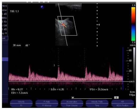
The peak systolic velocity (PSV), end diastolic flow velocity (EDV) and resistive index (RI) (RI = PSV-EDV/PSV) were measured in the central retinal artery () and the intraorbital continuation of ophthalmic artery (). Examinations took about 10 minutes on average. Each measurement was repeated three times and the mean of each measurement was calculated.
Figure 1 Color Doppler ultrasonogram showing central retinal artery spectral Doppler waveform in a cataract patient. The resistive index was 0.61.
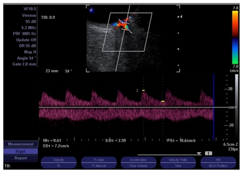
Figure 2 Color Doppler ultrasonogram showing central retinal artery spectral Doppler waveform of the ophthalmic artery in a normal subject. The resistive index was 0.77.
Statistical significance was set at P < 0.05. SPSS for Windows (v 16.0; SPSS Inc, Chicago, IL) was used for all data analyses. Continuous variables were reported as mean ± (standard deviation [SD]) and categorical variables were reported as frequencies. Independent t-tests were used for analysis and a P value of less than 0.05 was considered statistically significant.
Results
Our study included 112 patients with 224 eyes (53 female, 59 male) and 38 controls with 76 eyes (17 female, 21 male). The mean ± SD age of patients with cataracts was 67.11 ± 9.6 years and for the control group was 69.86 ± 10.84 years. There was no significant difference between the groups in terms of sex and age distribution (P = 0.6).
The mean ± SD PSV of the ophthalmic artery in patients with cataracts (34.59 ± 22.49 cm/second) was significantly different to that in the controls (52.11 ± 14.01 cm/second) (P < 0.001).
The mean ± SD PSV of the central retinal artery in patients with cataracts (15.31 ± 4.93 cm/second) was significantly different to that in the controls (9.61 ± 5.64 cm/second) (P < 0.001).
The mean ± SD RIs in OA and CRA in patients with cataract were 0.67 ± 0.07 and 0.61 ± 0.09, respectively and in controls were 0.81 ± 0.04 and 0.78 ± 0.05, respectively. Both differences were statistically significant (P < 0.001). All duplex indices are summarized in .
Table 1 Duplex indices in the central retinal and ophthalmic arteries in cases and controls
Discussion
Cataract is defined as any opacity in the lens and is the leading cause of visual impairment worldwide. Because of recent advances in treatment of infectious causes of blindness, the percentage of age-related causes of blindness is increasing.Citation21 The prevalence of cataracts is 50% in the 65–74 years age group and 70% after 75 years of age.Citation1 Apart from aging, there are factors in developing cataract such as; trauma, toxins, diabetes mellitus, corticosteroid use, smoking, and hereditary cataracts.Citation1
The pathophysiology of cataract development is not clearly defined. Some factors include oxidative damage from free radicals, ultraviolet radiation from sunlight, and malnutrition.Citation22 Age-related cataracts are classified as nuclear sclerosis, cortical cataracts, and posterior subcapsular cataracts. Citation1 Glare and visual impairment are common symptoms of age-related cataracts.
For the past 40 years, ultrasound has been used as a diagnostic tool for diseases of the eyeball and orbit. The earliest application of ultrasound in orbital pathology was amplitude (A) mode devices in which echoes appeared as spikes, the magnitude of which were dependent on the density of the reflecting tissue. Next, ultrasound was developed to detect intraocular tumors, vitreous hemorrhages, retinal detachment, and foreign bodies.Citation4 Color and duplex Doppler imaging are used for the evaluation of normal vascular anatomy, vascular flow in ocular tumors, traumatic injury, and detection of vascular malformations and arteriovenous fistulae. Modern ultrasound devices have been improved regarding image resolution and are an important part of ophthalmologic evaluations.
Peak systolic velocity, end diastolic velocities, and RI that are independent of the Doppler angle are commonly measured. RI provides quantitative measurement of flow patterns for evaluation and for comparing the resistance of the vascular bed.
Because aging has a major role in the development of age-related cataractsCitation4,Citation8 we enrolled age-matched control subjects in order to have a homogenous study population.
According to previous studies, smoking has a direct relationship with the development of nuclear cataracts, and this correlation is dose dependent.Citation4,Citation7,Citation9 A previous study has revealed that the effect of smoking on the formation of cataracts is limited to those with nuclear cataracts rather than non-nuclear cataracts.Citation8 Because of controversies about the role of smoking in cataract formation, we selected patients and controls without a history of smoking to omit the possible role of this confounding factor.
Based on our study results, the mean RI of the ophthalmic and central retinal arteries in patients with cataracts is lower compared to the control group. In this report, we measured RI as an indication of the quality of blood flow to the orbit and not ocular blood volume, which is a quantitative assessment.
An earlier study showed that changes in ocular blood flow could be a risk factor in the development of glaucoma.Citation23 We know that one of the main theories about the etiology of age-related cataracts is oxidative damage, and some recent studies have suggested using carotenoids for protection.Citation1 According to this theory, hypoperfusion and reduction in oxygen and delivery of nutrients to the eyes may have an important role in the formation and elimination of free radicals.
The results of PSV in the ophthalmic artery in our study are in agreement with the results of another study. Grieshaber et alCitation17 performed a study on a small series of patients with cataracts (confounding factors were not excluded and patients and controls were not age-matched) and showed that the mean PSV was significantly lower in cataract patients than in controls. The mean RI of the ophthalmic artery in both groups was not statistically different.Citation17 In contrast, our study showed that the mean RI in the two groups was statistically different (P < 0.001). One of the limitations of our study is the absence of grading of the cataracts and its correlation with the Doppler study.
In conclusion, the mean PSV and RI of the ophthalmic and central retinal arteries in patients with cataracts were lower than in normal subjects. This suggests that ocular hypoperfusion and changes in ocular hemodynamics may play roles in the formation of age-related cataracts.
Disclosure
The authors report no conflicts of interest in this work.
References
- WestSKLooking forward to 20/20: a focus on the epidemiology of eye diseasesEpidemiol Rev2000221647010939008
- KeeffeJETaylorHRCataract surgery in Australia 1985–1994Aust N Z J Ophthalmol19962443133178985542
- BrianGTaylorHCataract blindness – challenges for the 21st centuryBull World Health Organ200179324925611285671
- WestSKMunozBIstreJMixed lens opacities and subsequent mortalityArch Ophthalmol2000118339339710721963
- WangJJMitchellPSimpsonJMCummingRGSmithWVisual impairment, age-related cataract, and mortalityArch Ophthalmol200111981186119011483087
- HammondCJSniederHSpectorTDGilbertCEGenetic and environmental factors in age-related nuclear cataracts in monozygotic and dizygotic twinsN Engl J Med2000342241786179010853001
- LeskeMCSperdutoRDThe epidemiology of senile cataracts: a reviewAm J Epidemiol198311821521656349331
- CummingRGMitchellPAlcohol, smoking, and cataracts: the Blue Mountains Eye StudyArch Ophthalmol199711510129613039338677
- RobmanLTaylorHExternal factors in the development of cataractEye (Lond)200519101074108216304587
- WestSKDuncanDDMunozBSunlight exposure and risk of lens opacities in a population-based study: the Salisbury Eye Evaluation projectJAMA199828087147189728643
- MunozBTajchmanUBochowTWestSAlcohol use and risk of posterior subcapsular opacitiesArch Ophthalmol199311111101128424707
- NirmalanPKRobinALKatzJRisk factors for age related cataract in a rural population of southern India: the Aravind Comprehensive Eye StudyBr J Ophthalmol200488898999415258010
- CummingRGMitchellPLeederSRUse of inhaled corticosteroids and the risk of cataractsN Engl J Med199733718149203425
- KleinBEKleinRLeeKEDiabetes, cardiovascular disease, selected cardiovascular disease risk factors, and the 5-year incidence of age-related cataract and progression of lens opacities: the Beaver Dam Eye StudyAm J Ophthalmol199812667827909860001
- TranquartFBergesOKoskasPColor Doppler imaging of orbital vessels: personal experience and literature reviewJ Clin Ultrasound200331525827312767021
- SteigerwaltRDJrLauroraGIncandelaLCesaroneMRBelcaroGVDe SanctisMTOcular and orbital blood flow in cigarette smokersRetina200020439439710950419
- GrieshaberMCKocakIDublerBFlammerJOrgulSRetrobulbar blood flow in patients with cataractBr J Ophthalmol200690121512151516885186
- ChangBYHeeWCLingRBroadwayDCBeigiBLocal anaesthetic techniques and pulsatile ocular blood flowBr J Ophthalmol200084111260126311049951
- HuberKKRemkyAEffect of retrobulbar versus subconjunctival anaesthesia on retrobulbar haemodynamicsBr J Ophthalmol200589671972315923508
- WatkinsRBeigiBYatesMChangBLinardosEIntraocular pressure and pulsatile ocular blood flow after retrobulbar and peribulbar anaesthesiaBr J Ophthalmol200185779679811423451
- GlynnRJRosnerBChristenWGEvaluation of risk factors for cataract types in a competing risks frameworkOphthalmic Epidemiol20091629810619353398
- HennisAWuSYNemesureBLeskeMCRisk factors for incident cortical and posterior subcapsular lens opacities in the Barbados Eye StudiesArch Ophthalmol2004122452553015078670
- FlammerJOrgulSCostaVPThe impact of ocular blood flow in glaucomaProg Retin Eye Res200221435939312150988