Abstract
Purpose
To report a case of a patient receiving tamoxifen with visual deterioration and describe the unusual optical coherence tomography (OCT) findings.
Method
Observational case report.
Results
A 55-year-old female patient was referred to our department complaining of gradual visual deterioration in both eyes. Medical history was unremarkable apart from breast cancer for which she had received tamoxifen for 10 years (mean dosage 20 mg/day). Best corrected visual acuity was 20/400 in her right eye and 20/40 in her left eye. Fundoscopy in both eyes was without any obvious signs of maculopathy. However, Fourier-domain OCT demonstrated bilateral extensive areas of disruption in inner retinal layers without any signs of crystalline retinopathy. Six months after the cessation of tamoxifen, the situation remains unchanged.
Conclusion
Patients receiving tamoxifen should be monitored with high-resolution OCT for fundoscopically invisible changes in the inner retinal layers, the progression of which may seriously affect the patient’s vision and subsequently their quality of life.
Keywords:
Introduction
Optical coherence tomography (OCT) is an interferometric imaging technique which produces cross-sectional images of the retina and in recent years has been widely used in the study of macular diseases.Citation1,Citation2 Tamoxifen is a nonsteroidal antiestrogen, which belongs to the family of selective estrogen receptor (ER) modulators, drugs that have the ability to occupy ERs, acting as ER antagonists in breast tissue. As a result, tamoxifen is widely used as adjuvant endocrine therapy for women with hormone-responsive breast cancer.Citation3
Incidence of ocular toxicity among patients receiving tamoxifen is rare (0.6%); cataract, vortex keratopathy, optic neuritis and retinopathy are the most common manifestations.Citation3–Citation5
We report, herein, a case of a 55-year-old woman who presented with bilateral, severe visual deterioration after treatment with tamoxifen and describe the Fourier-domain OCT findings.
Case report
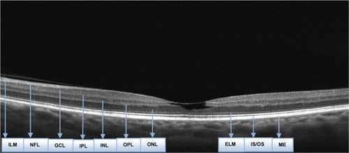
A 55-year-old female patient was referred to our department complaining of gradual visual deterioration in both eyes. She had been seen by her local optician and ophthalmologist who had failed to explain the reduction in her vision. Medical history was unremarkable apart from breast cancer, diagnosed in 2002, for which she received tamoxifen for 10 years (mean dosage 20 mg/day), which was withdrawn only 15 days prior to her visit. The reason for the prolonged reception of tamoxifen is not known, since her oncological medical record was not available. The patient’s best corrected visual acuity at presentation was 20/400 in her right eye and 20/40 in her left eye. Intraocular pressure was 17 and 13 mmHg in her right and left eye, respectively. Examination of the anterior segment was unremarkable. Fundoscopy in both eyes was without any obvious signs of maculopathy (). Posterior vitreous was attached to the retina in both eyes. Fourier-domain OCT was used for examination of the fovea; it demonstrated bilateral extensive areas of disruption in inner retinal layers ( and ). The OCT type that was used for the diagnosis was RTVue model RT-100 by Optovue (Fremont, CA, USA). Through the OCT, the dimensions of the foveal cystoid spaces were measured and found to be 78 μm × 823 μm in the right eye and 74 μm × 607 μm in the left eye. Measurement was achieved by using the manual program of the instrument. Fluorescein angiography did not show any leakage and excluded the diagnosis of type 2 retinal telangiectasia. Six months after the cessation of tamoxifen, the situation remains unchanged.
Figure 1 Optical Coherence Tomography of the right eye.
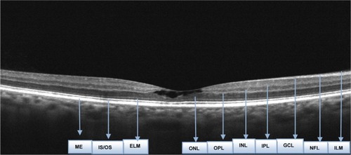
Figure 2 Optical Coherence Tomography of the left eye.
Discussion
OCT is an interferometric imaging technique which produces cross-sectional images by mapping the depth-wise reflections of low-coherence laser light from tissue. Fourier- or spectral-domain OCT refers to Fourier transformation of the optical spectrum of the low-coherence interferometer. Imaging of multilayer objects, such as retina, results in various modulation periodicities representing the depth of each layer, with amplitude of the spectrum modulation proportional to the reflectivity of the layer. The greatest advantage of spectral-domain OCT, over conventional time-domain OCT is the increase in scan speed. With the spectral-domain OCT, imaging with 25,000–100,000 axial scans per second is routinely possible. This is more than 100 times faster than the time-domain technique.Citation1,Citation2
Tamoxifen is a nonsteroidal antiestrogen, which belongs to the family of selective ER modulators, drugs that have the ability to occupy ERs (ERα and ERβ), acting as ER antagonists in breast tissue. As a result, tamoxifen is widely used as adjuvant endocrine therapy for women with breast cancer. ERα and ERβ are also present within the neural retina and the pigment epithelium of men and women, where tamoxifen is reported to decrease glutamate uptake.Citation3
Incidence of ocular toxicity among patients receiving tamoxifen is rare, approximately 0.6%. Ocular manifestations of tamoxifen include tamoxifen retinopathy, corneal epithelial deposits which can lead to vortex keratopathies, induction of posterior subcapsular cataract, and clinically evident optic neuritis.
Tamoxifen retinopathy is typically characterized by the presence of small refractile deposits in the nerve fiber and inner plexiform layers, primarily in the perifoveal area. Refractile deposits do not cause significant visual impairment; histopathologically, they are suggested to represent axonal degeneration, mainly in the nerve fiber layer in the parafoveal region. Due to tamoxifen’s amphiphilic nature, it is suggested that it binds to polar lipids, accumulates in lysosomes, and causes cell oxidative damage.Citation4–Citation7 Numerous researchers have suggested that tamoxifen retinopathy is not caused by actions of tamoxifen on ERs, but stems instead from tamoxifen’s cationic amphiphilic properties, which resemble those of chloroquine.Citation3
All cases of tamoxifen maculopathy studied with spectral-domain OCT in the literature include crystalline maculopathy with refractile deposits, accompanied by unilateral foveal cysts of small size.Citation7–Citation10 In all cases, the described lesions consisted of hyperreflective substances in combination with outer retinal atrophy, photoreceptor disruption, and defects in the outer retinal layers which caused the decrease in the visual acuity of the patients. In our case, interestingly, the retinal damage concerned only the inner layers, without any refractile deposits or outer retinal defects, giving the impression of a pseudohole from which tamoxifen maculopathy can be differentiated by the absence of posterior vitreous detachment and the normal foveal contour. The absence of refractile deposits, posterior vitreous detachment, and outer retinal defects was responsible for the almost “normal appearance” of the macula in the fundoscopy. However, the permanent bilateral reduction in the vision of our patient associated only with subtle fundoscopic changes emphasizes the role of early detection of inner foveal damage with high-resolution OCT. Patients receiving tamoxifen should be monitored with high-resolution OCT for fundoscopically invisible changes in the inner retinal layers, the progression of which may seriously affect a patient’s vision and subsequently their quality of life.
Disclosure
The authors report no conflicts of interest. The authors alone are responsible for the content and writing of the paper.
References
- KiernanDFMielerWFHariprasadSMSpectral-domain optical coherence tomography: a comparison of modern high-resolution retinal imaging systemsAm J Ophthalmol20101491183120103039
- WolfSWolf-SchnurrbuschUSpectral-domain optical coherence tomography use in macular diseases: a reviewOphthalmologica2010224633334020453539
- EisnerALuohSWBreast cancer medications and vision: effects of treatments for early-stage diseaseCurr Eye Res2011361086788521819259
- HagerTHoffmannSSeitzBUnusual symptoms for tamoxifen-associated maculopathyOphthalmologe20101078750752 German20393723
- SrikantiaNMukeshSKrishnaswamyMCrystalline maculopathy: a rare complication of tamoxifen therapyJ Cancer Res Ther20106331331521119261
- ChungHKimDAhnSHEarly detection of tamoxifen-induced maculopathy in patients with low cumulative doses of tamoxifenOphthalmic Surg Lasers Imaging2010915 Epub392010
- TolerSMOxidative stress plays an important role in the pathogenesis of drug-induced retinopathyExp Biol Med (Maywood)2004229760761515229354
- Mauget-FaÿsseMGambrelleJQuaranta-ElMaftouhiMOptical coherence tomography in tamoxifen retinopathyBreast Cancer Res Treat200699111711816541311
- GianniLPanziniILiSInternational Breast Cancer Study Group (IBCSG)Ocular toxicity during adjuvant chemoendocrine therapy for early breast cancer: results from International Breast Cancer Study Group trialsCancer2006106350551316369994
- GualinoVCohenSYDelyferMNSahelJAGaudricAOptical coherence tomography findings in tamoxifen retinopathyAm J Ophthalmol2005140475775816226541