Abstract
Purpose
To study the anatomical and visual outcomes and prognostic factors that may predict the outcomes of rhegmatogenous retinal detachment (RRD) in children.
Methods
A retrospective chart review was performed for patients 16 years of age or younger who underwent retinal reattachment surgery for RRD at the King Abdulaziz University Hospital from 1996 to 2005 and the King Khalid Eye Specialist Hospital from 2002 to 2006, Riyadh, Saudi Arabia. Good visual outcome was defined as ≥20/200. The association between two categorical variables was evaluated with the Chi-squared test or the exact test, as appropriate. Predictors for RRD and good final visual acuity were identified by conducting stepwise logistic regression analysis. P < 0.05 was statistically significant.
Results
The study population comprised 148 patients (166 eyes). There were 104 (70%) males and 44 (30%) females. Mean age at presentation was 8.33 ± 3.26 years (range 1.5–16 years). The retina was reattached after one surgical procedure in 106 (63.8%) eyes and reattached in 130 (78.3%) eyes after multiple surgeries. Factors predicting recurrence after the first surgery were myopia (P = 0.028), proliferative vitreoretinopathy (PVR) at presentation (P = 0.024), and total retinal detachment (P = 0.032). Good final visual outcome was achieved in 60 (44.4%) eyes. Predictors of good visual acuity were: good visual acuity at presentation (P < 0.001); absence of PVR at presentation (P < 0.001); one quadrant of retinal detachment (P = 0.0024); macula on (P = 0.0107); absence of primary repair of a ruptured globe (P = 0.0059); no pars plana vitrectomy (PPV) (P = 0.0123); clear phakic lens at follow-up (P < 0.001); absence of postoperative complications (P < 0.001); absence of recurrence of RRD (P < 0.001); and absence of epiretinal membrane (P = 0.0088). Logistic regression analysis indicated that recurrence of RRD was associated with myopia and previous congenital cataract surgery; good final visual outcome was associated with macula on detachment and poor visual outcome was associated with recurrence of RRD and occurrence of postoperative complications and previous repair of a ruptured globe.
Conclusion
RRD in children is usually associated with a predisposing factor, a high rate of PVR, and total retinal detachment. Despite late diagnosis and the presence of PVR, favorable anatomical and visual outcomes can be achieved.
Background
Retinal breaks are present in 5%–10% of the general public; however, very few lead to rhegmatogenous retinal detachment (RRD). In the general population, the estimated incidence of RRD is one in 10,000. After an RRD in one eye, the risk of RRD is approximately 10% in the fellow eye. Pediatric retinal detachment has been reported in numerous studies.Citation1–Citation38 The incidence of pediatric retinal detachments is low, accounting for approximately 1.7%–5.9% of all retinal detachments.Citation2–Citation8 There is a greater preponderance of pediatric retinal detachment in males (70%–79% of all cases). The most common type of retinal break is a horseshoe tear.Citation7
Etiological factors for RRD in children include congenital or structural abnormalities, trauma, myopia, previous ophthalmic surgery, and a positive family history.Citation13 Congenital and developmental structural abnormalities play a major role in the pathogenesis of pediatric RRD, and may be more common than previously reported.Citation7–Citation16,Citation35–Citation38 Associations have been reported for familial exudative vitreoretinopathy, congenital retinoschisis, Coats’ disease, retinopathy of prematurity,Citation26–Citation28,Citation38 Marfan syndrome, Stickler syndrome,Citation29–Citation31 morning-glory disk anomaly,Citation24 retrolental fibroplasia, X-linked retinoschisis, CHARGE (syndrome of colobomatous microphthalmia, heart defects, choanal atresia, retarded growth, genital abnormalities, and ear abnormalities), Sturge–Weber syndrome, retinoblastoma, microspherophakia, buphthalmos, neurofibromatosis type I,Citation9 and mental and growth retardation were reported.Citation1–Citation16 Traumas, as well as congenital and developmental anomalies, are also associated with myopia.Citation1–Citation12
Trauma has been reported as the major cause of RRD in children worldwide. Blunt, penetrating, or high-velocity objects, and surgical iatrogenic trauma or surgical procedures for congenital cataract, cryotherapy, or laser photocoagulation in retinoblastoma, retinopathy of prematurity,Citation24 and other procedures have also been implicated as a cause of RRD.Citation7–Citation16 Retinal detachment secondary to child abuse was first reported by Kiffney.Citation17
The outcome of surgical repair in pediatric RRD depends on etiology, the chronic nature of the RRD, and the intended procedure. Selection of the surgical procedure can be limited, especially when considering the type of tamponade. Silicone oil is more reliable than gas, because of the difficulties in keeping a child in one position for a prolonged period of time. Reoperation may be dependent on etiology, surgical procedure technique, and intra- or postoperative complications.
The most common complication after surgical repair of RRD is proliferative vitreoretinopathy (PVR). Khvatova et alCitation34 reported on 55 eyes that had undergone reoperation for RRD, and stated that the efficacy of the surgical treatment depends on the origin of the disease and the severity of the PVR. In the current study, we investigate the anatomical and visual outcomes and prognostic factors that may predict outcomes of RRD in children.
Patients and methods
Approval to conduct the study from the Research and Medical Ethics Committees at King Khalid Eye Specialist Hospital was granted for this study. The patient charts were identified for the 5 consecutive years (January 2001–December 2005) under study and reviewed. At King Abdul-Aziz University Hospital, ethics approval from the Ophthalmology Department was obtained. The patient charts and the surgical logbooks for 10 consecutive years spanning January 1995 to December 2004 were reviewed.
To be included in the study, patients had to be 16 years old or younger and had undergone surgical repair for RRD and completed 1 year of follow-up. The data-collection sheet was designed to collect parameters based on a current literature review. The data collected included patient age, sex, complaints, duration of symptoms, previous ophthalmic history, history of trauma, history and details of previous ophthalmic surgery, eye affected, ocular family history, visual acuity (VA) at presentation (preoperatively) and at last postoperative visit, intraocular pressure (IOP) at presentation, refractive error or spectacle prescription, strabismus, lens and vitreous status, extent of retinal detachment and status of the macula, presence of PVR, type and location of retinal holes and breaks, dialysis, and peripheral retinal changes. Data were collected for both eyes. The type of surgical procedure and operative notes were reviewed, including examination under anesthesia, type of procedure, and tamponade.
Data were collected for postoperative complications and management, with specific attention to the recurrence of RRD. Final anatomical and visual outcomes were analyzed and evaluated for presenting symptoms, procedures, complications, and number of surgeries performed.
Statistical methods
All data were manually entered into the data-collection sheet, then digitally entered and analyzed. The association between two categorical variables was investigated using either the Chi-squared test or the exact test as appropriate. Two proportions from the same sample were compared using Student’s t-test. P < 0.05 was considered statistically significant.
Predictor variables for RRD and good final VA of 20/200 or better were identified by conducting stepwise logistic regression analysis. In this analysis, all variables that were investigated as risk factors for RRD and final VA in univariate analysis were included as significance of a selected predictor variable and judged by computation of 95% confidence intervals (CIs) around the odds ratio (OR) for that variable. CIs that did not include a value of 1.0 indicated statistical significance. Statistical analyses were performed with SPSS version 13 (IBM, Armonk, NY, USA), StatsDirect (Altrincham, UK) and BMDP 2007 (Statistical Solutions, Saugus MA, USA) software.
Results
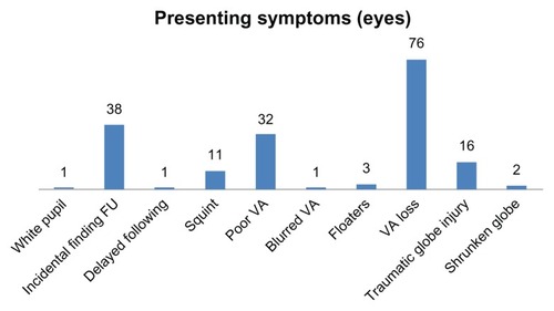
A total of 668 patient charts were reviewed, of which only 166 eyes of 148 patients fitted the inclusion criteria. There were 104 (70%) males and 44 (30%) females (M:F = 2.36) (). The age at presentation was 8.33 ± 3.26 years (range 1.25–16 years). Right and left eyes were equally affected. Mean follow-up was 40.14 ± 27.06 months (range 12–156 months). The mean duration of symptoms was 5 ± 277 days (range 1–1460 days), and the median was 17 days. Presenting symptoms varied from being asymptomatic (ie, incidental finding in routine exam or follow-up) in 38 (23%) eyes, floaters in three (1.8%) eyes, decrease or loss of vision in 76 (45.7%) eyes, traumatic globe injury in 16 (9.6%) eyes, and shrunken globe in two (1.2%) eyes ().
Figure 1 Patient demographics of children who presented with rhegmatogenous retinal detachment.
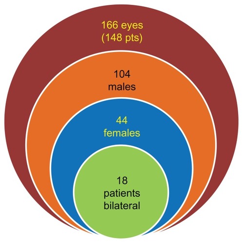
Figure 2 Presenting symptoms of children with rhegmatogenous retinal detachment.
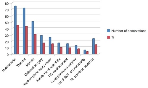
Ocular history showed 45.18% of patients with multiple predisposing factors. Seventy-two (43.3%) eyes had a history of trauma, 51 (30.7%) eyes were myopic, 29 eyes (17.5%) had a history of cataract surgery, and 13 eyes (7.8%) had a history of surgery for congenital glaucoma. There was a history of globe-rupture repair in 26 (15.7%) eyes, retinopathy of prematurity and history of preterm delivery in six (3.6%) eyes, previous retinal reattachment surgery in 16 (9.6%) eyes, and no previous ocular history in 24 (14.45%) eyes. Seventeen (10.2%) eyes had a family history of retinal reattachment surgery ().
Figure 3 Predisposing factors of rhegmatogenous retinal detachment in children.
Ophthalmic, congenital, and developmental associations were found in 31% of the patients. These associations included Stickler syndrome in 26 (15.6%; bilateral in four patients) eyes, choroidal coloboma in five (3%) eyes, and microphthalmia in three (1.8%) eyes (). One (0.6%) eye presented with RRD associated with high IOP (Schwartz’s syndrome), and one (0.6%) eye presented with RRD, choroidal detachment, and severe hypotony ().
Table 1 Ophthalmic and congenital/developmental associations of rhegmatogenous retinal detachment in children
At presentation, VA was measured in 151 (91%) eyes. Presenting VA ranged from 20/20 in two (1.2%) eyes to light perception (LP) in 27 (16.27%) eyes. Presenting VA was grouped into VA ≥ 20/200, Counting Fingers (CF), and no light perception (NLP)/LP-hand motion (HM). Thirty-six (23.7%) eyes had ≥20/200 at presentation, 52 (31%) eyes presented with CF, and 58 (35%) eyes had VA of LP-HM. In 15 eyes of younger children where VA charts were not applicable, ten (6%) eyes were able to fixate and follow, and five (3.3%) eyes had poor fixation. Twenty-five (15%) eyes had strabismus preoperatively.
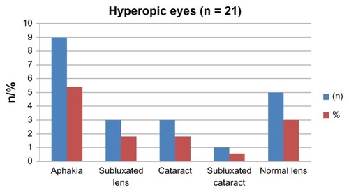
Refraction was documented in 73 (44%) eyes, with mean spherical equivalent preoperatively −5.07 ± 10.20 D (range −24 to 14.5 D); the median was −7.25 D. Fifty-one (30.7%) eyes were myopic (), and 21 (12.6) eyes were hyperopic. Myopic eyes were divided into eyes with a spherical equivalent of −6 D or less (low myopia; eleven [6.6%] eyes) and spherical equivalent greater than −6 D (high myopia; 40 [24%] eyes). In the high-myopia group, the mean spherical equivalent was −13.1 ± 4.49 D (range −6 to −24 D), with a median of −13 D. highlights some features of the myopia group. shows the lens status in the hyperopic group.
Figure 4 Manifest refractive spherical equivalent in children with rhegmatogenous retinal detachment.
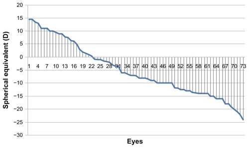
Figure 5 Features of rhegmatogenous retinal detachment in myopic children.
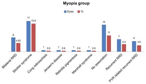
Figure 6 Lens status in hyperopic children.
Seven (46.7%) of the myopic eyes with no other associated pathology had a recurrence of RRD postoperatively, and seven (46.7%) eyes developed PVR, with six (86%) progressing to RRD.
Preoperatively, complete retinal detachment was present in 109 (65.4%) eyes, one-quadrant detachment was present in 20 (12%) eyes, a detachment in two quadrants was present in 17 (10%) eyes, and a detachment in three quadrants was present in 19 (11.7%) eyes. Macula-off retinal detachments were present in 130 (78.35%) eyes. Lattice degeneration was found in 18 (10.8%) eyes ().
Figure 7 Presenting retinal features of the affected eye of children with rhegmatogenous retinal detachment.
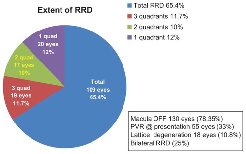
PVR at presentation occurred in 55 (33%) eyes. Eighteen of these eyes (33%) were grade C or worse, and in four eyes the PVR was located in the posterior pole.
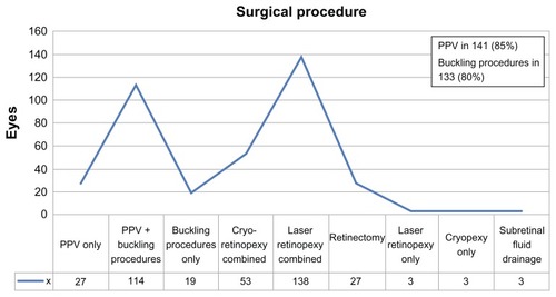
The primary (first) surgery was pars plana vitrectomy (PPV) in 141 (85%) eyes. Twenty-seven (16.26%) eyes underwent PPV only. PPV combined with encircling band was performed in 114 eyes (80% of eyes that underwent PPV and 68% of the total primary procedures). Buckling procedures alone as primary surgery were performed in 19 (11.5%) eyes, seven (4.2%) eyes underwent an encircling band procedure only, nine (5.4%) eyes underwent band and segmental buckle, and three (1.8%) eyes underwent a segmental buckle only. Six patients with localized RRD were treated by either laser alone (three [1.8%] eyes) or cryotherapy alone (three [1.8%] eyes). Subretinal fluid drainage was performed in three (1.8%) eyes, and 27 (16.3%) eyes underwent a retinectomy ().
Figure 8 Primary (first) surgical intervention in children with rhegmatogenous retinal detachment.
Abbreviation: PPV, pars plana vitrectomy.
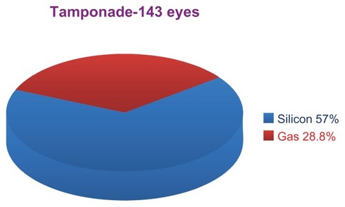
Silicone oil intravitreal tamponade during primary surgery was performed in 96 (57.8%) eyes, and gas (C3F8 or SF6) tamponade was performed in 48 (28.8%) eyes, including two (1.2%) eyes that underwent a buckling procedure and an injection of a gas bubble ().
Figure 9 Intraocular tamponade.
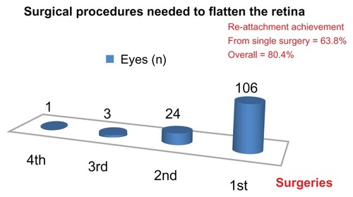
Intraoperatively, the retina was completely flattened in 156 eyes (ten operative reports did not note retinal status). Retinal reattachment after the first procedure occurred in 106 (63.8%) eyes. Overall, retinal reattachment after multiple surgeries occurred in 134 (80.4%) eyes. ().
Figure 10 Number of surgeries required to flatten the retina in children with rhegmatogenous retinal detachment.
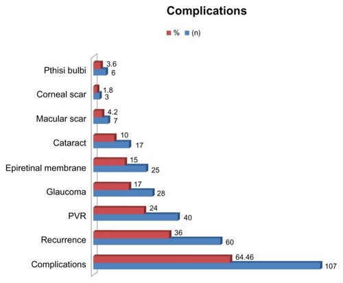
Postoperative complications () after the first retinal reattachment surgery occurred in 107 (64.46%) eyes. Postoperatively, recurrent RRD was present in 60 (36%) eyes, of which PVR was the cause in 40 (24%) eyes; eleven (6.6%) eyes with postoperative PVR did not have PVR preoperatively. Postoperative cataract was present in 17 (10%) eyes, and glaucoma in 28 (17%) eyes (three eyes had undergone previous glaucoma surgery). Postoperatively, epiretinal membranes were present in 25 (15%) eyes; in twelve (7.2%) of these eyes, the membranes were part of PVR. Postoperatively, seven (4.2%) eyes had a macular scar, three (1.8%) eyes had a corneal scar, and six (3.6%) eyes became phthisical.
Figure 11 Rates of complications in children who underwent retinal reattachment surgery for rhegmatogenous retinal detachment.
Abbreviation: PVR, proliferative vitreoretinopathy.
Modalities to treated postoperative glaucoma included medical therapy (topical drops) for ten (6%) eyes, surgical intervention for two (1.2%) eyes, and medical and surgical intervention for 16 (9.6%) eyes. Surgical management for glaucoma included cyclophotocoagulation, removal of silicone oil when indicated, and tube or filtering procedures. Lensectomy was performed during the primary procedure (PPV) in 85 (51.2%) eyes. Of these eyes, 40 (24.1%) had cataract, six (3.6%) had a subluxated lens, two (1.2%) had phacodonesis, one (0.6%) had a lens coloboma, and 32 (19.3%) had a clear physiologic lens preoperatively.
The outcomes of univariate analysis for predisposing factors for recurrence of RRD after the first reattachment surgery are presented in . Predictors of recurrence after successful primary surgery were myopia (P = 0.028), PVR at presentation (P = 0.024), and total RRD rather than partial RRD (P = 0.032).
Table 2 Risk of recurrence of rhegmatogenous retinal detachment
Predictors of good final VA (≥20/200) included good initial vision (P < 0.001), absence of PVR at presentation (P < 0.001), one-quadrant RRD (P = 0.005); macula on (P = 0.0107), absence of primary globe repair (P = 0.0059), no PPV (P = 0.0123), and clear phakic lens postoperatively (P < 0.001) (). Predictors of poor final VA included poor vision at presentation (P = 0.0033), total RRD (P = 0.0479), macular involvement (P = 0.0229), PPV (P = 0.0115), and macular scarring (P = 0.0204) ().
Table 3 Factors affecting final visual acuity
Comparison of initial and final VA is presented in . Paired data were available on 135 eyes. Sixty-eighty (50.4%) eyes had no change in VA from preoperatively to postoperatively (). VA improved in 46 (34.1%) eyes (). VA decreased in 21 (15.5%) eyes postoperatively (). There was a statistically significant increase in the prevalence of 20/200 or better VA from 23.7% at presentation to 44.4% of eyes at last follow-up (P = 0.0031, Student’s t-test for two proportions of the same sample). The prevalence of worst vision of HM-LP/NLP decreased from 37.3% of eyes at presentation to of 27.4% eyes at last follow-up (P = 0.195).
Table 4 Comparison of initial and final visual acuity
Multivariate stepwise logistic regression analysis indicated variables associated with recurrence of RRD after first surgery and for good final VA. Recurrence of RRD was associated with cataract surgery (OR = 3.25, 95% CI 0.95–11.1) and myopia (OR = 2.19 95% CI 1.06–4.56). For good final VA (≥20/200), the positive predictor was macula on (OR = 7.13, 95% CI 1.55–32.9), and negative predictors were globe repair (OR = 0.043, 95% CI 0.006–0.312), recurrence of RRD (OR = 0.124, 95% CI 0.039–0.398), and postoperative complications (OR = 0.409 95% CI 0.149–1.13).
Fellow eye
Sixty-nine (41.4%) fellow eyes were free of ocular pathology at presentation, and 97 (58.6%) eyes had multiple pathologies. At presentation, retinal detachment was documented in 47 (28.2%) eyes. These detachments were deemed inoperable in 16 eyes (9.6%); the remaining 13 eyes (7.8%) were included in this study. Four eyes had coloboma components, and six eyes had lattice degeneration, with one associated with a giant tear. Five eyes showed myopic changes, and two eyes were microphthalmic, one associated with coloboma and one with pseudo-retinitis pigmentosa. Retinitis pigmentosa associated with RRD was documented in one eye. X-linked retinoschisis was present in two eyes, vitreoretinal degeneration was present in one eye, white without pressure in one eye, empty vitreous in two eyes, one of which was associated with a giant-tear inoperable RRD, and cystic changes were found in two eyes, one of them associated with a break.
Nine fellow eyes had retinal reattachment surgeries documented on presentation. One eye had a history of recurrent tractional RD. Tears/holes were found in 17 fellow eyes (multiple breaks in four eyes, atrophic holes in one eye, one found to have a break and one with open holes, horseshoe tear found in one eye). Two giant tears (as above) were observed, one of them associated with lattice degeneration and one with empty vitreous. Three eyes underwent glaucoma surgeries; one had NLP vision, and one fellow eye was eviscerated.
Discussion
In this study of RRD in children, we found the majority of fellow eyes (approximately 60%) were also affected and could be treated early. We found a preponderance of males (70%) compared to females in the current study. This observation is consistent with previous studies that reported between 60% and 80% of males with RRD compared to females.Citation5,Citation11,Citation12 Separate studies by Yokoyama et alCitation8 and Weinberg et alCitation9 reported even higher rates of 86% and 97%, respectively. Differing enrollment criteria such as age and much smaller study sample sizes compared to our study may account for the differences in gender preponderance in our study compared to the two previous studies.Citation8,Citation9 In the trauma group, 70% were males in our study, which is similar to the 80% male figure in the trauma group reported by Winslow and Tasman.Citation2 The greater number of males in the trauma group is likely a result of greater risk-taking behavior in male children compared to females.
The presenting symptoms of RRD in children differ in nature from adult RRD. For example, pediatric patients may not be able to complain of decreased vision or loss of vision, and a significant number tend to be discovered incidentally due to chronic retinal detachments. In the current study, the most common presenting symptoms were a decrease or loss of vision in 45.7% of eyes and incidental findings in routine exam or follow-up in 38 eyes (23%). These outcomes concur with previous literature. Fivgas and CaponeCitation7 found the most frequent presenting symptom was poor vision (62%). Wang et alCitation14 reviewed 278 patients and found blurring of vision was the most common complaint. Gonzales et alCitation37 reported that 46% of patients have presenting symptoms pointing to RD.
The general consensus in the published literature is that trauma, myopia, previous ophthalmic procedures, and congenital or developmental anomalies play a major role in the etiology of RRD in children.Citation5–Citation7,Citation10–Citation12,Citation21–Citation23,Citation35–Citation38 Idiopathic or unknown cause of detachment is still a common finding in most studies. Classification as idiopathic disease could be because this age-group does not expresses the full features of the commonly associated diseases, could be related to an unwitnessed blunt trauma, or there is really is no predisposing factor. Strong evidence that there is an underlying etiology is the observation that the vast majority of cases have retinal breaks or holes found intraoperatively (in 86.75% of eyes in the current study). Additionally, if the patient had no ocular history prior to presentation, there may be at least one predisposing factor elicited via a complete examination. In the current study, eight (4.8%) eyes had no associated predisposing factor preoperatively. Of these eyes, one had a macular hole, another eye had a giant tear, two eyes had associated cataract changes, and one eye had peripheral degenerative and cystic changes in the fellow eye. Hence, only three (1.8%) of patients had an uncertain etiology of RRD. The low rate of idiopathic RRD in our study is similar to that reported by Wang et al,Citation14 who found 4.7% of 296 eyes with no predisposing factor. Similarly, Gonzales et alCitation37 found underlying retinal conditions in 98% of the 46 eyes they evaluated.
We found almost half the eyes (43.3%) had a previous history of trauma. This observation is similar to previous studies that reported between 42% and 45% of eyes with RRD had a history of trauma.Citation2,Citation7,Citation33,Citation37 The highest rate of eyes with RRD with a history of trauma is 53%.Citation36 However, lower rates than ours have been reported for eyes with RRD with a history of trauma ranging from 22% to 36%.Citation3,Citation9,Citation11,Citation14,Citation16 Differences in the sample size, sex ratios (females prone to more risk-averse behavior), and enrollment criteria likely explain the differences between our study and studies reporting a lower history of trauma. Of note, even with the lowest rate of 22%, trauma is clearly a significant factor for RRD in children.
Over half (50.6%) of eyes with RRD had undergone previous ophthalmic surgery in the current study. This observation falls well within the range reported by previous studies of 34% to 60%.Citation9,Citation7,Citation37 However, Yokoyama et al,Citation8 Wang et al,Citation14 and Chang et alCitation16 reported much lower rates of 2%, 5%, and 6%, respectively. Similar to trauma, differences in the sample size, sex ratios (females prone to more risk-averse behavior), and enrollment criteria likely explain the differences between our study and the previous three studies.Citation8,Citation14,Citation16
Thirty-one percent of the patients in the current study had developmental or ophthalmic/systemic associations with RRD. There is a wide variation in the reported association of developmental, ophthalmic, or system association that varies between 3.3% and 56%.Citation3,Citation4,Citation8,Citation9,Citation14,Citation16,Citation33,Citation37 This wide range is due to the diversity in defining these variables. For example, some studies include myopia and Stickler syndrome, whereas others class them separately. We included all the contributing developmental or congenital ophthalmic or systemic associations in one group. The variation in outcomes of our study and previous studies indicated further investigation is required with strictly established criteria of developmental, ophthalmic, or systemic associations with RRD.
Thirty percent of eyes in the current study had myopia, which is within the range of previous studies (20%–38%).Citation7,Citation11,Citation14 In 1978, Winslow and TasmanCitation2 reported 15% of 179 eyes with myopia. Similar to our study, Winslow and Tasman evaluated children 16 years of age or younger. A more recent study by Gonzales et alCitation37 reported 17% of children with RRD had myopia as a predisposing factor. In the current study, isolated myopia accounted for only 9% of eyes with RRD. The refractive error ranged from −6 to −24 D (mean −13.1 ± 4.49 D median −13 D).
We found the most common breaks were round holes and tears in 62% of eyes with RRD. These data concur with Chen et al,Citation11 who reported 71%, Butler et alCitation4 who reported 73%, and Akabane et al,Citation3 who reported 68%. Retinal dialysis was found in 20.8% of the eyes with RRD in our study population and in 27% of the eyes studied by Yokoyama et al.Citation8 However, Winslow and TasmanCitation2 reported 40% of eyes with retinal dialysis, and Akabane et alCitation3 reported a rate of 63%. Giant tears were found in 22.9% of eyes in the current study population, whereas the range in the literature varies from 6.3% to 15%.Citation3,Citation9,Citation36 Posterior breaks were documented in 17.5% of eyes in the current study and multiple holes/breaks in 22.3% of eyes. Multiple giant breaks were rare in the current study (1.2%), and no breaks or holes were identified in twelve eyes (7.22%). Macular holes were also rare, occurring in only two eyes in the current study.
PPV with or without a buckling procedure (encircling band or segmental buckle or both) was the most common primary surgical procedure (85% of cases) in the current study. Scleral buckle or band as the primary procedure was uncommon, with only 11.5% eyes undergoing this procedure. Overall scleral buckling with or without PPV was common (78% of cases) in the current study. We reported the highest rate of PPV as primary surgical procedure for pediatric RRD. This could be due the high incidence of giant tears, dialysis, and associated lens pathology (34%) at presentation. Scleral buckle seems to be the most common procedure in previous studies. For example, Winslow and TasmanCitation2 performed scleral buckle on all their patients. However, this was before the PPV era. Kocaoglan et alCitation33 reported 100% buckling surgery. Over time, there has been an increasing adoption of PPV. In order of earliest to most recent studies documenting PPV for RRD, Butler et alCitation4 reported 21% of cases, Yokoyama et alCitation8 reported 24% of cases, Chang et alCitation16 reported 38% of cases, Soheilian et alCitation35 reported 63% of cases, and Gonzales et al reported 74% of cases.Citation37 The success rate after the first surgery was 63.8%, which is well within the range reported in previous studies (25%–96%).Citation33,Citation35 The final retinal attachment rate was 80.4%, which compares favorably to the 72%–96% reported in previous literature.Citation7,Citation33 Reported overall anatomical retinal reattachment is encouraging despite late initial presentation and high rates of macular detachment and PVR at presentation. Citation3,Citation15 Vision improved from 20/200 or better at presentation in 23.7% to 44.4% at last follow-up visit. This is comparable to previous studies by Soheilian et al,Citation35 Akabane et al,Citation3 and Wang et al.Citation14
We found predictors of recurrence on univariate analysis were myopia (P = 0.028), PVR at presentation (P = 0.024), and total RRD (P = 0.032) (). Predictors of good final VA on univariate analysis were presenting VA of ≥20/200 (P < 0.001), absence of PVR at presentation (P < 0.001), and attached macula at presentation (P = 0.0107) (). The negative predictors of final VA were development of complications after first surgery (P < 0.001), specifically recurrence of RRD (P < 0.001), postoperative PVR (P < 0.001), epiretinal membrane (P < 0.001), and ruptured globe repair (P = 0.0059). Total retinal detachment found statistically significant for final vision of CF (P = 0.0479) and also for final visual acuity ≥20/200 (P = 0.0005). This outcome could be related to the high percentage of total RRD in our series.
Risk factors for poor surgical outcomes have been extensively studied. Winslow and TasmanCitation2 found that chronic RRD and the presence of PVR are predictors of poor outcome. Yokoyama et alCitation8 linked poor final outcomes to initial VA and preoperative PVR. Weinberg et alCitation9 found that predictors of poor outcome were LP, macula off, need for PPV, PVR grade C or worse, and the use of silicone oil. Wang et alCitation14 found poor surgical outcome was associated with congenital anomalies, previous intraocular surgery, PVR grade C or worse, macula off, total RRD, and the use of silicone oil. Gonzales et alCitation37 found that poor outcomes were related to younger age, worse initial vision, greater extent of RRD, and grade C PVR or worse. Chang et alCitation16 performed logistic regression analysis and reported that nonmyopic RRD, macular involvement, and PVR are risk factors for poor surgical outcomes. The poorer outcome with silicone oil could be related to the severity of the detachment itself, for which silicone is used as a tamponade.Citation9 PVR incidence in children is 29.8%–37.5%, while in adults it is 5%–10%, possibly because of delayed diagnosis.Citation3
Our model of logistic regression related recurrence of RRD to congenital cataract surgery and myopia. Logistic regression indicating good final VA was associated with macula-on retinal detachment. Negative predictors were ruptured globe repair, recurrence of RRD, and postoperative complications.
In conclusion, pediatric RRD is a multifactorial condition in which late presentation and recurrence after surgical repair significantly affect both anatomical and visual outcomes. There is a recent trend toward PPV combined with scleral buckling, possibly due to better surgical results in cases that were deemed inoperable in the past. Finally, a good number of patients will have VA better than 20/200 if they undergo timely surgery.
Acknowledgments
Delia Pilapil, Dustan Kangave, and Rich Bains, for their contribution to data entry and analysis.
Disclosure
The authors report no financial conflicts in this project. The data were presented in a resident’s thesis in June 2009 at the King Khaled Eye Specialist Hospital in Riyadh, Saudi Arabia and in the Saudi Ophthalmology society annual meeting; February 2013, Riyadh, Saudi Arabia.
References
- TassmanWRetinal detachment in childrenTrans Am Acad Ophthalmol Otolaryngol1967714554606067885
- WinslowRLTasmanWJuvenile rhegmatogenous retinal detachmentOphthalmology197885607618580955
- AkabaneNYamamotoSTsukaharaISurgical outcomes in juvenile retinal detachmentJpn J Ophthalmol20014540941111485775
- ButlerTKHKielAWOrrGMAnatomical and visual outcome of retinal detachment surgery in childrenBr J Ophthalmol2001851437143911734516
- HaimannMHBurtonTCBrownCKEpidemiology of retinal detachmentArch Ophthalmol19821002892927065947
- RosnerMTreisterGBelkinMEpidemiology of retinal detachment in childhood and adolescenceJ Pediatr Ophthalmol Strabismus19872442443494113
- FivgasGDCaponeAJrPaediatric rhegmatogenous retinal detachmentRetina20012110110611321134
- YokoyamaTKatoTMinamotoACharacteristics and surgical outcomes of paediatric retinal detachmentEye (Lond)20041888989214752507
- WeinbergDVLyonATGreenwaldMJMetsMBRhegmatogenous retinal detachments in childrenOphthalmology20031101708171313129866
- HudsonJRRetinal detachments in childrenTrans Ophthalmol Soc UK19658579914960103
- ChenSNJiunn-FengHTe-ChengYPediatric rhegmatogenous retinal detachment in TaiwanRetina20062641041416603959
- MadanatASMustafaTAAPediatric retinal detachment: is it a real clinical challengeMiddle East Journal of Family Medicine200533
- GoSLHoyngCBKlaverCCGenetic risk of rhegmatogenous retinal detachment: a familial aggregation studyArch Ophthalmol20051231237124116157805
- WangNKTsaiCHChenYPPediatric rhegmatogenous retinal detachment in East AsiansOphthalmology20051121890189516271317
- NagpalMNagpalKRishiPNagpalPNJuvenile rhegmatogenous retinal detachmentIndian J Ophthalmol20045229730215693321
- ChangPYYangCMYangCHClinical characteristics and surgical outcomes of pediatric rhegmatogenous retinal detachment in TaiwanAm J Ophthalmol20051391067107215953438
- KiffneyGTJrThe eye of the “battered child.”Arch Ophthalmol19647223123314162948
- MushinASOcular damage in the battered-baby syndromeBr Med J197134024045566619
- OberRHemorrhagic retinopathy in infancy: a clinicopathologic reportJ Pediatr Ophthalmol Strabismus19801717207365643
- WeidenthalDTLevinDBRetinal detachment in a battered infantAm J Ophthalmol197681725727937424
- LevinAVOcular manifestations of child abuseOphthalmol Clin North Am19903249264
- ShuklaMAhujaOPJamalNTraumatic retinal detachmentIndian J Ophthalmol19863429323443496
- SadehADDotanGBrachaRLazarMLoewensteinACharacteristics and outcomes of paediatric rhegmatogenous retinal detachment treated by segmental scleral buckling plus an encircling elementEye (Lond)200115313311318290
- GrevenCMTasmanWRhegmatogenous retinal detachment following cryotherapy in retinopathy of prematurityArch Ophthalmol1989107101710182751455
- LeeJWSongSGParkYHClinical features and surgical results of rhegmatogenous retinal detachment in childrenJ Korean Ophthalmol Soc200344830835
- ParkKHHwangJMChoiMYYuYSChungHRetinal detachment of regressed retinopathy of prematurity in children aged 2–15 yearsRetina20042436837515187658
- TerasakiHHiroseTLate-onset retinal detachment associated with regressed retinopathy of prematurityJpn J Ophthalmol20034749249712967866
- JandeckCKellnerUFoersterMHLate retinal detachment in patients born prematurely: outcome of primary pars plana vitrectomyArch Ophthalmol2004122616414718296
- SticklerGBHughesWHouchinPClinical features of hereditary progressive arthro-ophthalmopathy (Stickler syndrome): a surveyGenet Med2001319219611388760
- ParkeIDWStickler syndrome: clinical care and molecular geneticsAm J Ophthalmol200213474674812429253
- AngAPoulsonAVGoodburnSFRichardsAJScottJDSneadMPRetinal detachment and prophylaxis in type I Stickler syndromeOphthalmology200811516416817675240
- AbeysiriPBunceCda CruzLOutcomes of surgery for retinal detachment in patients with stickler syndrome: a comparison of two sequential 20-year cohortsGraefes Arch Clin Exp Ophthalmol20072451633163817579881
- KocaoglanHUnlüNAcarMASarginMAslanBSDumanSThe efficacy of conventional rhegmatogenous retinal detachment surgery in the pediatric populationJ Pediatr Ophthalmol Strabismus2003404512580263
- KhvatovaAVZakharovaGIMosinIMKiselevVVRemote results of surgical treatment rhegmatogenous retinal detachment in childrenVestn Oftalmol1997113712 Russian9381646
- SoheilianMRamezaniAMalihiMClinical features and surgical outcomes of pediatric rhegmatogenous retinal detachmentRetina20092954555119174726
- LeeRWMayerEJMarkhamRHThe etiology of pediatric rhegmatogenous retinal detachment: 15 years experienceEye (Lond)20082263664017293792
- GonzalesCRSinghSYuFKreigerAEGuptaASchwartzSDPediatric rhegmatogenous retinal detachment: clinical features and surgical outcomesRetina20082884785218536601
- OonoYUeharaKHarutaMYamakawaRCharacteristics and surgical outcomes of pediatric rhegmatogenous retinal detachmentClin Ophthalmol2012693994322791976