Abstract
We report the case of a 13-year-old boy who had a bilateral macular injury after playing with a green laser pointer for a duration of 1 minute. Clinical examination revealed a decrease in visual acuity and macular injury in both eyes, and imaging investigations revealed a bilateral macular lesion due to exposure to the laser pointer. At 3 months’ follow up, visual function had improved but remained partially impaired. This case emphasizes the importance of cautious and appropriate use of laser pointer devices because of the potential vision-threatening hazards induced by mishandling of these devices.
Introduction
Lasers produce a beam of light that is coherent, monochromatic, and unidirectional, and can converge most of its radiant power over small areas, even at great distances. Laser pointers are useful ubiquitous devices used in everyday situations, especially in the educational environment. They are also used frequently by children as toys. Their potential to cause retinal damage is a matter of concern, and manufacturers warn against injudicious ocular exposure to laser light. Low-energy green laser pointers are generally considered to be safe devices and their potential to cause retinal damage is questionable. Here we report a case of macular damage caused by a green laser pointer in a teenager, along with a brief review of the literature.
Case report
A 13-year-old boy presented to our clinic complaining of decreased vision in both eyes 1 day after having intentionally gazed directly into the beam of a green laser pointing device (wavelength 532 nm), that had a maximum power rating of 5 mW (US Food and Drug Administration class 3A or IEC class 3R) stated on its labeling. He held the laser 5 cm away from his eyes for an estimated 30–60 seconds. Prior to this incident, the boy had reported no visual complaints. His last ocular examination had revealed visual acuity of 20/20 in both eyes.
Current examination revealed best-corrected visual acuity of 20/50 in the right eye and 20/30 in the left eye. Anterior segments were normal in both eyes. Fundus examination showed bilateral, yellowish, oval-shaped, drusenoid-like lesions with attenuation of the foveal reflex ( and ).
Figure 1 (A and B) Fundus photographs showing yellowish, oval-shaped drusenoid-like lesion with attenuation of the foveal reflex in both eyes. (C and D) Red-free fundus photographs showing heterogeneous foveal lesions. (E and F) Fluorescein angiogram showing early foveal hyperfluorescence with late ill-defined leakage in right eye and left eye, respectively. (G and H) Autofluorescence photos showed heterogeneous hyperfluorescence in the macula of both eyes.
Note: A,C,E, and G are right eye; B,D,F, and H are left eye.
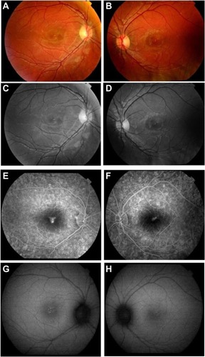
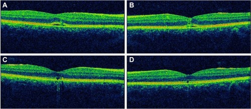
Imaging studies were done on presentation to our practice 18 hours after exposure to the laser device. Optical coherence tomography (3D OCT; Topcon, Tokyo, Japan) of both eyes showed disruption of the outer retinal layer with nonspecific retinal thickening ( and ); red-free photographs demonstrated hypopigmented foveal dots bilaterally ( and ); fluorescein angiography showed early foveal hyperfluorescence in both eyes with late ill-defined leakage ( and ); and autofluorescence images showed heterogeneous hyperfluorescence in the macula of both eyes ( and ). Finally, a computerized 10–2 visual field threshold test (Humphrey Automated Perimeter; Humphrey Instruments, San Leandro, CA, USA), showed small pericentral scotomata in the right eye and a normal field in the left eye.
Figure 2 Optical coherence tomography. (A and B) Disruption of the outer retinal layers with nonspecific retinal thickening in both eyes at presentation. (C and D) Disruption of the inner segment/outer segment junction in both eyes, at 3 months after presentation.
Note: A and C are right eye; B and D are left eye.
The patient was treated with an oral steroid (Prednisone; H.J. Harkins Company, Inc., Grover Beach, CA, USA) 1 mg/kg for 4 weeks then tapered over 2 months. At 3 months, visual acuity remained impaired but improved to 20/30 in the right eye and 20/25 in the left eye.
At 3 months, optical coherence tomography showed improvement of the retinal thickening in both eyes. The hyperreflective line representing the inner segment/outer segment junction was disrupted in the right eye and the left eye ( and ). The visual field improved and no scotoma was detected in the right eye.
Discussion
Laser pointer devices are a common and essential part of everyday life. This may lead to an increasing number of exposures to this type of laser device. However, there is debate about the ocular risks posed by inadvertent exposure to standard laser pointer devices, with the presence of an actual laser-induced injury often inconclusive or entirely absent in some studies.Citation1–Citation3 Literature supporting laser pointer-induced retinal injury has been limited to only a few articles on class 3A red laser pointers, and even less literature exists on the retinal hazards of class 3A green laser pointers. In the literature, retinal lesions induced by laser pointers (both green and red devices) include foveal granularity, perifoveal drusenoid-like deposits, or foveal ring-shaped hypopigmented lesions, subretinal hemorrhage, vitreous or chorioretinal hemorrhage, retinal edema, scars in the pigment epithelium, and rarely choroidal neovascularization.Citation4 Comparison of the retinal findings in our patient with those of other reported cases in the literature caused specifically by green lasers are listed in .
Table 1 Reported cases in the literature of retinal damage caused specifically by green lasers
According to Barkana and Belkin, several factors contribute to laser-related retinal damage, and these can be divided into two categories, ie, laser-related factors (wavelength of the radiation; pulse duration; and energy level of the beam) and patient-related factors (size of the pupil, with injury being more severe in larger pupil sizes; degree of retinal pigmentation, with dark-skinned individuals suffering more severe injury than light-skinned ones; proximity of incident beam to the fovea; and refraction status, with damage being more severe in emmetropic eyes due to the laser beam being more focused on the retina).Citation5 Also, experimental studies that have evaluated the clinical and histopathologic effects of laser pointers in eyes undergoing enucleation for melanoma concluded that green laser pointers (490–575 nm) are more damaging to the retina than red laser pointers (635–750 nm).Citation1,Citation6
Laser-induced damage to the retina is even more concerning in children and infants than in adults. Whereas adults terminate accidental laser pointer exposure in less than 0.25 seconds by pupil, blink, and aversion responses,Citation7 children have been reported to display “unusual” behavior, ie, staring for a prolonged period of time into the laser beam without blinking or averting the eye.Citation8
Current medical therapy for retinal injury is mainly limited to corticosteroids on an undetermined regimen and has variable results.Citation5 Final visual acuity ranges from 20/20 to 20/60 vision, and this depends on the size and location of the macular lesion.Citation4
In recent years, cheap laser pointers are increasingly being used as toys for children. While shrinking in size, handheld laser pointers are becoming increasingly more powerful, and safety is becoming a public health issue.Citation9
The potential vision-threatening hazards caused by mishandling laser pointers, even class 3A lasers, emphasize the importance of cautious and appropriate use of these devices. Recommendations regarding the purchase and use of these devices are being reconsidered. Further restrictions on their sale and use by the general public will require more than simple recommendations; legislation will have to be passed and enforced by governmental bodies.
Disclosure
The study did not receive any external funding. None of the authors has any proprietary, commercial, or financial interest in any of the products mentioned in this paper.
References
- RobertsonDMLimTHSalomaoDRLinkTPRoweRLMcLarenJWLaser pointers and the human eye: a clinicopathologic studyArch Ophthalmol2000118121686169111115266
- MensahEVafidisGMarshallJLaser pointers: the facts, media hype, and hysteriaLancet1998351911112919643781
- Van NorrenDKeunenJEVosJJThe laser pointer: no demonstrated danger to the eyesNed Tijdschr Geneeskd19981423619791982 Dutch9856195
- TurakaKBryanJSGordonAJReddyRKwongHMJrSellCHLaser pointer induced macular damage: case report and mini reviewInt Ophthalmol201232329329722466425
- BarkanaYBelkinMLaser eye injuriesSurv Ophthalmol200044645947810906379
- RobertsonDMMcLarenJWSalomaoDRLinkTPRetinopathy from a green laser pointer: a clinicopathologic studyArch Ophthalmol2005123562963315883281
- MainsterMATimberlakeGTWarrenKASlineyDHPointers on laser pointersOphthalmology19971048121312149261305
- FujinamiKYokoiTHiraokaMNishinaSAzumaNChoroidal neovascularization in a child following laser pointer-induced macular injuryJpn J Ophthalmol201054663163321191730
- Silver SpringConsumer health information: illuminating the hazards of powerful laser productsFood and Drug Administration2009 Available from: http://www.fda.gov/ForConsumers/ConsumerUpdates/ucm166649.htmAccessed September 22, 2013
- WyrschSBaenningerPBSchmidMKRetinal injuries from a handheld laser pointerN Engl J Med2010363111089109120825327
- ZiahosseiniKDorisJPTurnerGSLaser eye injuries. Maculopathy from handheld green diode laser pointerBMJ2010340c298220530564
- HosseinMBonyadiJSoheilianRSoheilianMPeymanGASD-OCT features of laser pointer maculopathy before and after systemic corticosteroid therapyOphthalmic Surg Lasers Imaging Off J Int Soc Imaging Eye201142135e138
- PollithySAchTSchaalKBDithmarSAcute bilateral impaired vision with central scotoma in an 11-year-old boyOphthalmol Z Dtsch Ophthalmol Ges20121099907910 German